
Zelotibia, Russell-Smith & Murphy, 2005
|
publication ID |
https://doi.org/10.3897/zookeys.13.145 |
|
publication LSID |
lsid:zoobank.org:pub:060B6F4D-830F-443E-8E5A-F9DE9ECB7289 |
|
DOI |
https://doi.org/10.5281/zenodo.3792136 |
|
persistent identifier |
https://treatment.plazi.org/id/3B2B8794-2C15-1925-FF64-8F09A3578C0D |
|
treatment provided by |
Plazi (2020-04-27 14:51:37, last updated 2021-10-29 03:02:17) |
|
scientific name |
Zelotibia |
| status |
|
Key to the species of Zelotibia
(illustrations modified from Russell-Smith & Murphy, 2005)
Males:
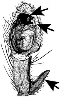
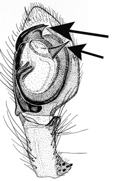
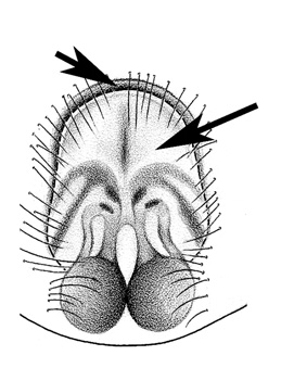
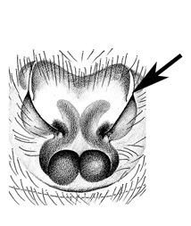
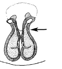
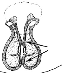
1 Male palpal femur clearly curved (A2); retrolateral margin of cymbium with triangular extension (A1) View Figure ............................................................................ 2
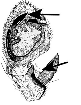
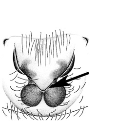
– Male palpal femur straight (B1), retrolateral margin of cymbium straight or slightly curved (except in Z. dolabra ) (B2) View Figure ................................................. 10
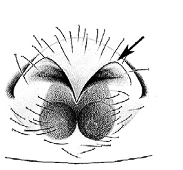
2 Tibial apophysis of palp originating on basal part of tibia, scoop-shaped in lateral view, reflexed forward so that it lies parallel to the long axis of the tibia. (A3) ............................................................................................................ 3
– Tibial apophysis not scoop-shaped in lateral view and originating midway of tibia (B3) View Figure .................................................................................................... 8
3 Tibial apophysis bifid, with two strong prongs at tip (C1) View Figure ........... Z. lejeunei
– Tibial apophysis not bifid (D2) View Figure ................................................................... 4
4 Embolus broad, bifid or indented at tip (D1) ............................................. 5
– Embolus tapered to sharp tip (E1) View Figure .............................................................. 6
5 Tibial apophysis almost straight as seen from below (D2); embolus flat, with shallow indentation at tip (D1)...................................................... Z. kaibos
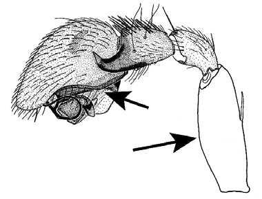
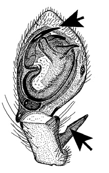
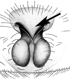
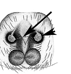
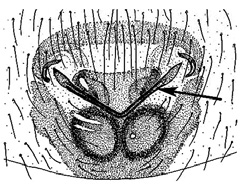
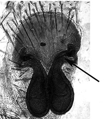
– Tibial apophysis slightly curved outward as seen from below (F1); embolus twisted with deep indentation, appearing bifid (F2) View Figure ............... Z. curvifemur
6 Extremity of embolus beak-shaped (G1); median apophysis small (G2) View Figure ........ ..................................................................................................... Z. mitella
– Extremity of embolus sharp (E1); median apophysis large (E2) or with two prongs (H2)................................................................................................ 7
7 Tegulum with semitransparent distal extension (H1); median apophysis with two prongs (H2) View Figure ............................................................................. Z. major
– Tegulum without distal extension; median apophysis occupying half the bulbus (E2)...................................................................................... Z. simpula
8 Embolus blade shaped in ventral view (I2) View Figure .................................... Z. cultella
– Embolus thin and needle-shaped (K2) View Figure ........................................................ 9
9 Tibial apophysis with minute basal tooth (I1)............................... Z. acicula
– Tibial apophysis without such a basal tooth (K1) View Figure ......................... Z. scobina
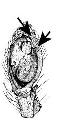
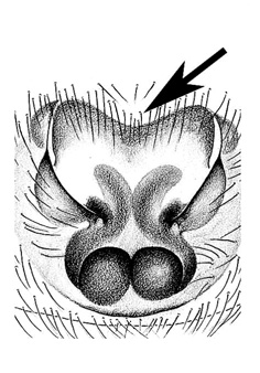
10 Tibial apophysis of palp, viewed laterally, large and robust, the tip reflexed dorsally through 90 o (L1) View Figure ............................................................ Z. flexuosa
– Tibial apophysis of palp otherwise ............................................................ 11
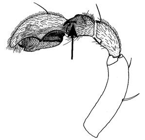
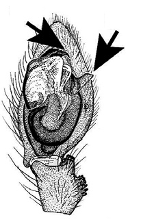
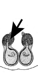
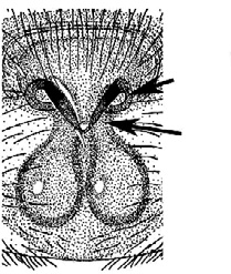
11 Tibial apophysis of palp small and semi-translucent in lateral view (M1). Palpal tibia with a group of small dark papillae on a boss behind apophysis (M2) View Figure ......................................................................................................... 13
– Palpal tibia lacking a group of dark papillae on a boss............................... 12
12 Tibial apophysis of palp minute, triangular (J1), the tip slightly downcurved View Figure .................................................................................................. Z. filiformis
– Palpal tibia with two large apophyses (retro-lateral and pro-lateral) (N1) View Figure ...... ................................................................................................ Z. bicornuta
13 MA in ventral view clearly truncate at tip (Q1, P1) View Figure .................................. 14
– MA in ventral view sharply pointed at tip (Q1) View Figure ........................................ 15
14 In ventral view, MA almost twice as long as broad (O1), tip of embolus smoothly curved.(O2)................................................................ Z. papillata
– In ventral view, MA only slightly longer than broad (P1), tip of embolus sinuous (P2).................................................................................... Z. supercilia
15 In ventral view, embolus narrow, tip undivided (Q1) View Figure ........ Z. paucipapillata
– In ventral view, embolus broad, the tip divided ( R 1) View Figure .................... Z. dolabra
Females:
1 Epigyne with large hood-shaped atrium anteriorly, lacking a clearly defined scape (a1) View Figure .................................................................................................... 2
– Epigyne without a clearly defined atrium and with a scape (a2) View Figure .................. 7
2 With area of ridged cuticle anterior to epigyne (a3) View Figure .................... Z. scobina
– Without area of ridged cuticle anterior to epigyne ..................................... 3
3 Anterior margin of atrium broadly rounded (a4)......................................... 4
– Anterior margin incurved (a5) View Figure ....................................................................6
4 Copulatory ducts reflexed outwards at anterior end and opening at base of atrium (b1) View Figure ................................................................................................. 5
– Copulatory ducts not reflexed outwards at anterior end, openings surrounded by blackish area (b2) View Figure ................................................................... Z. simpula
5 Copulatory ducts at frontal curve with short diverticulum (b3) View Figure .................... .............................................................................................. Z. curvifemur
– Copulatory ducts at frontal curve without diverticulum ............... Z. mitella
6 Atrium heart-shaped (a5); copulatory openings long lateral slits (b5) View Figure ............ ................................................................................................... Z. angelica
– Atrium kidney-shaped (b6); copulatory openings oval (b7) View Figure ........................... ................................................................................................... Z. subsessa
7 Epigynal scape tongue-shaped, the tip smoothly rounded (c1) View Figure .... Z. bicornuta
– Epigynal scape not tongue-shaped, the tip pointed (a2) .............................. 8
8 Epigynal scape large, reaching spermathecae (c2) View Figure ......................... Z. lejeunei
– Epigynal scape much smaller, not overhanging part of spermathecae (a2)..... 9
9 Copulatory openings large (c3), connected by transverse groove (c4) View Figure ............ ....................................................................................................... Z. major
– Copulatory openings smaller, not connected by groove............................. 10
10 Spermathecae globular (c5), copulatory ducts S-shaped (c6) View Figure ..................... 11
– Spermathecae oval or flask-shaped (d1) View Figure ..................................................... 12
11 Posterior margin of epigynal scape with sinuous sides (d2 View Figure ); copulatory ducts strongly curved (c6) ..................................................................... Z. fosseyae
– Posterior margin of epigynal scape with straight sides (d3) View Figure ; copulatory ducts less strongly curved (d4) View Figure ............................................................. Z. flexuosa
12 Epigynal scape a narrow small triangle; copulatory ducts longer than spermathecae (e1) View Figure ................................................................................ Z. kibira
– Epigynal scape a larger triangle; copulatory ducts shorter than spermathecae (e2) View Figure ........................................................................................................... 13
13 Tip of epigynal scape forming an obtuse angle, spermathecae located imme- diately posterior to tip of scape (e3) View Figure .......................................... Z. supercilia
– Tip of epigynal scape forming an acute angle (e4) View Figure ..................................... 14
14 Spermathecae not touching (f1) View Figure ................................................. Z. papillata
– Spermathecae adjacent (f2) View Figure ...................................................................... 15
15 Convex median part of spermathecae touching (f2).................................. 16
– Spermathecae touching in front and behind concave median part (f3) View Figure .......... ......................................................................................... Z. paucipapillata
16 Copulatory ducts curved at right angle between copulatory opening and spermathecae..................................................................................... Z. kanama
– Curve of copulatory ducts more obtuse (f5) View Figure .............................................. 17
17 Curve delimiting copulatory openings at base of scape narrow (e5)............... .................................................................................................. Z. filiformis
– Curve delimiting copulatory openings at base of scape wide (f5) View Figure ................... .................................................................................................. Z. johntony
Russell-Smith A, Murphy JA (2005) Zelotibia, a new zelotine spider genus from central Africa (Araneae, Gnaphosidae). Journal of Afrotropical Zoology 2: 103 - 122.

Male palpal femur clearly curved (A2); retrolateral margin of cymbium with triangular extension (A1)
Male palpal femur straight (B1), retrolateral margin of cymbium straight or slightly curved (except in Z. dolabra ) (B2)


Tibial apophysis slightly curved outward as seen from below (F1); embolus twisted with deep indentation, appearing bifid (F2)


Tibial apophysis of palp, viewed laterally, large and robust, the tip reflexed dorsally through 90 o (L1)

Tibial apophysis of palp small and semi-translucent in lateral view (M1). Palpal tibia with a group of small dark papillae on a boss behind apophysis (M2)


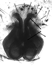
Tip of epigynal scape forming an obtuse angle, spermathecae located imme- diately posterior to tip of scape (e3)


| R |
Departamento de Geologia, Universidad de Chile |
No known copyright restrictions apply. See Agosti, D., Egloff, W., 2009. Taxonomic information exchange and copyright: the Plazi approach. BMC Research Notes 2009, 2:53 for further explanation.
|
Kingdom |
|
|
Phylum |
|
|
Class |
|
|
Order |
|
|
Family |
1 (by plazi, 2020-04-27 14:51:37)
2 (by felipe, 2020-05-07 14:50:39)
3 (by felipe, 2020-05-07 20:00:55)
4 (by felipe, 2020-05-07 20:17:03)
5 (by felipe, 2020-05-07 20:46:55)
6 (by admin, 2020-05-08 00:12:05)
7 (by ExternalLinkService, 2020-05-08 00:21:56)
8 (by ExternalLinkService, 2021-10-29 00:24:42)
9 (by ExternalLinkService, 2021-10-29 03:02:17)
10 (by plazi, 2023-10-31 10:02:41)