
Speleonectes emersoni, Lorentzen, Dörte, Koenemann, Stefan & Iliffe, Thomas M., 2007
|
publication ID |
https://doi.org/ 10.5281/zenodo.177940 |
|
DOI |
https://doi.org/10.5281/zenodo.6250529 |
|
persistent identifier |
https://treatment.plazi.org/id/03FA8780-7857-F62E-21EB-FD82FD6ED8A1 |
|
treatment provided by |
Plazi |
|
scientific name |
Speleonectes emersoni |
| status |
sp. nov. |
Speleonectes emersoni new species
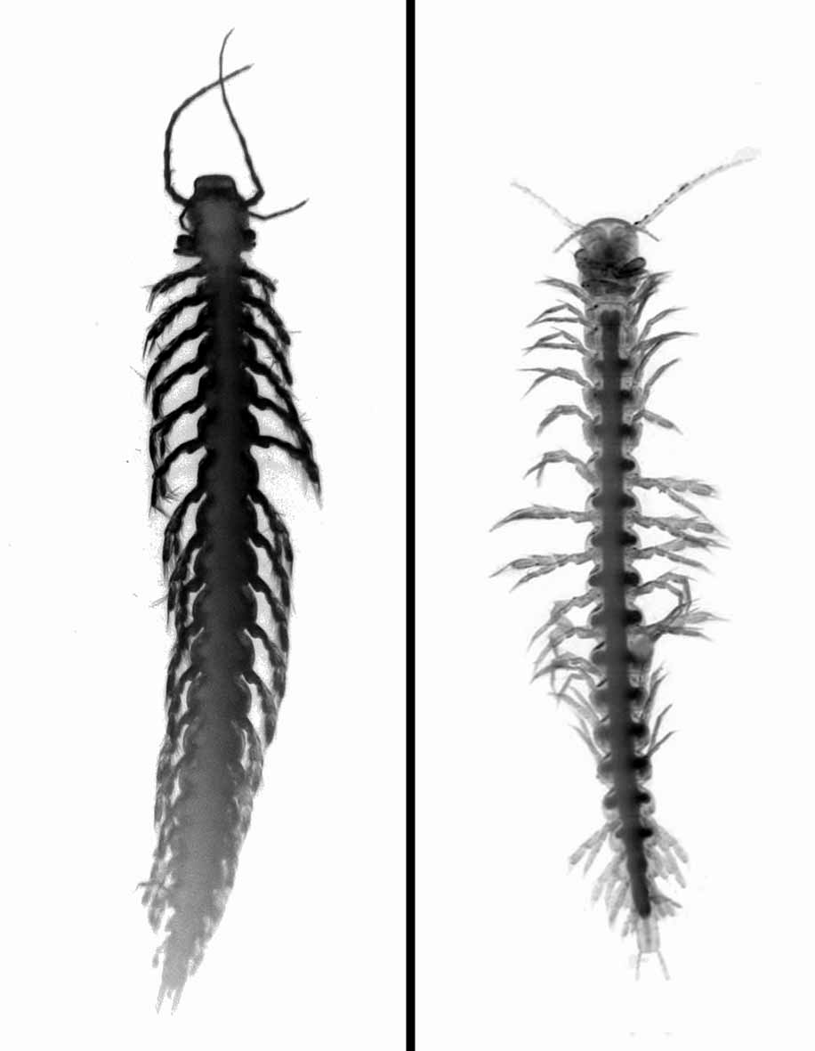
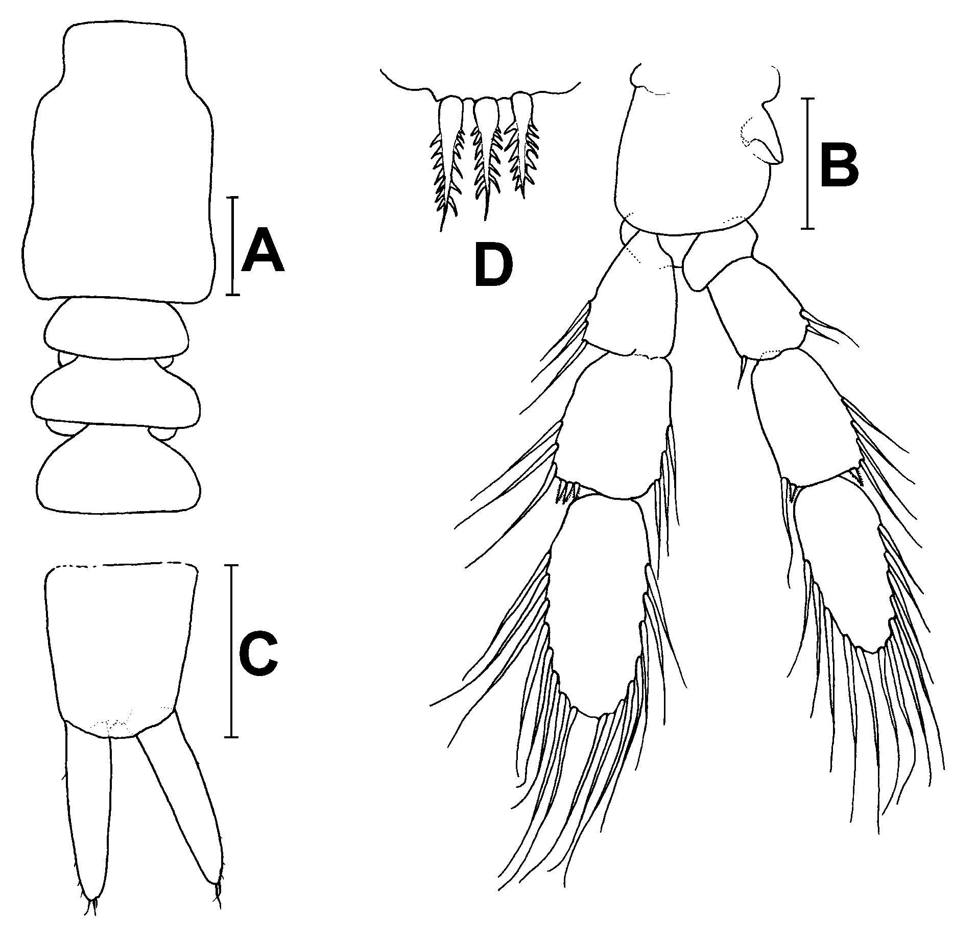
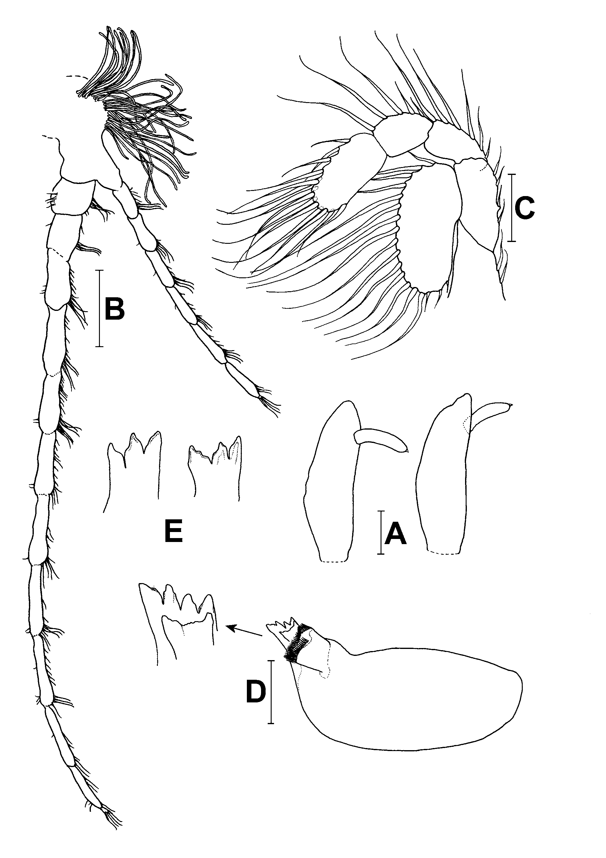
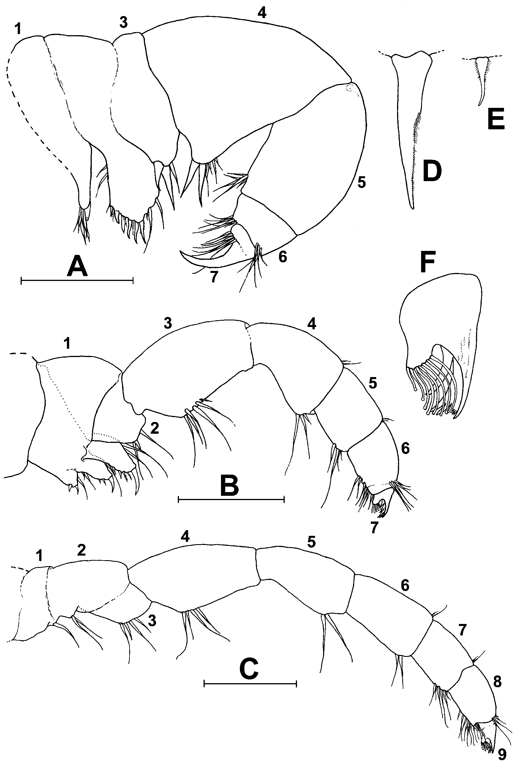
( Figs. 1–4 View FIGURE 1 View FIGURE 2 View FIGURE 3 View FIGURE 4 )
Type locality. Cueva Los Jardines Orientales, south coast near Santo Domingo, Dominican Republic.
Material examined. Holotype (deposited at Biozentrum Grindel und Zoologisches Museum, Hamburg; catalogue number K - 41270) (12.5 mm, 21 trunk segments), five paratypes (9.5 mm, 19 trunk segments; 10.5 mm, 20 trunk segments; 11 mm, 20 trunk segments; 11 mm, 20 trunk segments; 11.5 mm, 20 trunk segments; all in research collection of SK) from Cueva Los Jardines Orientales. One paratype (10 mm, 20 trunk segments) from Cueva Taina. Specimens were collected on May 1, 2005 (Cueva Taina) and May 5, 2005 (Cueva Los Jardines Orientales) at or below the halocline in 15–37 m depth by Thomas Iliffe. The holotype and one paratype (whole specimens) are preserved in alcohol; two paratypes were dissected and preserved in glycerine; three paratypes were used for molecular analyses.
Etymology. The epithet emersoni is in honor of the biologist Michael James Emerson (8 May 1954 – 22 March 1990). Michael worked as a research assistant on the “Remipede Project” at the San Diego Natural History Museum from 1982 to 1990. His experience as a skillful scientific illustrator and attentive observer resulted in several publications that set benchmarks in the systematics of Remipedia.
Diagnosis. A small-sized, slender species, up to 12.5 mm, adult specimens composed of 19–21 segments; pleural tergites weakly developed, with rounded lateral margins in anterior part of trunk, becoming slightly pointed in posterior trunk; dorsal ramus of antennule 11-segmented, ventral ramus with 6 segments; arcshaped, horseshoe-like claw of maxilla and maxilliped composed of 7–10 denticles; anal segment longer than wide; caudal rami approximately as long as anal segment.
Description. Based on holotype and paratypes. Body slender with a maximum length of 12.5 mm and 21 trunk segments ( Fig. 1 View FIGURE 1 ). Pleural tergites narrow, with rounded distolateral corners on trunk segments 1–7 ( Figs. 1 View FIGURE 1 , 2 View FIGURE 2 A), becoming slightly acuminate in posterior part of trunk, tergite on first trunk segment reduced. Sternal bars sublinear and isomorphic. Male gonopores on trunk limb 14 cylindrical, with rounded pointed lobes ( Fig. 2 View FIGURE 2 B). Head shield subrectangular, tapering at anterior end, approximately as long as trunk segments 1–3 ( Fig. 2 View FIGURE 2 A). Frontal filaments with relatively short distomedial processes, extending beyond tip of main filament ( Fig. 3 View FIGURE 3 A).
Antennule ( Fig. 3 View FIGURE 3 B): Peduncle with small bulbus bearing relatively few aesthetascs. Dorsal flagellum 11- segmented, almost twice as long as head shield, reaching 9% of length of body. Ventral flagellum with 6 segments, nearly half as long as dorsal flagellum, as long as head shield.
Antenna ( Fig. 3 View FIGURE 3 C): Proximal segment of protopod with 2 marginal setae; distal segment with 4 marginal setae. Exopod longer and wider than adjacent distal segment of protopod, bearing 18–20 long setae. Endopod bent in a semicircular arc; first two proximal segments with 5–6 and 4–5 setae, respectively; distal segment with about 15 setae arranged in two rows composed of 9 and 5–6 setae. All setae faintly plumose.
Mandible ( Fig. 3 View FIGURE 3 D, E): Right incisor process and lacinia mobilis with three large denticles. Left incisor process with four large denticles; left lacinia mobilis crescent-shaped, apical margin serrate. Molar process prominent.
Maxillule ( Fig. 4 View FIGURE 4 A, D, E): Segment 1 with slender endite, distal margin terminating in 4 long stout setae (one of which prominent) and about 3 simple setae. Endite of second segment long and broad, with 2 setae at inner proximal margin; 6 stout, relatively large setae (all of which simple, except one setulose seta ( Fig. 4 View FIGURE 4 E)) and 5–6 fine setae arranged in a second row on distomedial and -lateral margins and two long setae on distolateral margin. Segment 3 short; endite tapering, slightly rounded, bearing 2 long, robust setulose setae and 1 simple seta. Segment 4 (lacertus) subtriangular, with obliquely expanded medial margin; proximal corner bearing 2 long, robust setae ( Fig. 4 View FIGURE 4 D) and 4–5 simple setae. Segment 5 as long as lacertus, but narrower, with a cluster of distomedial setae. Sixth segment equipped with separate clusters of long and shorter setae on distal margins. Claw slender, well-developed ( Fig. 4 View FIGURE 4 A).
Maxilla ( Fig. 4 View FIGURE 4 B): First endite of segment 1 bearing 2 short apical stout setae. Endites 2 and 3 of first segment each with 1 prominent apical stout seta accompanied by 2–4 short setae on subapical margins. Endite of segment 2 equipped with a single, short stout seta and a few long setae. Segment 3 (lacertus) slightly pearshaped, medial margin bearing 4–6 long and short setae. Segment 4 shorter than segment 3; distomedial margin rounded, with a cluster of 2–3 setae. Segment 5 shorter than segment 4, subquadrangular; distomedial corner with a cluster of 5 long and shorter setae. Segment 6 nearly as long as segment 5, with a row of about 7 setae along distomedial margin and a row of about 6 setae on distoposterior margin. Arc-shaped, horseshoetype claw serrated, composed of 7–10 small denticles flanked by 2 stronger, separate denticles. All setae simple.
Maxilliped ( Fig. 4 View FIGURE 4 C, F): Distinctly longer than maxillule, slender. Segments 1 and 2 bearing a few long medial setae. Segment 3 rather long, with 4 long setae. Segments 4–6 gradually decreasing in length; fourth segment with a row of 4 medial setae. Segment 5 with distomedial margin slightly expanded bearing 2 long setae. Segment 6 bearing 2 setae on distomedial margin. Segment 7 subquadrangular, with a row of about 6 long and shorter setae at distomedial margin. Eighth segment approximately as long as segment 7, equipped with a row of about 13 setae at distomedial margin and 7 short setae on distolateral margin. Claw subequal to that of maxilla ( Fig. 4 View FIGURE 4 F). All setae simple.
Trunk appendages ( Fig. 2 View FIGURE 2 B, D): Segment 1 of exopod equipped with 3–6 long setae and up to 2 serrate stout setae on distolateral corner. Segment 2 with setae on lateral and medial margins, and 3–4 serrate stout setae on distolateral corner. Segment 3 ovate, bearing 14–17 long marginal setae. Endopod slightly shorter and narrower than exopod; distribution of long plumose setae like those on exopod, with the following exceptions: basal segment naked; segment 3 with 2 serrate stout setae on distolateral corner and 1 serrate stout setae on distomedial corner; segment 4 with 10–12 setae ( Fig. 2 View FIGURE 2 B). All setae plumose. Anal segment ( Fig. 2 View FIGURE 2 C): 1.2 times longer than wide; caudal rami about 1.1 times longer than anal segment, bearing a few very fine setae along margins and about 3 fine, apical setae.
No known copyright restrictions apply. See Agosti, D., Egloff, W., 2009. Taxonomic information exchange and copyright: the Plazi approach. BMC Research Notes 2009, 2:53 for further explanation.