
Rhynchothorax swir, Staples, 2019
|
publication ID |
https://doi.org/ 10.11646/zootaxa.4567.3.1 |
|
publication LSID |
lsid:zoobank.org:pub:0AEFAF80-B001-4A18-88AC-5B6A189F6DCD |
|
DOI |
https://doi.org/10.5281/zenodo.5944900 |
|
persistent identifier |
https://treatment.plazi.org/id/03895C33-2909-4F1C-FF01-FE0AFBA2FF28 |
|
treatment provided by |
Plazi |
|
scientific name |
Rhynchothorax swir |
| status |
sp. nov. |
Rhynchothorax swir View in CoL sp. nov.
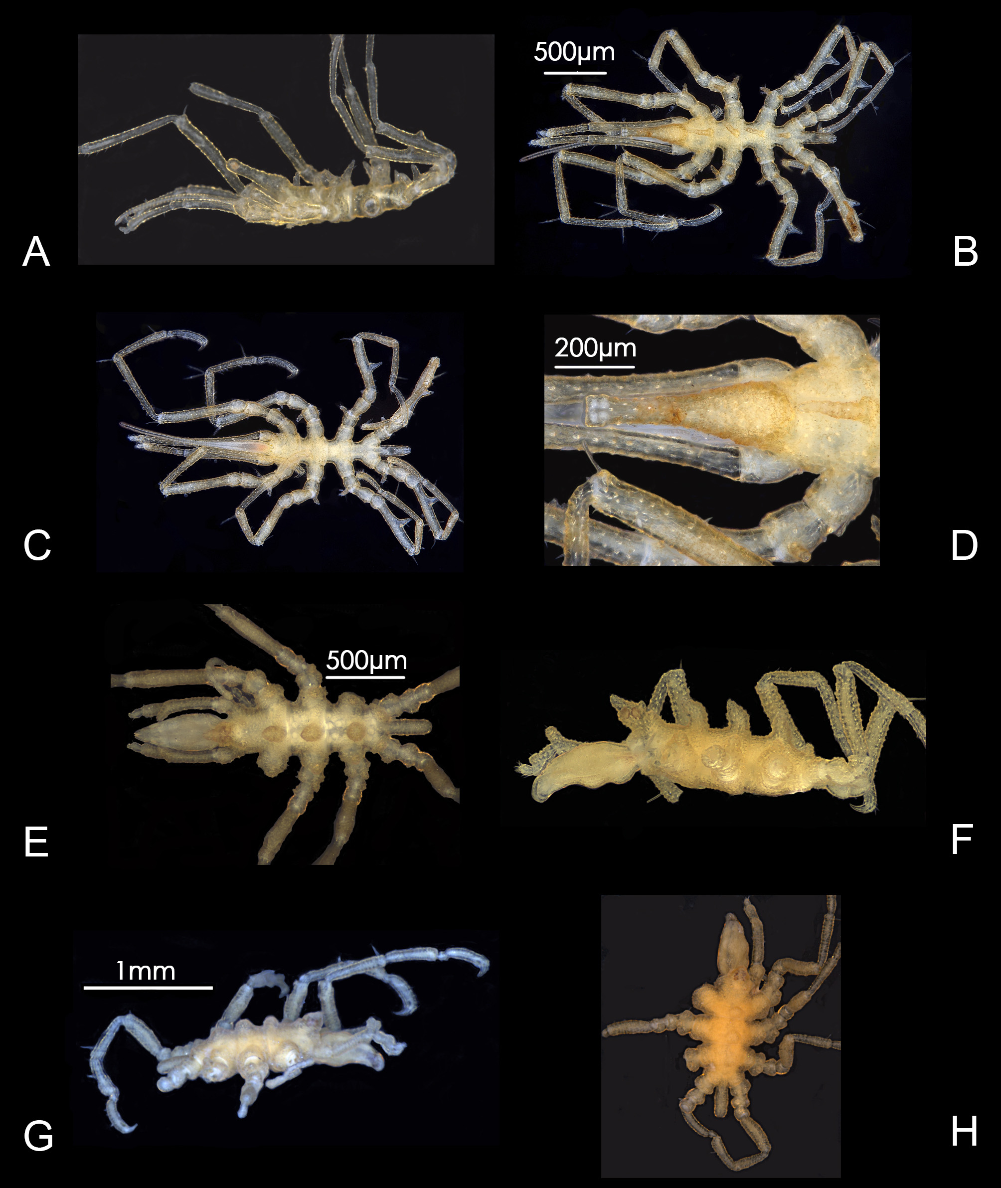
Figure 13 View FIGURE 13 A–L; Plate 4E, F View PLATE 4
Material examined. Holotype, female (gravid) ( NHMUK 2018.27 View Materials ), Southwest Indian Ocean, Coral Seamount, 41° 21.02´S, 42° 55.15´E, ROV, 1117 m, specimen JC066-143, stn 4.9, rubble, 14 November 2011. GoogleMaps
Paratypes. One female (gravid.), ( NHMUK 2018.28 View Materials ), Southwest Indian Ocean, Melville Bank, 38° 29.89´S 46° 43.39´E, HYBIS, 905 m, specimen JC066-2715, stn 5.18, coral rubble/pebbles, 25 November 2011. Southwest Indian Ocean, three females (gravid), one dissected, two specimens of undetermined sex ( NHMUK 2018.29 View Materials ), Coral Seamount, 41° 22.31´S, 42° 54.57´E, ROV, 732 m, specimen JC066-1296D, stn 4.38, mooring site, on Mango wood net, 20 November 2011. Left palp, right and left ovigers mounted in Euparal on glass slides GoogleMaps .
Other material. One specimen (NHMUK 2018.30), Southwest Indian Ocean, Middle of What Seamount, 37° 57.92´S, 50° 24.43´E, ROV net, 1008 m, specimen JC066-2743, stn 6.4, coral rubble and sediment, 1 December 2011. One female (gravid), one undetermined sex ( NHMUK 2018.31 View Materials ), Southwest Indian Ocean, Coral Seamount, 41° 21.03´S, 42 55.15´E, ROV net, 1100 m, specimen JC066-4313, stn 4.9, coral framework/rubble, 14 November 2011 GoogleMaps . One specimen ( NHMUK 2018.32 View Materials ), Southwest Indian Ocean, Middle of What Seamount, 37˚ 56.795'S, 50˚ 27.240'E, ROV, 1414 m, specimen JC066-004, 20 November 2011 . One specimen (juvenile?) ( NHMUK 2018.33 View Materials ), Southwest Indian Ocean, Atlantis Bank , 32° 42.86´S, 57° 16.34´E, mooring site, on net containing Mango wood, 750 m, specimen JC066-4213C, stn 8.29, 14 December 2011 GoogleMaps .
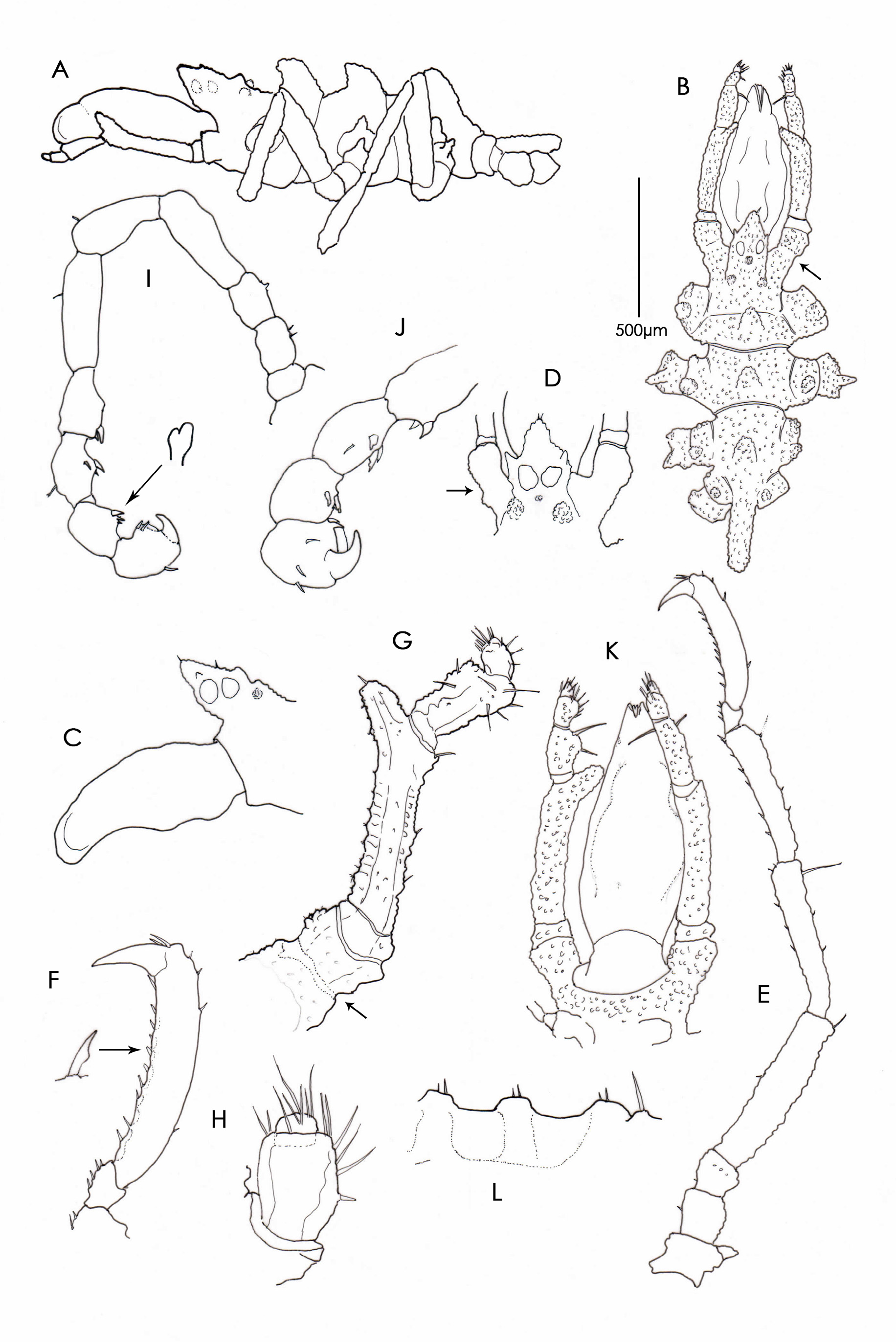
Description. Female holotype. Trunk ( Fig.13A, B View FIGURE 13 . Plate 4E,F View PLATE 4 ) and leg surfaces papillose, granular, papillae tipped with two to three tiny setae, papillae interspaced randomly by a stronger seta ( Fig. 13L View FIGURE 13 ); first two segmentation lines complete, third lacking, segments 1 to 3 each with prominent conical mid-dorsal process curved forward, anterior margin of cephalon without projections, cephalic lateral extensions clearly separated from, and diverging away from, the first lateral processes, divided at mid-length by shallow constriction giving appearance of segmentation, dorsodistal surface raised, rugose; lateral processes narrowly separated and shorter than basal width, each process with distal transverse granular rim and prominent conical dorsodistal tubercle. Ocular tubercle ( Figs 13C, D View FIGURE 13 ) close to anterior margin of cephalon, over-reaching proboscis base, conical dorsal part about equal to height of basal part but variable, terminal spinule present, broad basal part bearing two raised lateral sense organs and a single short process on the median posterior surface about level with bottom of eyes. Two granular processes on either side near the base; four eyes, pigmented.
Proboscis surface smooth, glabrous, with two dorsolateral bulges at about mid-length and two lesser, more distal dorsolateral bulges, ventral surface with strong bulge in mid-region and with a similar more proximal bulge most evident when the proboscis is extended ( Fig. 13C View FIGURE 13 ); distal part strongly down-curved, jaws vertically bilateral, with inner and outer antimeres, oral surface extending almost entire length of down-curved portion. Arthrodial membrane between proboscis base and the cephalon broad when extended ( Fig. 13K View FIGURE 13 ).
Abdomen unarticulated at base, horizontal, cylindrical, with blunt tip, about 3.5 times as long as wide, reaching slightly beyond distal margin of coxa 2, cleft distally with anal opening extending partially on to ventral surface.
Chelifores absent.
Palp ( Fig. 13G View FIGURE 13 ) five-segmented, segments 1and 5 very short, segment two longest, curvature corresponding to the lateral surface of the proboscis, with tall thumb-like dorsodistal process inclined upward and inward, segment 3 about half length of segment 2 and 2.5 times longer than segment 4. Segment 3 expanded dorsodistally, tuberculose, with few spines and setae, single prominent inwardly curved spine on distal margin reaching to oral antimeres, segment 5 at most one–quarter length of segment 4, but may be tiny, button-like or not evident ( Fig. 13H View FIGURE 13 ). Segments 3 to 5 with strong setae.
Oviger ( Fig. 13I, J View FIGURE 13 ) ten-segmented, segment 6 longest, segment 4 longer than segment 5, segment 5 widening distally, segment ten swollen, distal margin with lamina-like blade preceded by two simple spines at base, spines on segments 7 to 9 curved forward, simple, bi-dentate or tri-dentate, spine formula unclear but appears to be 3:3:3:2, terminal claw strong, curved, closing against lamina.
Third leg ( Fig. 13E View FIGURE 13 ) slender, first coxae legs 1 to 3 with single pronounced dorsodistal tubercle flanked by granular rim, tubercle height about twice own width, tubercle absent on fourth pair of legs. Coxa 2 longest, femur wider than tibiae, longer than combined length of coxae 1 to 3 and longer than tibia 1, tibia 2 shorter than tibia 1, longer segments each with long dorsodistal seta, tarsus short, less than one-sixth propodus length, with four to six ventral spines, propodal heel absent, sole gently and evenly curved, mostly with nine spines, several spines with node on inner margin ( Fig. 13F View FIGURE 13 ); terminal claw about one-third length of propodus, auxiliary claws almost onethird length of main claw. Gonopores not evident.
Measurements of holotype (mm). Length trunk (frontal margin of cephalic segment to tip of 4 th lateral processes), 1.008; length cephalon, 0.384; trunk width across second lateral processes, 0.520; proboscis length (ventral), 0.664; greatest diameter proboscis (ventral), 0.232; abdomen length, 0.280; lateral cephalic extension, 0.152. Palp: seg. 1, 0.032; seg. 2, 0.328; seg. 3, 0.144; seg. 4, 0.056; seg. 5, 0.024. Oviger: seg. 1,.032; seg. 2, 0.064; seg. 3, 0.056; seg. 4, 0.152; seg. 5, 0.096; seg. 6, 0.144; seg. 7, 0.096; seg. 8, 0.072; seg. 9, 0.072; seg. 10, 0.072; claw, 0.048. Third leg: coxa 1, 0.080; coxa 2, 0.120; coxa 3, 0.096; femur, 0.456; tibia 1, 0.428; tibia 2, 0.352; tarsus, 0.056; propodus, 0.328; claw, 0.112; aux claws, 0.032.
Etymology. Named for the Southwest Indian Ridge based on its acronym SWIR; a noun in apposition.
Remarks. This species is the most wide-ranging species in the collection being found on the southern-most and northern-most seamounts in the study area. The partial extension and withdrawal of the proboscis from an excavation of the cephalon is accompanied by wide arthrodial membrane. Withdrawal is seemingly limited by the ventral swellings on the proboscis and the size of the cephalon opening which forms a collar around the retracted proboscis. The proboscis has the appearance of being more spindle-shaped when extended from the cephalon ( Fig. 13K View FIGURE 13 ).
The ovigers are now mounted under glass cover slips and it is neither possible to get a clear understanding of the spine shapes or numbers.
Gravid females have developing oocytes in trunk only; none in legs. Oocytes 0.048 µm in diameter.
There is variation in the shape and height of the ocular tubercle whereby the apical cone is taller in some specimens and the mid-dorsal trunk tubercles also vary in shape; in one instance being almost molar-form.
There is a slight transverse constriction of the cephalic lateral extension in line with the anterior margin of the cephalon and in some specimens it is accompanied by a faint suture line ( Figs 13B, G, D View FIGURE 13 ). This is indicated by dotted lines in Fig. 13G View FIGURE 13 . This division is made more conspicuous by the absence of surrounding papillae found elsewhere on the integument. The strong inwardly curved spines on the fourth palp segments are capable of being in contact with the oral antimeres as illustrated by Zago (1970, Fig. 2 View FIGURE 2 ) in R. mediterraneus Costa, 1861 where presumably they assist with manipulation of food. The distal-most palp segment is variably developed and sometimes not evident. At most it appears to be one-quarter the length of the preceding segment but in some cases it forms a barely perceptible terminal dome or button. In three specimens this segment appears to be completely absent although there is no indication that the distal segments have fused or coalesced. Under strong back-lighting the base of segment 5 can sometimes be seen partially recessed into the distal margin of segment 4 which may explain the variation in its size and in some instances perhaps, its apparent absence ( Fig. 13H View FIGURE 13 ).
Several individuals have solitary ectoproct/kamptozoan-like zooids attached to the body surface similar to that on R. voxorinus illustrated by Stock (1966a, Fig. 3d View FIGURE 3 ) which he supposed may be a hydroid.
Using Clark’s (1976) key, this specimen can be followed to couplet 6 where it identifies as R. percivali from New Zealand waters with which it shares possession of auxiliary claws, similar mid-dorsal processes and welldeveloped rugose tubercles on all lateral processes. Rhynchothorax percivali differs in having a segmented abdomen, a characteristic long, slender anterior pointing extension on the ocular tubercle and very much taller digitiform tubercles on the first coxae. The proportions of the legs are additional points of difference.
Female specimens in this collection were principally identified by the presence of oocytes in their trunks. Gonopores were not evident. No males were identified.
Because of the risk of destroying the tiny holotype, a paratype from stn JC066-1296D was partly dissected for illustration of the palp and oviger.
Specimen JC066-4213C may be juvenile. It has a lower, ocular tubercle and lower mid-dorsal tubercles on the trunk and lateral processes compared to other specimens assigned to this species.


No known copyright restrictions apply. See Agosti, D., Egloff, W., 2009. Taxonomic information exchange and copyright: the Plazi approach. BMC Research Notes 2009, 2:53 for further explanation.
|
Kingdom |
|
|
Phylum |
|
|
Class |
|
|
Order |
|
|
Family |
|
|
Genus |