
Rhombophryne vaventy, Scherz, Mark D., Ruthensteiner, Bernhard, Vences, Miguel & Glaw, Frank, 2014
|
publication ID |
https://doi.org/ 10.11646/zootaxa.3860.6.3 |
|
publication LSID |
lsid:zoobank.org:pub:8119C95F-0BBB-4960-B06E-72725F2ECC74 |
|
DOI |
https://doi.org/10.5281/zenodo.6138352 |
|
persistent identifier |
https://treatment.plazi.org/id/03951971-361D-FFF5-FF5D-BBC2FAEF2B0D |
|
treatment provided by |
Plazi |
|
scientific name |
Rhombophryne vaventy |
| status |
sp. nov. |
Rhombophryne vaventy View in CoL sp. nov.
( Figs. 1–3 View FIGURE 1 View FIGURE 2 View FIGURE 3 )
Rhombophryne serratopalpebrosa —Glaw & Vences 2007: pp. 118–119, Fig. 3 View FIGURE 3 a. (depicts the paratype, FGZC 2842) Rhombophryne sp. 4— Wollenberg et al. 2008
Rhombophryne sp. 6— Vieites et al. 2009: Suppl. Material
Rhombophryne sp. aff. serratopalpebrosa —D’Cruze et al. 2010
Rhombophryne sp. Ca6 Marojejy—Perl et al. 2014: Suppl. Material
Holotype. ZSM 357/2005 ( FGZC 2876), adult male, collected by F. Glaw, M. Vences, R.D. Randrianiaina on 17 February 2005 at Marojejy National Park, “Camp Simpona”, 14°26.199'S, 49°44.601'E, 1326 m above sea level), northeastern Madagascar.
Paratype. UADBA uncatalogued ( FGZC 2842), probably adult, sex unknown, collected by F. Glaw, M. Vences, R.D. Randrianiaina on 16 February 2005 at Marojejy National Park, “Camp Simpona”, 14°26.199'S, 49°44.601'E, 1326 m above sea level), northeastern Madagascar. GenBank Accessions Numbers: KF611595 View Materials , EU341107 View Materials .
Diagnosis. A microhylid frog assigned to the genus Rhombophryne on the basis of molecular data (see discussion below). Currently there are no morphological characters to distinguish between Rhombophryne and Plethodontohyla ( Andreone et al. 2005; Wollenberg et al. 2008; Glaw et al. 2010). This species differs from all other Rhombophryne species and from all Plethodontohyla species by the combination of the following characters: large size, rough and tubercular dorsal skin, smooth ventral skin, fingers and toes without enlarged terminal discs, tibiotarsal articulation reaching beyond snout tip, second finger shorter than fourth, fifth toe shorter than third toe, a series of four soft superciliary spines, anterior-most two being most distinct, absence of dorsolateral folds, dark colouration beneath chin fading to lighter posteriorly, dorsal brown colouration with distinct black spots arranged almost symmetrically, a series of faint but distinct crossbands on the legs, stepped columellar footplates ( Fig. 2 View FIGURE 2 e), curved prevomers and vomerine teeth ( Fig. 2 View FIGURE 2 d), and nasals with a posterior but no anterior lateral process ( Fig. 3 View FIGURE 3 c). It differs from all other sequenced Malagasy microhylid frogs by a pairwise genetic divergence in its 16S rDNA sequence of ≥6.6%, being most closely related to R. sp. ‘Ambolokopatrika’ as discussed above ( Vieites et al. 2009).
Additionally, R. vaventy may be distinguished from its congeners by the following characteristics: from R. mangabensis by larger size (53 mm vs. 20–24 mm), tibiotarsal articulation reaching beyond the snout tip (vs. reaching the tympanum), and the presence of four superciliary spines (vs. absence); from R. alluaudi by tibiotarsal articulation reaching beyond the snout tip (vs. reaching the tympanum), and the presence of four superciliary spines (vs. absence); from R. testudo by larger size (53 mm vs. 33–45 mm), absence of barbels on the lower lip (vs. presence), tibiotarsal articulation reaching beyond the snout tip (vs. reaching the tympanum), and the presence of four superciliary spines (vs. absence); from R. coudreaui by larger size (53 mm vs. 28 mm), absence of webbing between digits (vs. traces of webbing), tibiotarsal articulation reaching beyond the snout tip (vs. reaching the tympanum), and the presence of four superciliary spines (vs. absence); from R. guentherpetersi by larger size (53 mm vs. 32–35 mm), absence of porous glandular formation in the latero-dorsal region (vs. presence), tibiotarsal articulation reaching beyond the snout tip (vs. reaching the eye), and the presence of four superciliary spines (vs. absence); from R. laevipes by larger size (53 mm vs. 45–47 mm), granular and tubercular dorsal skin (vs. smooth), tibiotarsal articulation reaching beyond the snout tip (vs. reaching the eye or nostril), and the presence of four superciliary spines (vs. absence); from R. minuta by larger size (53 mm vs. 16–22 mm), tibiotarsal articulation reaching beyond the snout tip (vs. reaching the eye), and the presence of four superciliary spines (vs. absence); from R. matavy by larger size (53 mm vs. 39–49 mm), tibiotarsal articulation reaching beyond the snout tip (vs. not reaching the insertion of the arms), and the presence of four superciliary spines (vs. absence); from R. coronata by much larger size (male SVL 53 mm vs. 21–23 mm), coarsely granular and tubercular dorsal skin (vs. slightly granular), presence of a distinct supratympanic fold (vs. indistinct supratympanic fold), smaller relative tympanum diameter (TDH/ED 41% vs. 51%), tibiotarsal articulation reaching beyond the snout tip (vs. reaching the tympanum), and longer relative tibia length (TIBL/SVL 56% vs. 39–41%).
Rhombophryne vaventy may be distinguished from R. serratopalpebrosa by larger size (53 mm vs. 28 mm), distinct supratympanic fold not extending beyond the anterior edge of the tympanum (vs. strong, almost straight supratympanic fold extending to the supraocular region), presence of almost symmetrical dark dorsal spots (vs. absence), and much smaller relative tympanum size (TDH/ED 41% vs. 78%). Furthermore, the following osteological differences were noticed ( Figs. 2 View FIGURE 2 and 3 View FIGURE 3 ): shape of the columellar footplate (stepped vs. domed), relative length of the prepollex (61% vs. 28% of first metacarpal), relative extension of the ilium (reaching the eighth presacral vertebra vs. reaching the seventh presacral vertebra), lateral processes of the nasal bone (just posterior vs. anterior and posterior), postchoanal prevomerine palate (curved vs. straight), the size order of the breadths of the vertebrae, measured from the tips of the transverse processes (L3 <L2 <L4 <L1 <T1 <S <T3 <T2 vs. L2 <L3 <L1 <L4 <T3 <T1 <S <T2; S = Sacrum), shape of the posterior vertebral articulations (slender vs. rounded), ossification of the sternum (present vs. absent), and the angle of the medial process of the pterygoid. However, due to the extremely low number of specimens available (one male of R. vaventy and one female of R. serratopalpebrosa ), we cannot exclude that some of these apparently diagnostic features are due to sexual dimorphism, individual variation, or differences in resolution of the CT scans.
Rhombophryne vaventy may be distinguished from all species of Plethodontohyla by the presence of superciliary spines (vs. absence), and from all Plethodontohyla except P. t uberata by its roughly tubercular skin (vs. smooth to granular skin). Additionally, it may be distinguished from P. notosticta , P. guentheri , P. mihanika , and P. inguinalis by the lack of enlarged terminal discs on its digits (vs. presence) and the lack of a sharp border between the dorsal and lateral colouration (vs. presence); from P. t ub e r a t a by larger size (53 mm vs. 35–45 mm), tibiotarsal articulation reaching beyond the snout tip (vs. the insertion of the arms); from P. ocellata by absence of large black spots bordered with white in the inguinal region (vs. presence), and tibiotarsal articulation reaching beyond the snout tip (vs. reaching the tympanum); from P. bipunctata by larger size (53 mm vs. 25–32 mm), and tibiotarsal articulation reaching beyond the snout tip (vs. reaching the insertion of the arm, the tympanum, or the eye); and from P. brevipes by larger size (53 mm vs. 36 mm), and tibiotarsal articulation reaching beyond the snout tip (vs. reaching the insertion of the arms).
Description of the holotype. Specimen in a very good state of preservation. A piece of muscle was taken from the right thigh as a tissue sample for genetic analyses. An incision was made in the right flank to check the sex and examine the contents of the intestines.
Body robust. Head wider than long. Pupil very small and roundish. Snout rounded in dorsal and lateral view. Canthus rostralis slightly concave. Loreal region slightly concave. Nostrils closer to tip of snout than to eyes; directed laterally; not protuberant. Eye-nostril distance smaller than the internarial distance. Tympanum distinct, rounded, 41% of eye diameter. A series of four short but distinct soft dermal spines present above each eye, not equally spaced; anterior two most distinct, the third almost imperceptible. Distinct supratympanic fold, curved over and behind the tympanum, not extending anteriorly beyond the tympanum. Vomerine teeth present, curving posteriorly toward the medial line of the palate. Tongue broad and disc-like, without distinct lobes.
Arms stout. Fingers without webbing, relative lengths 1<2<4<3. Fourth finger distinctly longer than second. Finger tips distinct but not enlarged; nuptial pads absent; prepollex not externally visible; inner metacarpal tubercle present; outer metacarpal tubercle absent. Hindlimbs long and muscular; tibiotarsal articulation reaches slightly beyond the snout tip; tibial length 56% of SVL. Large inner metatarsal tubercle present, outer metatarsal tubercle absent; no webbing between toes; relative toe lengths 1<2<5<3<4. Dorsal skin very granular and tubercular, with shallow ridges above the shoulders; ventral skin smooth. No dorsolateral folds.
Measurements: The holotype measurements (in mm) are: SVL 52.9, HW 25.05, HL 13.5, ED 7.0, END 4.0, NSD 3.3, NND 5.2, TDH 2.9, TDV 2.8, HAL 16.7, FORL 36.9, HIL 94.5, FOL 29.0, FOTL 42.8, TIBL 29.8, IMCL 3.0, IMTL 4.2.
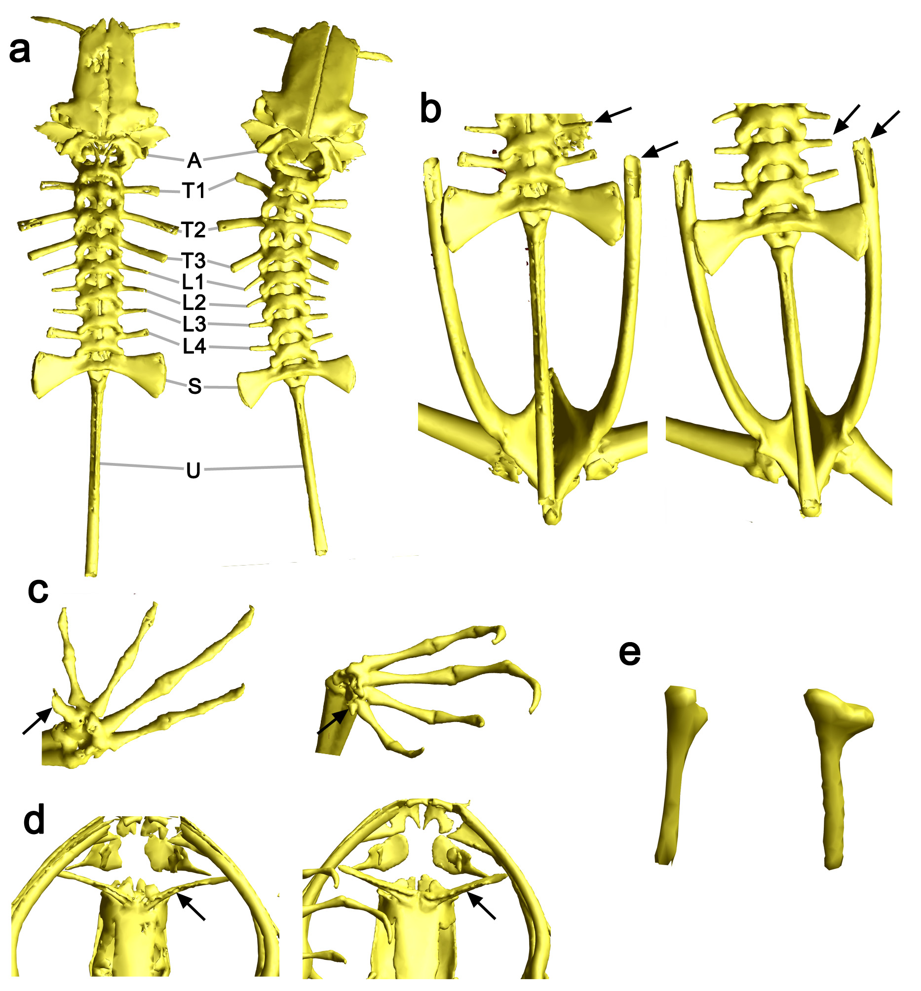
Osteology of the holotype ( Figs. 2 View FIGURE 2 , 3 View FIGURE 3 and Supplementary Fig. S1): Skull triangular in dorsal view. Prevomer divided, the postchoanal portion long, slightly posteriorly curved, nearly meeting medially, extending anteroventrolaterally from the sphenethmoid; overlapping, fused with, or replacing the palatine; possessing a serrated ventral ridge herein referred to as ‘vomerine teeth’. Teeth are present on the maxilla and premaxilla. Premaxilla without a distinct dorsal ramus visible in anterior view, rounded and without deeply separated rami in dorsal view. Septomaxilla U-shaped in dorsal view, consisting of an anterior plate with a small dorsal apophysis and two posterior-jutting rami, one on its ventromedial edge, and one on its ventrolateral edge; the ventrolateral ramus possessing a medial and a lateral apophysis, the medial apophysis extending ventromedially, toward, but not reaching, the medial ramus of the anterior plate. Squamosal broad, straight, and Y-shaped. Nasal broad and fairly rectangular, with a long process extending posterolaterally from the posterior end of its lateral edge. Columellar footplate with a stepped structure, reminiscent of a flush toilet in ventral view ( Fig. 2 View FIGURE 2 e).
Humerus with one humeral crista (crista ventralis), beginning 17% from the proximal end of the humerus, and extending to 45% of the way along it. Caput humeri with a distinct postero-dorsal apophysis on its distal edge, arcing distally, and a ventral ridge in line with the humeral crista, but not as high (81% of crista ventralis height). Ulna and radius fused. Finger phalangeal formula 2,2,3,3. Terminal phalanges of all fingers with distal knobs. Prepollex present; 61% of first metacarpal. Clavicles slim and curved anteriorly, approximately in parallel to the anterior curve of the coracoid. Sternum ossified anteriorly.
Toe phalangeal formula: 2,2,3,4,3. Terminal phalanges of all toes with distal knobs. Ilia rather short, extending beyond the sacrum to the level of the transverse process of the eighth presacral vertebra; ilial shafts laterally compressed, not cylindrical; fused synostotically with the ischia and pubes, which are ossified. Ten vertebrae are present: atlas, three thoracic vertebrae (T), four lumbar vertebrae (L), the sacrum and the urostyle. Urostyle possessing a dorsal ridge extending two thirds of the way down its length, beginning at its anterior. Transverse process breadths relative to the breadth of the sacral processes are T1 (89.3%), T2 (118.5%), T3 (102.9%), L1 (80.1%), L2 (71.3%), L3 (70.3%), L4 (76.2%). Posterior articular processes slender.
Colouration of the holotype: After almost nine years in preservative, dorsum light brown, with strong black mottling in a nearly symmetrical pattern. Each shoulder has on it a pair of dark oblong markings separated by a light brown bar. Venter dark brown beneath the chin, fading posteriorly to cream mottled with light brown.
Tympanic and supralabial regions are a lighter brown. A series of one distinct and two indistinct crossbands are present on the hindlimbs, which, in sitting position, line up across the thighs, shanks, and feet; two crossbands are present on the lower forelimbs. Feet dorsally strongly mottled white and brown. Hands dorsally white on the inner half, but brown as the rest of the dorsum on the outer. Toes of the left foot have light and dark crossbands, while the right foot is marbled light and dark; fingers of both hands banded, but less distinctly than toes; finger and toe tips light brown. No distinct border between dorsal and ventral colouration; white mottling extends up to the lateral regions. Tympanic region slightly lighter, with faint small dark spots.
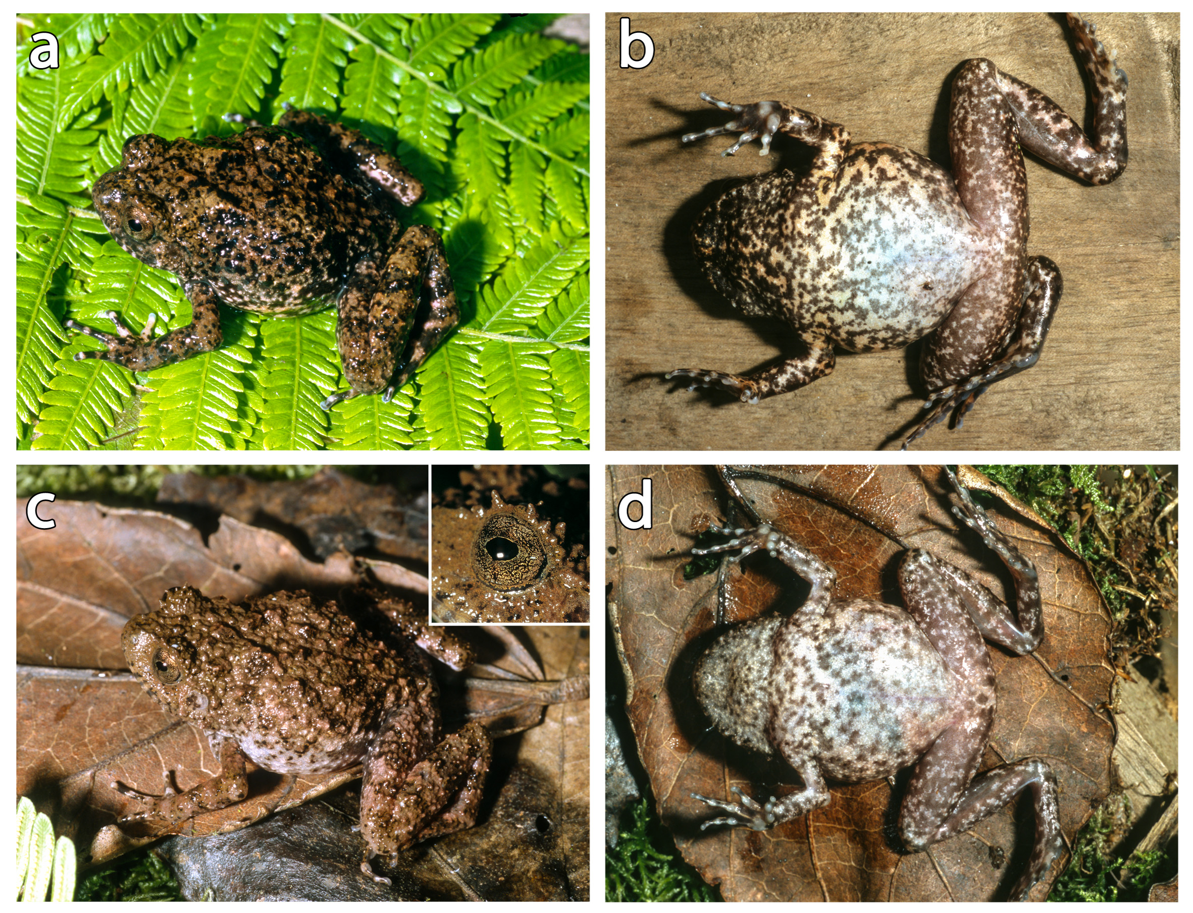
In life ( Fig. 1 View FIGURE 1 ), dorsally similar in colouration though a darker brown, with blackish markings strongly distinguished from the background colouration. Dark oblong markings in shoulder region distinct. Iris goldencopper with black vermiculation.
Variation. The paratype was not available for this study. However, based on colour photographs of it ( Fig. 1 View FIGURE 1 ), its four superciliary spines are more strongly pronounced than those of the holotype, and it has a more strongly tuberculated dorsum. Its colouration is generally similar to the holotype, with the following notable differences: lighter overall colouration; less distinct black mottling of the dorsum; partially faint dark crossbands on the lower legs, and almost indistinct crossbands on the lower forelimbs; less distinct toe banding; ventrally lighter in colour, especially in the chin region.
Etymology. The specific epithet is the Malagasy word ‘vaventy’ which translates as ‘huge’ or ‘enormous’, and refers to the rather large size of the new species in comparison to its congeners (the only comparably sized species is R. alluaudi ). It is used as a noun in apposition.
Natural history. The holotype and paratype were collected in the late afternoon close to dusk on the ground around a local campsite (Camp Simpona). The micro-CT scan revealed a millipede in the stomach of the holotype, which was identified as Zoosphaerium sp. by Thomas Wesener (pers. comm. 2013). It is recovered with a similar x-ray absorption to the bone of the frog ( Fig. 2 View FIGURE 2 ), probably due to the presence of both chitin and calcium deposits in its exoskeleton, which is typical for millipedes ( Blower 1951). The large intestine contained the remains of at least six ants, a spider, a dipteran, and at least three species of beetle (as judged by the presence of their heads).

No known copyright restrictions apply. See Agosti, D., Egloff, W., 2009. Taxonomic information exchange and copyright: the Plazi approach. BMC Research Notes 2009, 2:53 for further explanation.