
Pseudocandona falcula, Smith & Kamiya, 2015
|
publication ID |
https://doi.org/ 10.5852/ejt.2015.136 |
|
publication LSID |
lsid:zoobank.org:pub:530F395F-97A9-46F1-957C-8E57B9C3ACD9 |
|
DOI |
https://doi.org/10.5281/zenodo.3794659 |
|
persistent identifier |
https://treatment.plazi.org/id/0138AA0F-95A0-4AED-AF47-9DD1CCCB63C1 |
|
taxon LSID |
lsid:zoobank.org:act:0138AA0F-95A0-4AED-AF47-9DD1CCCB63C1 |
|
treatment provided by |
Carolina |
|
scientific name |
Pseudocandona falcula |
| status |
sp. nov. |
Pseudocandona falcula sp. nov.
urn:lsid:zoobank.org:act:0138AA0F-95A0-4AED-AF47-9DD1CCCB63C1
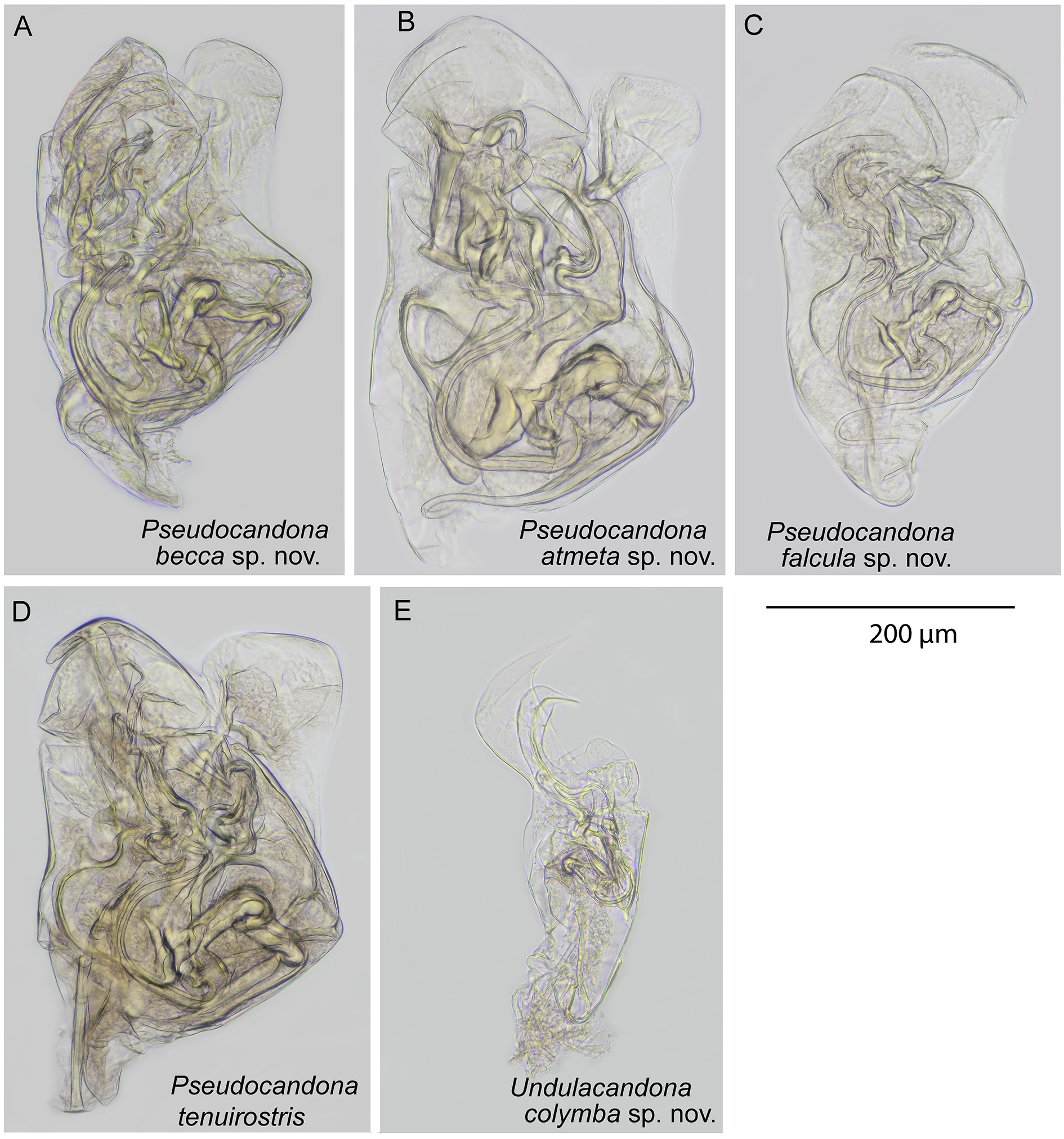
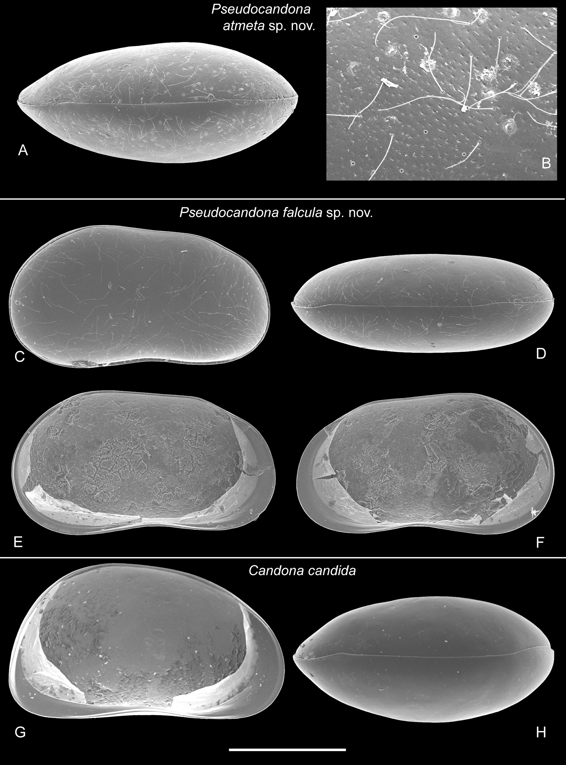
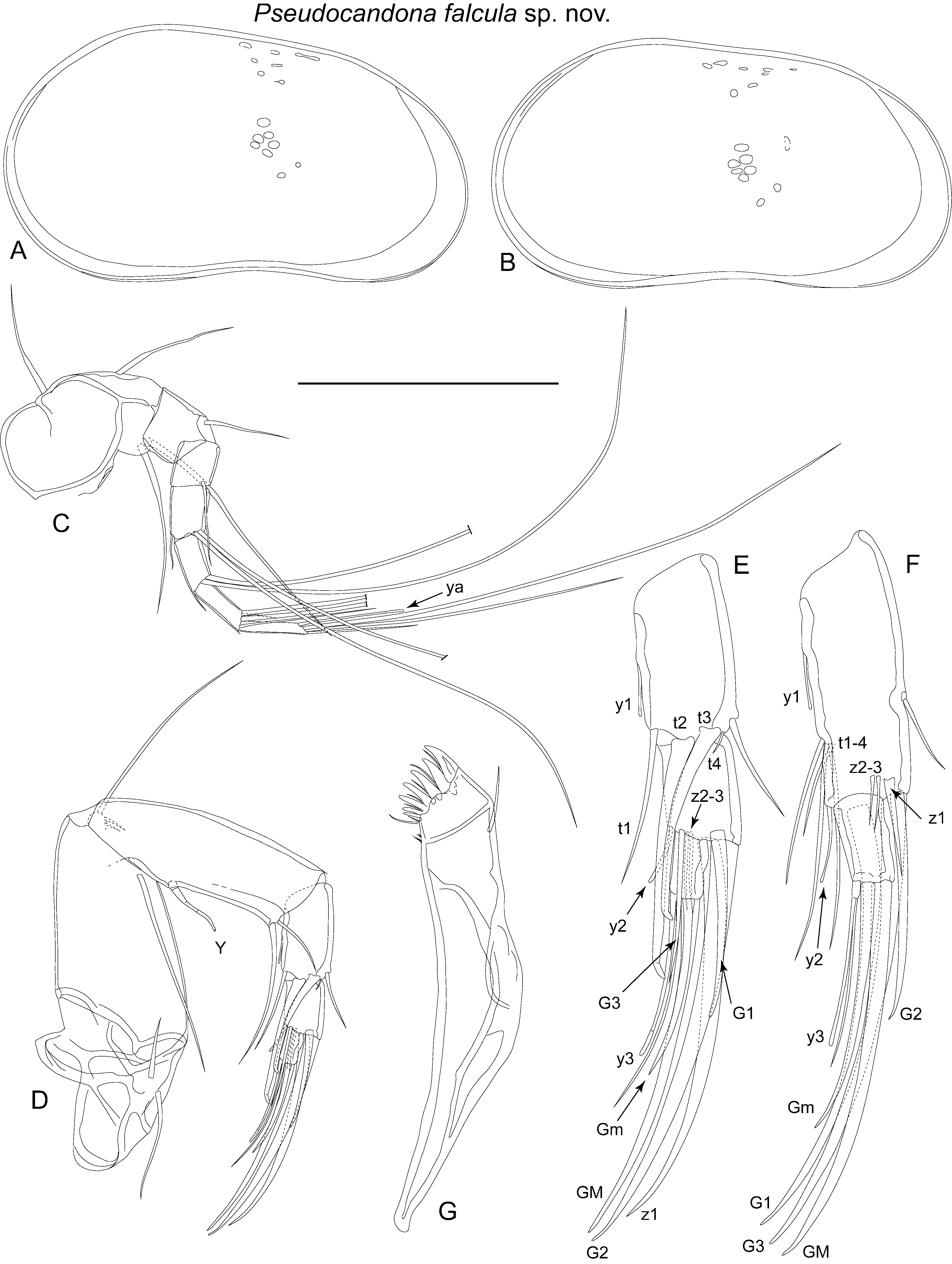
Figs 6C View Fig , 7 View Fig C–F, 11–13
Pseudocandona sarsi View in CoL – Smith & Janz 2008: 2909 View Cited Treatment , figs 17K–L, 25.
Diagnosis
Hinge straight, sloping to anterior region. Maximum height at posterior third. Posterior margin more inflated than anterior margin, both margins more or less evenly rounded. Calcified inner lamella narrow, wider anteriorly than posteriorly. Dorsal view compressed, with rounded anterior and posterior ends. Male antenna with sub-divided second endopodal segment, and with well-developed t2 and t 3 male bristles. Female antennal claw G2 approximately half length of claw G3. Mandible with 3+1+beta setae on second segment of palp, and with long gamma seta and very short alpha and beta setae; beta shorter than alpha. Walking leg with long d1 seta, and with e, and f setae shorter than next segment respectively. Seventh limb with five segments, terminating with long h2 and h3 setae of approximately equal length, and short, reflexed and curled h1 seta. Hemipenis with curved, relatively short M-process, outer lobe (a) large, and extending significantly beyond medial lobe (h), inner lobe (b) angular and folded.
Etymology
From the Latin falcula , meaning a “sickle” or “scythe”, and referring to the shape of the M-process of the male sexual organ.
Type material
Holotype
♂ ( LBM 1430006275 View Materials ), dissected with appendages sealed in a glass slide and valves stored dry in a micropalaeontological cavity slide. Collected from the type locality on 27 Sep. 2004.
Allotype
♀ ( LBM 1430006276 View Materials ), dissected with appendages sealed in a glass slide and valves stored dry in a micropalaeontological cavity slide. Collected from the type locality on 27 Sep. 2004.
Paratypes
1 ♀ ( LBM 1430006277), dissected with appendages sealed in a glass slide and valves stored dry in a micropalaeontological cavity slide. 1 ♀ ( LBM 1430006278), whole, stored dry in a micropalaeontological cavity slide. All collected from the type locality on 27 Sep. 2004.
Type locality
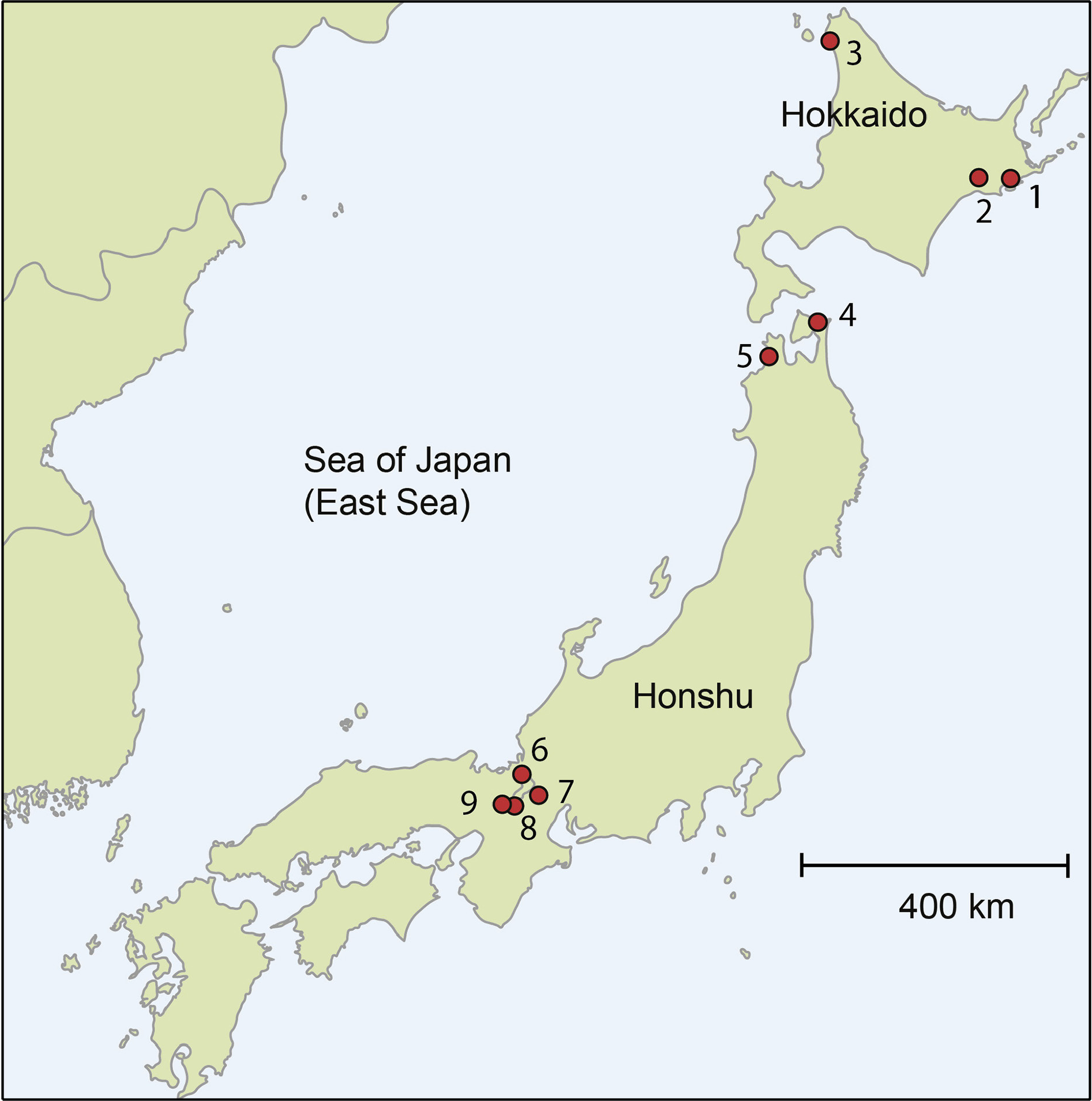
Sarugamori, Higashidori, Shimokita District, Aomori Prefecture, Japan (41º16’00.6” N, 141º22’38.4” E). Boggy, marshy area with little standing water, lots of reeds, and sodden ground. Locality 4 on Fig. 1 View Fig .
Other material examined
1 ♀, from the type locality, collected 27 Sep. 2004.
Description
Carapace ( Figs 7 View Fig C–F, 11A–B) length 918–937 µm, height 496–533 µm. Maximum height at posterior third, hinge straight, sloping towards evenly rounded anterior margin. Posterior margin more inflated than anterior margin, and evenly curved. Ventral margin slightly concave. Inner calcified lamella relatively narrow, wider anteriorly than posteriorly. Six small adductor muscle scars in tight formation
at mid-height, anterior of mid-length. Indistinct dorsal scars near dorsal margin. Female very similar to male. Colour, white. Surface covered with stiff setae.
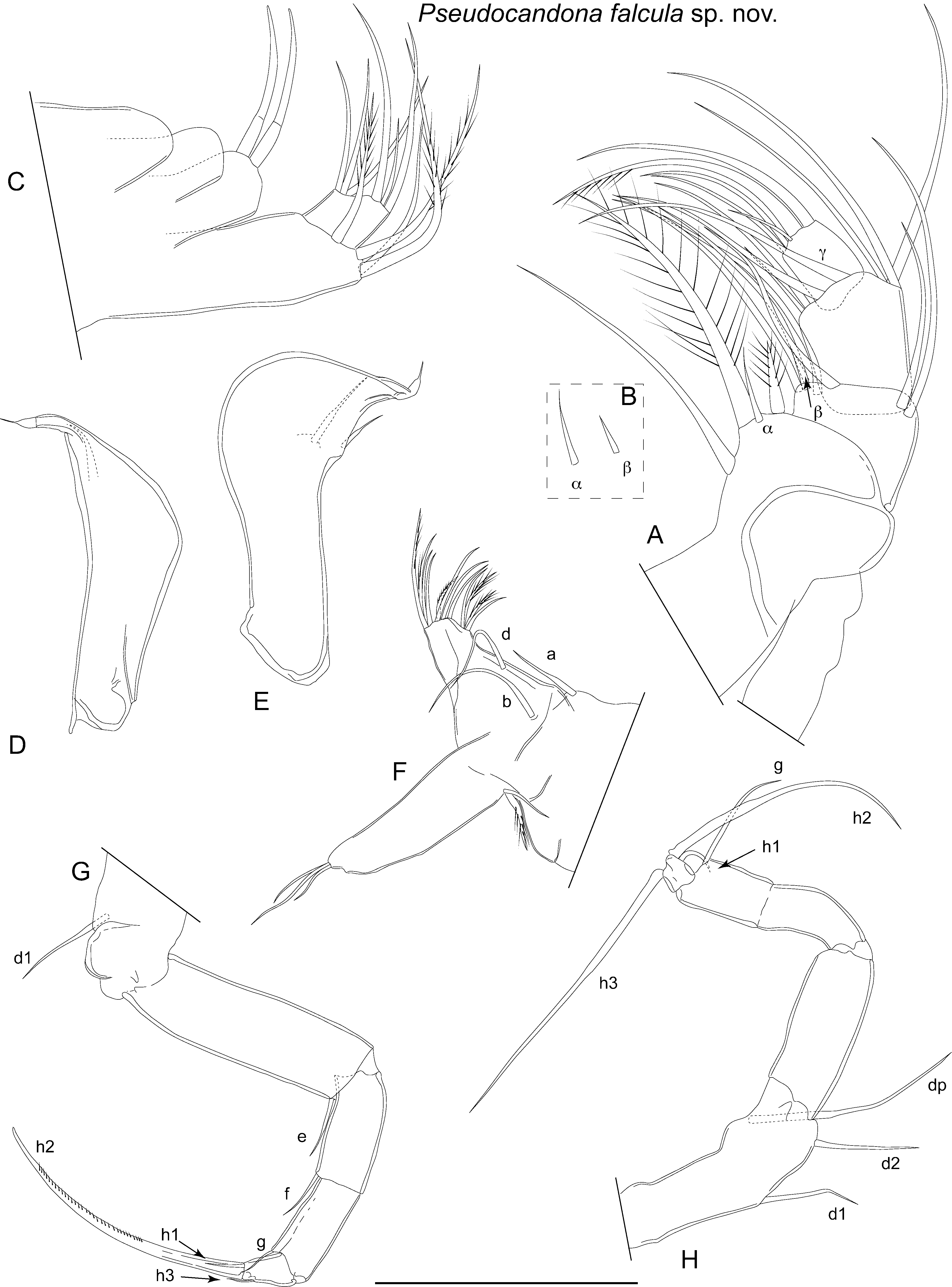
Antennule with seven articulated segments ( Fig. 11C View Fig ). First segment large, supporting two setae on dorsal edge and two long setae on ventral-apical corner. Second and third segments quadrate each with one dorsal-apical seta. Fourth and fifth segments each with two long dorsal-apical setae and one short ventral-apical seta. Sixth segment with two long, one mid-length and one short apical setae. Final segment with two long and one short setae, and aesthetasc ya.
Male antenna with second endopodite segment sub-divided ( Fig. 11 View Fig D–E). Setae t2 and t3 represented by male bristles, both similar in morphology, but with t2 longer than t3, both terminating with small, triangular process. Seta t4 tiny, protruding from near base of t3. Setae z2 and z3 very short, z1 represented by well-developed claw, slightly shorter than claw G2. Claw G1 less than half length of claw G2. Claw Gm on final segment half the length of claw GM. Female antennal claw G2 just under half length of G1, z1 stout and claw-like, less than half length of G2, setae z2 and z3 very short. Claw Gm more than half length of claw GM ( Fig. 11F View Fig ).
Mandibular palp ( Fig. 12 View Fig A–B) with four segments. Alpha seta of first segment short, and with very narrow, flagellum-like end. Inner edge of second segment with 3+1+beta arrangement of setae; seta beta shorter than alpha. Outer edge of second segment with two apical setae. Third segment with three long sub-apical setae on outer edge, and three long and one short setae arranged along apical edge; outermost gamma seta, long and without obvious setules. Final segment with robust seta on outer edge, thick claw-like seta in mid-apical position and three shorter setae on inner apical edge. Number of setae on branchial plate not observed. Mandibular coxa ( Fig. 11G View Fig ) with five well-developed teeth plus two much smaller, spine-like teeth.
Maxillula ( Fig. 12C View Fig ) palp first segment with three setae on apical outer margin, and one apical seta offset towards inner edge. Second segment with slightly stepped apical margin, with outer part more distal than inner part. Outer part of apical margin with two long and one short setae, and inner part with three mid-length setae. Branchial plate with morphology typical of subfamily.
Fifth limb male palps asymmetrical ( Fig. 12 View Fig D–E). Right palp rounded, helmet-shaped, more inflated than left. Left palp almost sub-triangular distally with angular outer margin, and almost straight inner margin.
Fifth limb of female ( Fig.12F View Fig ) with long b and d setae, and one long a seta on basis. Endite with approximately nine apical setae, and four sub-apical setae on inner edge. Palp (endopodite) terminating with three setae on unequal lengths. Branchial plate consisting of two rays, one shorter and narrower than other.
Sixth limb ( Fig. 12G View Fig ) five-segmented, with first segment bearing d1 seta. Setae e and f of second and third segments respectively, both about half length of next respective segment. Fourth segment with g seta reaching to end of fifth segment. Fifth segment with short h1 seta, tiny h3 seta and well-developed claw h2.
Seventh limb with five segments ( Fig. 12H View Fig ). First segment with long dp seta, and medium-length d1 and d2 setae. Second and third segments with no setae. Fourth segment with long g seta. Fifth segment approximately quadrate, h2 and h3 long and of approximately equal length, h1 short, hook-like and reflexed.
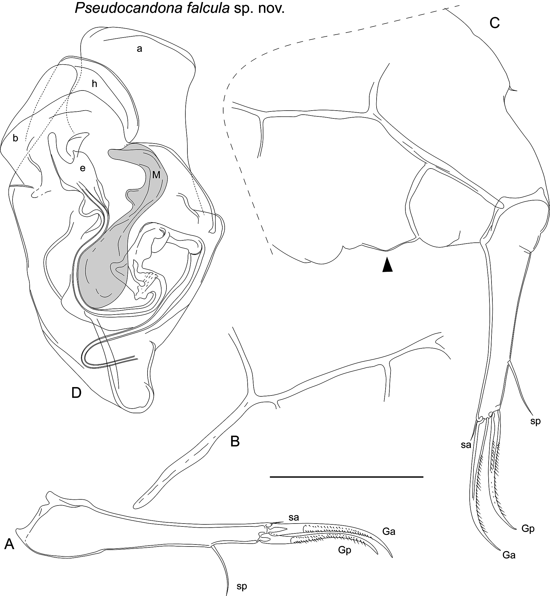
Caudal ramus ( Fig. 13 View Fig A–C) with inflated base, tapering distally, slightly curved to straight. Claw Ga straight proximally, curving distally. Claw Gp slightly shorter and thinner than Ga. Seta sa short, seta sp long, about 50% length of claw Gp. Caudal ramus attachment of male ( Fig. 13B View Fig ) with one posterior branch and two anterior branches on dorsal-most half. Female caudal ramus attachment ( Fig. 13C View Fig ) with one posterior branch and two anterior branches, lowest of which curves to posterior edge of female genital lobe.
Hemipenis ( Fig. 13D View Fig ) outer lobe (a) with truncated, sub-quadrate distal margin, extending beyond other two lobes. Medial lobe (h) with unevenly curved, rounded distal margin. Inner lobe (b) folded, triangular on inner edge, one part tongue-shaped, other quadrate. M-process small, rounded proximally, strongly curved and hook-like with lobe on inner edge. Bursa copulatrix (e) elongate with long finger distally.
Female genital lobe ( Fig. 13C View Fig ) with prominent projection with rounded apex. NB morphology noted before cover slip added. In material examined, lobes lightly sclerotized, with tendency to deform and break apart with addition of cover slip. In particular, rounded lobe (marked with triangle on Fig. 13C View Fig ) less prominent and more spread out laterally after addition of cover slip.
Remarks
As noted above, the female genital lobe was noticeably deformed in shape in dissection slides after the cover slip had been lowered into position. The female genital lobe of candonids is widely used for the discrimination of species, but as noted by Namiotko & Danielopol (2004), this feature is prone to deformation in dissection slides.
Pseudocandona falcula sp. nov. is similar to Pseudocandona sarsi (Hartwig, 1899) in the shape of the carapace in lateral view, although the dorsal view of P. sarsi is slightly wider and more pointed anteriorly and posteriorly. The male fifth limb palps are similar in both species, but the right palp of Pseudocandona falcula sp. nov. is more rounded on the outer edge than that of P. sarsi (which has a more angular outer edge). The seventh limbs of the two species are also similar, both with a short reflexed h1 seta on the final segment. Differences in the hemipenes include a larger outer lobe (a) in Pseudocandona falcula sp. nov., which clearly extends beyond the distal edge of the medial lobe (h). In P. sarsi the outer lobe is more offset to the outer edge of the hemipenes, has a more angular outer edge, and only slightly extends beyond the medial lobe. The M-processes of the two species are also different in shape.
The females of this species closely resemble the female specimens of P. sarsi reported from Lake Biwa (locality 8 on Fig. 1 View Fig ) by Smith & Janz (2008). As no males were found from Lake Biwa, Smith & Janz’s (op. cit.) identification was tentative. Further investigation indicates that the carapaces and appendages of females from Lake Biwa very closely match material from the type locality, and it is concluded that the specimens from Lake Biwa are Pseudocandona falcula sp. nov.
Ecology and distribution
The type locality of this species is the same as for Pseudocandona atmeta sp. nov. (see below Pseudocandona atmeta sp. nov. for a description of that site). In Lake Biwa, the specimens were found at the water’s edge in the root mat of a willow tree ( Smith & Janz 2008). To date, only a small number of females have been collected from Lake Biwa, in samples collected during Apr. 2005. It hasn’t been collected since this time despite numerous samples taken at the same locality, and this may indicate that it was a temporary occurrence or it is rare in Lake Biwa. So far, this species is only known from Lake Biwa and the type locality.
| LBM |
Lake Biwa Museum |
No known copyright restrictions apply. See Agosti, D., Egloff, W., 2009. Taxonomic information exchange and copyright: the Plazi approach. BMC Research Notes 2009, 2:53 for further explanation.
|
Kingdom |
|
|
Phylum |
|
|
Class |
|
|
Order |
|
|
Family |
|
|
Genus |
Pseudocandona falcula
| Smith, Robin James & Kamiya, Takahiro 2015 |