
Polymastigos javaensis, Abstract, Joko Pamungkas, 2015
|
publication ID |
https://doi.org/ 10.11646/zootaxa.3980.2.8 |
|
publication LSID |
lsid:zoobank.org:pub:BD6CE38A-4EB1-4932-B780-F9EAC0973AAC |
|
DOI |
https://doi.org/10.5281/zenodo.6106051 |
|
persistent identifier |
https://treatment.plazi.org/id/03D387B5-FFEF-FFCB-BC92-FF14FB69FECD |
|
treatment provided by |
Plazi |
|
scientific name |
Polymastigos javaensis |
| status |
sp. nov. |
Polymastigos javaensis View in CoL n. sp.
Material examined. SA-01 (holotype, MZB. Pol. 00132); SA-01 (1 paratype, MZB. Pol. 00133); SA-01 (13, MZB. Pol. 00134); SA-02 (6, MZB. Pol. 00135), SA-01 (1, MZB. Pol. 00136), SA-03 (7, MZB. Pol. 00137), SA- 0 4 (11, MZB. Pol. 00138).
Description. Holotype complete with about 197 chaetigers, 30 mm long by 1.0 mm wide in abdomen. Paratype complete with about 251 chaetigers, 35 mm long by 1.0 mm wide in abdomen. Other adult specimens complete or anterior fragments, ranging from 8 mm long by 0.8 mm wide (32 chaetigers), to 75 mm long by 1.2 mm wide (about 366 chaetigers). Less mature specimens complete, with 35 mm long by 0.8 mm wide (about 224 chaetigers) and 53 mm long by 0.8 mm wide (about 237 chaetigers). Original colour in field reddish pink, in alcohol light brown. Prostomium conical with palpode. Eyespots and nuchal organs not seen. Proboscis partially everted, globular, with papillae. Peristomium achaetous.
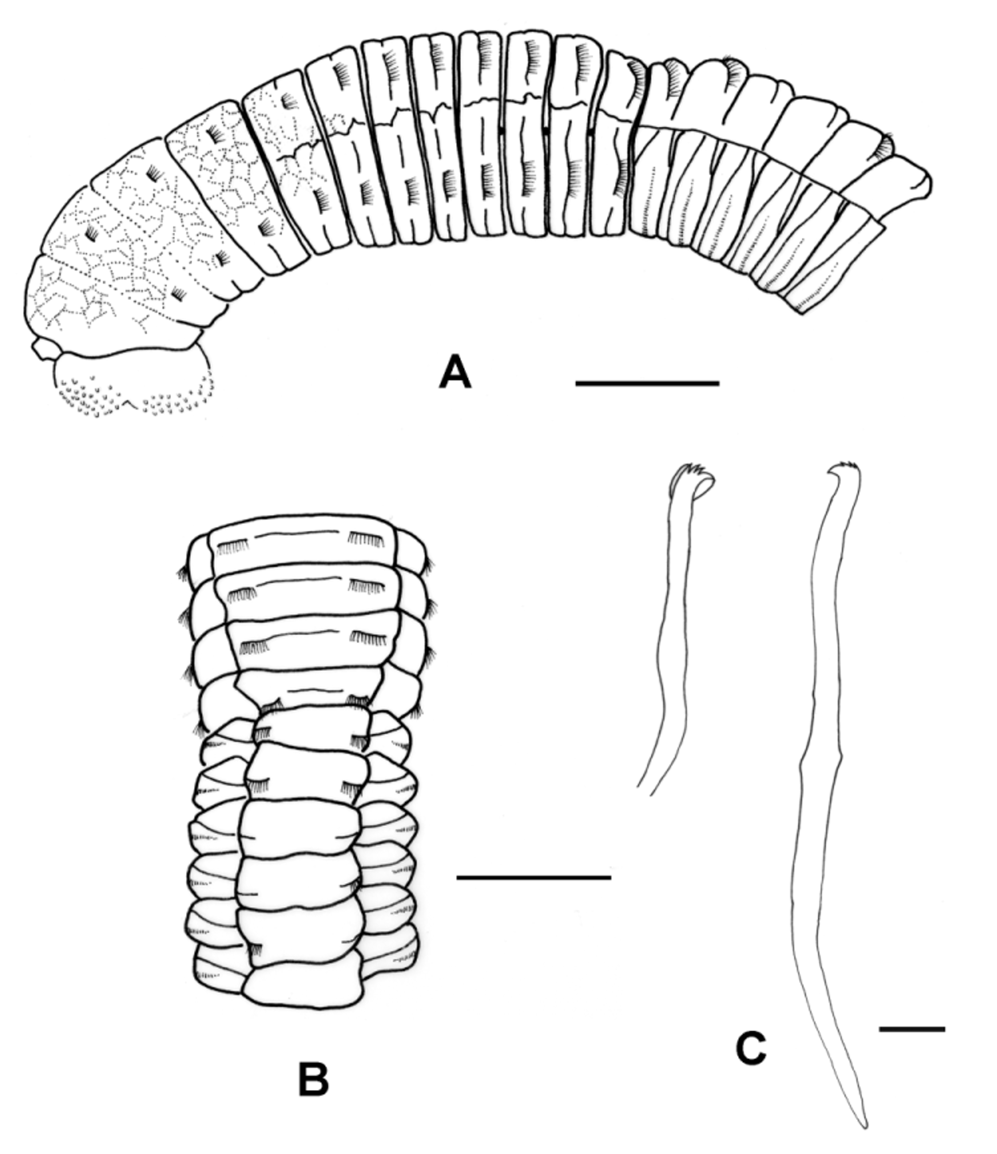
Thorax with 12 segments, including 1 achaetous peristomium followed posteriorly by 11 chaetigers with exclusively bilimbate capillary chaetae in both rami ( Fig. 2 View FIGURE 2 A). All chaetigers, including chaetiger 1, biramous. Notopodia oriented laterally in first chaetiger, gradually moving dorsally in subsequent segments. Parapodia reduced. Chaetae embedded in middle part of thoracic segments, except in chaetiger 11 embedded more posteriorwards.
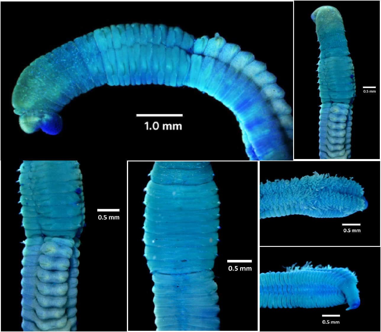
Peristomium to chaetiger 4 or 5 with areolated epithelium; remaining thorax smooth. Intersegmental groove between chaetiger 4 and 5 somewhat deeper than previous chaetigers. Based on these two characteristics, i.e. epithelium texture and intersegmental groove, thorax appears divided into two portions, i.e. areolated and smooth thorax ( Figs 2 View FIGURE 2 A & 3A–B, D). Thoracic segments biannulated. Areolated thorax less biannulated with obscure lateral line; smooth thorax more obviously biannulated with strong lateral line ( Figs 2 View FIGURE 2 A & 3A).
Lateral organs difficult to see. Genital pores lie on last four thoracic chaetigers, i.e. on the intersegmental grooves between chaetigers 7/8, 8/9, 9/10 and 10/11 ( Fig. 2 View FIGURE 2 A).
Transition between thorax and abdomen clearly marked by deep intersegmental groove between chaetiger 11 and 12 ( Fig. 3 View FIGURE 3 A–D), distinct dorsal abdominal tori ( Figs 2 View FIGURE 2 B & 3B–C), and changes to neurochaetae ( Fig. 2 View FIGURE 2 A–B) and methyl green staining ( Fig. 3 View FIGURE 3 A–F).
Dorsal surface of first several abdominal segments appear uni-annulate (sometimes with some weak furrows), changing posteriorly to more bi-annulate appearance in middle and posterior abdomen (sometimes with additional weak furrows), due to extension of ventral intra-annular groove dorsalwards. Ventral abdominal segments appear bi-annulate ( Figs 2 View FIGURE 2 B, 3D & 3F). Abdominal segments tapering posteriorly; width and length of posterior segments smaller than those of anterior ones. Holotype with 5 transitional abdominal segments with bilimbate capillary notochaetae and neurohooks (chaetiger 12 to 16). Paratype with 2 transitional abdominal segments (chaetiger 12 to 13); chaetiger 14 and 15 may have lost capillaries due to damage. Non-type materials vary, with 0 (less mature specimens) to 7 transitional abdominal segments. Remaining chaetigers with hooks in both rami.
In anterior abdomen, number of notohooks within a fascicle (about 20 hooks) fewer than neurohooks (about 43 hooks). Hooks within fascicle decrease in number posteriorly, about 5 notohooks and 23 neurohooks in posterior chaetigers. Abdominal hooks subequal. Notohooks with longer shaft than neurohooks, about two times. Length of anterior and posterior shaft nearly equal with an obvious node in middle of shaft. Hooks with 3 teeth above main fang, with narrow neck and shoulder. Short vague hood (hood with weak outline) observed in neurohooks, not seen in notohooks.
Branchiae simple digitate, may be retractable, observed on dorsal region of posterior chaetigers, arise from intersegmental grooves, evident posteriorly from about chaetiger 83 in holotype and about chaetiger 86 in paratype ( Fig. 3 View FIGURE 3 E). Pygidium retracted in holotype, everted in paratype, with mid-ventral caudal cirrus ( Fig. 3 View FIGURE 3 F). Middle and posterior part of body with eggs in coelom. Egg oval, dark grey, around 300 µm and 800 µm, minimum and maximum diameter, respectively.
Methyl green stain complete on thorax, including lateral line, intersegmental and intra-annular grooves. Dorsal region of proboscis, as well as prostomium to about half of chaetiger 2, stained greenish blue with medium intensity. Chaetiger 3, slightly extending to about half of chaetiger 2, stained dark blue. Chaetiger 4 with slightly brighter blue intensity than chaetiger 3. Ventral region of proboscis stained the darkest blue. In general, ventral side of areolated thorax stained darker than dorsal and lateral sides. Smooth thorax stained blue with rather more intensity than areolated thorax. Weakly stained areas on neuropodial lobes of chaetiger 4 to 11, forming oval appearance. Dorsal abdominal tori weakly stained with scattered blue spots. Area around notopodial lobes stained brightly, creating circle patterns; area around mid-dorsal line close to intersegmental areas stained darkly, forming longitudinal line. On abdominal ventrum, prechaetal annuli stained darkly. Mid-ventral region of posterior part with two dark longitudinal lines, interrupted by intersegmental ring. Pygidium rim stained darkly ( Fig. 3 View FIGURE 3 A–F).
Less mature specimens with 5 capillary chaetae (remaining chaetigers with hooks), smaller body, either absence or a few number of eggs in abdominal cavity, and show no branchiae. MGSP with similar appearance as in adult specimens.
Distribution. Eastern area of the Segara Anakan mangrove habitat (nearby industrial area), Cilacap, Central Java Province, Indonesia. All specimens were collected in sandy clay sediment at 0–30 cm depth.
Etymology. The species is named based on its locality, i.e. Segara Anakan mangroves, Java Island, Indonesia. It is also the place where I spent so much time studying polychaetes for the first time.
Remarks. The new species P. javaensis n. sp. resembles P. re i s h i in having more than four transitional abdominal segments and in the position of their genital pores on the intersegmental grooves between chaetigers 7/ 8, 8/9, 9/10 and 10/11, but the new species differs in the following features. First, chaetiger 1 of P. javaensis n. sp. is biramous, whereas it is uniramous in P. re i s h i. Further, Green did not mention segment width difference as an important character to define P. re i s h i, but in the author’s observation, the feature is a distinctive characteristic of P.javaensis n. sp. The character makes the whole body of P. javaensis n. sp. seem to be divided into four portions when seen from a dorsal view, i.e. areolated thorax (with weak lateral line), smooth thorax (with strong lateral line), anterior abdominal region (without branchiae) and posterior abdominal region (with branchiae and pygidium). Another difference is that dorsal surface of P. j a v a en s i s n. sp. first several abdominal segments look uni-annulate, changing to more bi-annulate appearance in mid and posterior segments, whereas Green described the dorsum of P. reishi ’s abdominal segments as multi-annulate. The annular pattern of the anterior abdominal ventrum between them is also different (this can be more clearly seen after staining with methyl green). Polymastigos javaensis n. sp. shows prechaetal annuli that are slightly similar to diamond (with a large angle around the mid-ventral line and a low angle around lateral sides—Fig.3D), whereas P. reishi has rectangular-like prechaetal annuli.
Next, the hooks of P. javaensis n. sp. are either without hood (notohooks) or with relatively small and short hood (seen in neurohooks), while P. reishi ’s hooks with long hood with relatively long fringe. In P. javaensis n. sp. the number of hooks within a fascicle decrease in number posteriorly, but they increase in P. reishi . Lastly, the MGSP between P. javaensis n. sp. and P. reishi is also very different as explained above.
| MZB |
Museum Zoologicum Bogoriense |
No known copyright restrictions apply. See Agosti, D., Egloff, W., 2009. Taxonomic information exchange and copyright: the Plazi approach. BMC Research Notes 2009, 2:53 for further explanation.