
Polydora cavitensis Pillai, 1965
|
publication ID |
https://doi.org/ 10.5281/zenodo.176375 |
|
DOI |
https://doi.org/10.5281/zenodo.6242399 |
|
persistent identifier |
https://treatment.plazi.org/id/03AF87BA-6E33-4D6C-FF7A-FC1A251BFE21 |
|
treatment provided by |
Plazi |
|
scientific name |
Polydora cavitensis Pillai, 1965 |
| status |
|
Polydora cavitensis Pillai, 1965 View in CoL ( Figs. 2–3)
Polydora cavitensis Pillai, 1965: 152 View in CoL –154, Figs. 16 E, F, 17A–G.
Material examined. Philippines, Bacoor, Cavite in Manila Bay, from mud tubes among live green mussels Perna viridis , 20 Jul 2000, coll. by researchers at the Marine Science Institute of the University of the Philippines (one complete specimen in alcohol, USNM 1096802; one anterior end on SEM stub, USNM 1096803).
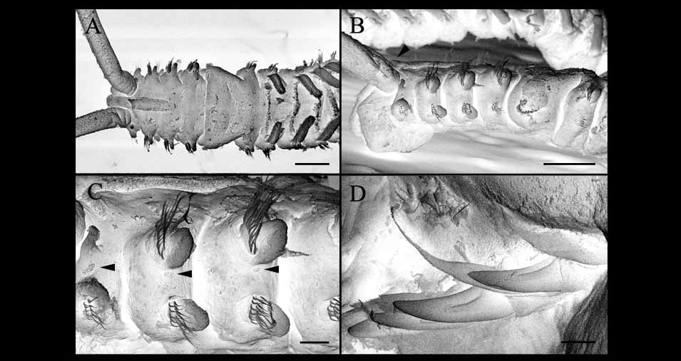
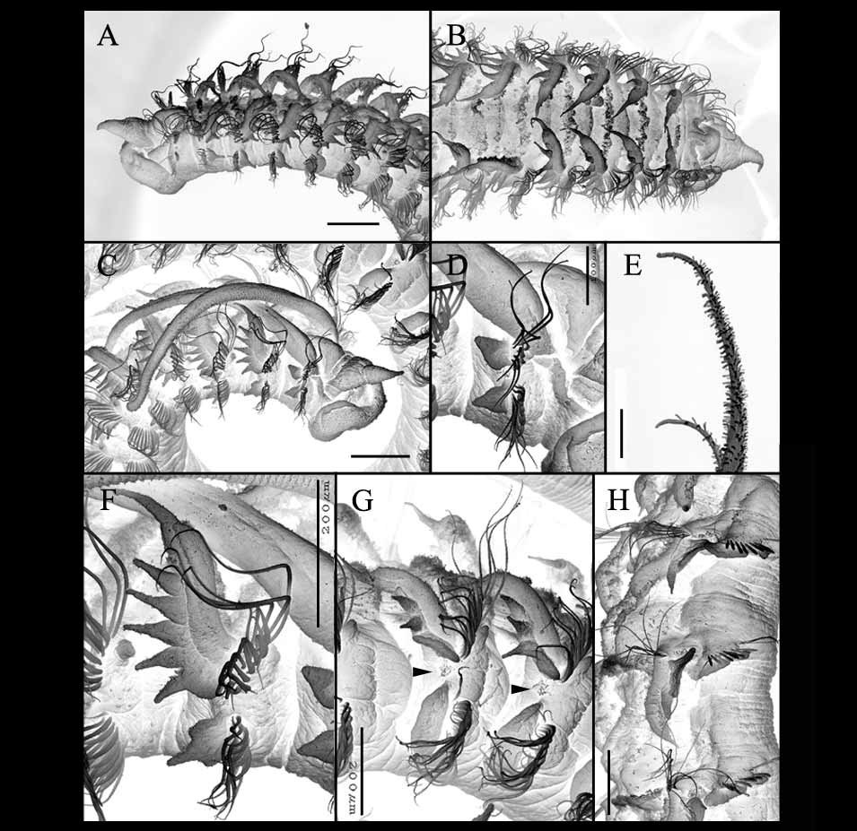
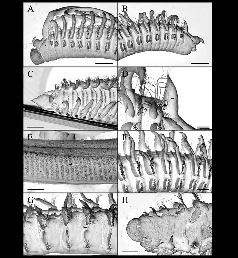
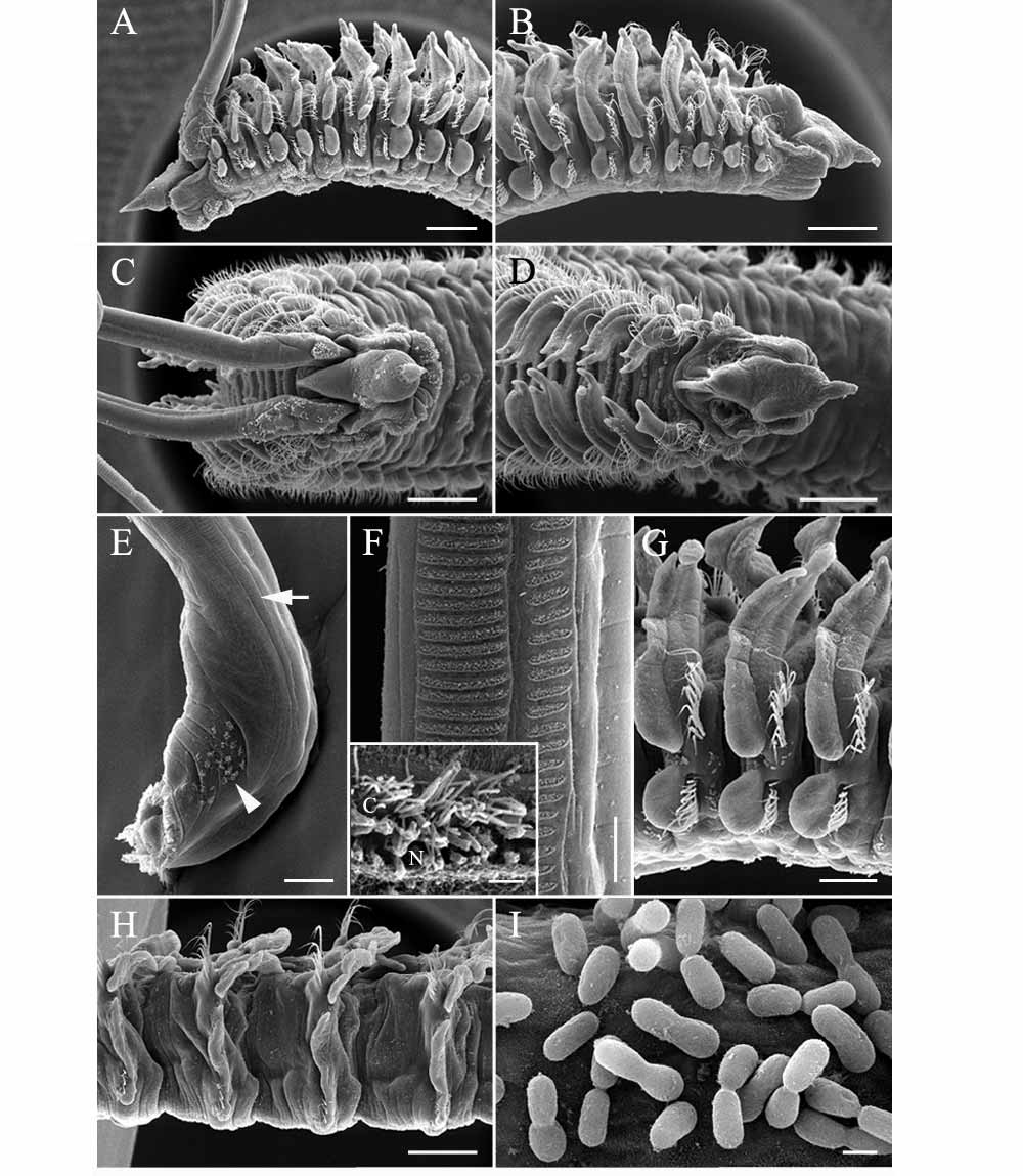
Description. Complete specimen of 67 setigers 17.42-mm long, and 0.7mm wide at setiger 7. Prostomium weakly incised anteriorly ( Fig. 2 A–B); caruncle extending posteriorly to near end of setiger 3; four round, black eyes present in a trapezoidal pattern; cirriform occipital tentacle present behind eyes and between base of palps ( Figs. 2 A, C, 3A–B). Palps extending posteriorly for about 12 setigers. Color in alcohol opaque white to light tan, with very slight patch of pigmentation on dorsal side of setiger 3, palps with irregular patches of black pigmentation ( Fig. 2 A).
Setiger 1 with neurosetae, without notosetae, with weakly developed notopodial lobes ( Figs. 2 A, B, 3C). Winged capillary notosetae of setigers 2–4, 6 and subsequent setigers arranged in three successive rows; no specialized posterior notosetae ( Figs. 2 H, 3C). Winged capillary neurosetae of setigers 2–4, 6 and subsequent setigers arranged in two vertical rows ( Figs. 2 I, 3C); six bidentate hooded hooks begin on setiger 7, not accompanied by capillaries, up to nine in series at setiger 9; hooks with near right angle between main fang and shaft, wide angle between main fang and apical tooth, with constriction on shaft ( Fig. 2 G).
Setiger 5 almost twice as large as setigers 4 and 6, with slightly curved row of six exposed major spines and two embedded spines, major spines alternating with pennoned companion setae exhibiting broom-like tips; with posterior ventral fascicle of six winged capillary neurosetae; anterior dorsal fascicle of 4–6 geniculate notosetae present. Major spines simple, falcate, with very shallow lateral flange ( Figs. 2 E–F, 3B, D).
Branchiae from setiger 7, free from notopodial postsetal lamellae, continuing to postgerior setigers; branchiae nearly meeting at middle from setigers 8–19, diminishing in length posteriorly ( Figs. 2 A–B, D, 3A). Lateral organs (=interramal organs, interramal ciliary organs, or lateral ciliated organs; see Radashevsky 2005; Purschke & Hausen 2007) present between notopodial and neuropodial lamellae on setigers 1-3, 6 and subsequent setigers ( Fig. 3 View FIGURE 3 C), lateral organs largest on setiger 1.
Pygidium broad, cup-shaped with dorsal gap, small patches of pigmentation present on sides ( Fig. 2 D).
D
C
A E
B
F No Ne G H I No gizzard-like structure present in digestive tract. Glandular pouches not observed in preserved specimens.
Remarks. The present specimens are similar to the original description of P. cavitensis from among oysters in the Manila Bay, Philippines based on the bifid prostomium, presence of an occipital tentacle, and presence of superior and inferior bundles of spines on setiger 5 ( Pillai 1965). P. c a v i t e n s i s was noted by Pillai (1965) to have two transverse brown bands of pigmentation on the palps, patches of reddish-brown pigmentation on the dorsal side of each setiger, and pigment patches on each side of the pygidium. The present specimens lack distinct bands on the palps but do exhibit diffuse pigmentation along the sides of the palps and patches of pigmentation on the pygidium; no distinct patches of pigmentation were found on the dorsal side of each setiger. However, such pigmentation can be variable within polydorids. The only other potential distinguishing feature between Pillai’s material and the present specimens is with the morphology of the major spines of setiger 5. Pillai (1965) described the spines as having a notched tip but the picture of the spines ( Fig. 17 View FIGURE 17 D in Pillai (1965)) shows a flange-like structure, slightly more pronounced but similar to that found in the present specimens. For these reasons, the newly collected specimens are referred to P. c a v i t e n s i s.
Identification of this species is difficult because it is very similar to Polydora cornuta Bosc, 1802 , a widely distributed species found to construct tubes in soft sediments of estuarine waters, often attaining large population sizes and acting as a pollution indicator species ( Rice & Simon 1980). The species are similar in possession of a bifid prostomium, occipital tentacle, and length of caruncle. Radashevsky (2005) recently completed extensive morphological and larval studies of P. c o r n u t a based on specimens collected worldwide (east and west coasts of North America, Mexico, Brazil, Argentina, Germany, Romania, Russia, Korea, Taiwan, and China). While the species are similar in many respects, Polydora cavitensis exhibits dorsal superior capillaries present on setiger 5, whereas according to Radashevsky (2005) these are invariably absent in P. cornuta . However, Radashevsky (2005) did not mention that dorsal superior capillaries were present on setiger 5 in populations of P. c o r n u t a collected from Florida (see Figs. 9 View FIGURE 9 and 15 View FIGURE 15 in Rice & Simon 1980) although these specimens were listed among those in synonymy with P. c o r n u t a. Additional characters that distinguish Polydora cavitensis and P. cornuta are the prostomial morphology and nature of the major spines of setiger 5. The prostomium of Polydora cavitensis is weakly bifid but could be confused as rounded unless specimens are viewed from the ventral side where the split nature of the prostomium is best observed (see Fig. 2 B); in contrast, the prostomium of P. cornuta is widely bifurcated with flaring lateral sides. The spines of the two Polydora cavitensis specimens examined lacked a lateral tooth, even in the unworn spines, and exhibited only a slight lateral flange; in P. cornuta the spines exhibit both a large lateral tooth and subterminal flange.
In the Philippines Polydora cavitensis was found in Manila Bay inhabiting mud tubes constructed among masses of the green mussel Perna viridis . Perna viridis are commercially harvested mussels that have been transported to the Caribbean and Gulf of Mexico ( Agard et al. 1992; Benson et al. 2001; Ingrao et al. 2001). While Benson et al. (2001) indicated Perna viridis was likely transported to the Caribbean and Florida in ballast water, it is possible that the mussels and associated fauna such as Polydora cavitensis have been transported to other islands in the Indo-West Pacific where they are cultured. Similarly, Polydora cornuta is believed to have been widely transported through transport of aquacultural products and uptake of larvae in ballast water ( Carlton 1975; Gordon & Read 1991; Radashevsky & Hsieh 2000; Çinar et al. 2005). Such introductions are common among spionids (e.g., Bailey-Brock 1990, 2000; Carlton & Geller 1993; Röhner et al. 1996; Blake 1996; Williams 2001; Radashevsky & Olivares 2005) and can have both negative ecological and commercial impacts.
Distribution. From mud tubes between shells of the green mussel Perna viridis in Manila Bay, Philippines; subtidal.
| USNM |
Smithsonian Institution, National Museum of Natural History |
No known copyright restrictions apply. See Agosti, D., Egloff, W., 2009. Taxonomic information exchange and copyright: the Plazi approach. BMC Research Notes 2009, 2:53 for further explanation.