
Plesiocolochirus dispar ( Lampert, 1889 )
|
publication ID |
https://doi.org/ 10.5281/zenodo.172917 |
|
DOI |
https://doi.org/10.5281/zenodo.5674058 |
|
persistent identifier |
https://treatment.plazi.org/id/1E5A87CB-0A61-5747-FF37-93C2F48BFE53 |
|
treatment provided by |
Plazi |
|
scientific name |
Plesiocolochirus dispar ( Lampert, 1889 ) |
| status |
|
Plesiocolochirus dispar ( Lampert, 1889) View in CoL
Figure 9 View FIGURE 9
Colochirus dispar Lampert, 1889:820 View in CoL , fig. 4; Ekman, 1918:32. pl. 3, fig. 25, pl. 4, fig. 26. Pentacta dispar Clark & Rowe, 1971:180 View in CoL (dist.); Cherbonnier, 1988:164, fig. 68. Colochirus gravieri Vaney 1905:187 .
Pentacta gravieri Cherbonnier, 1955:162 View in CoL , pl. 48, figs. k–s.
Pentacta trimorpha Clark, H.L. 1921:171 View in CoL , pl. 37 (1–8).
Plesiocolochirus dispar Rowe View in CoL (in Rowe & Gates 1995):278 (syn.)
Type
Syntypes perhaps in Stuttgart, Germany.
Type locality
Mermaid Strait, northwest Australia.
Previous southern African record
None.
Material examined
SAM A23175, Coconut Bay, Mozambique, 17.v.1973, 1 spec.
Description
Form barrelshaped, with dorsal surface arched and ventral flattened, solelike, but sole not clearly defined; anterior end broad, posterior narrow and turned upwards. Length about 10 mm, diameter of anterior and posterior ends 5 mm and 2.5 mm respectively.
Colour uniformly white, including podia and tentacles. Podia restricted to ambulacra in double rows but most of those of midventral ambulacrum retracted, except at anterior and extreme posterior ends; those of ventrolateral ambulacra mostly well extended; dorsal podia indistinct because of scales which envelope most of dorsum. Ventral podia with welldeveloped suckers. Mouth starshaped, boarded by five, distally calcified valves, especially the midventral one which appears as a toothlike structure. Tentacles 10, midventral two reduced. Anus terminal, surrounded by five teeth, each flanked by subterminal podia. Skin thin and rough to the touch.
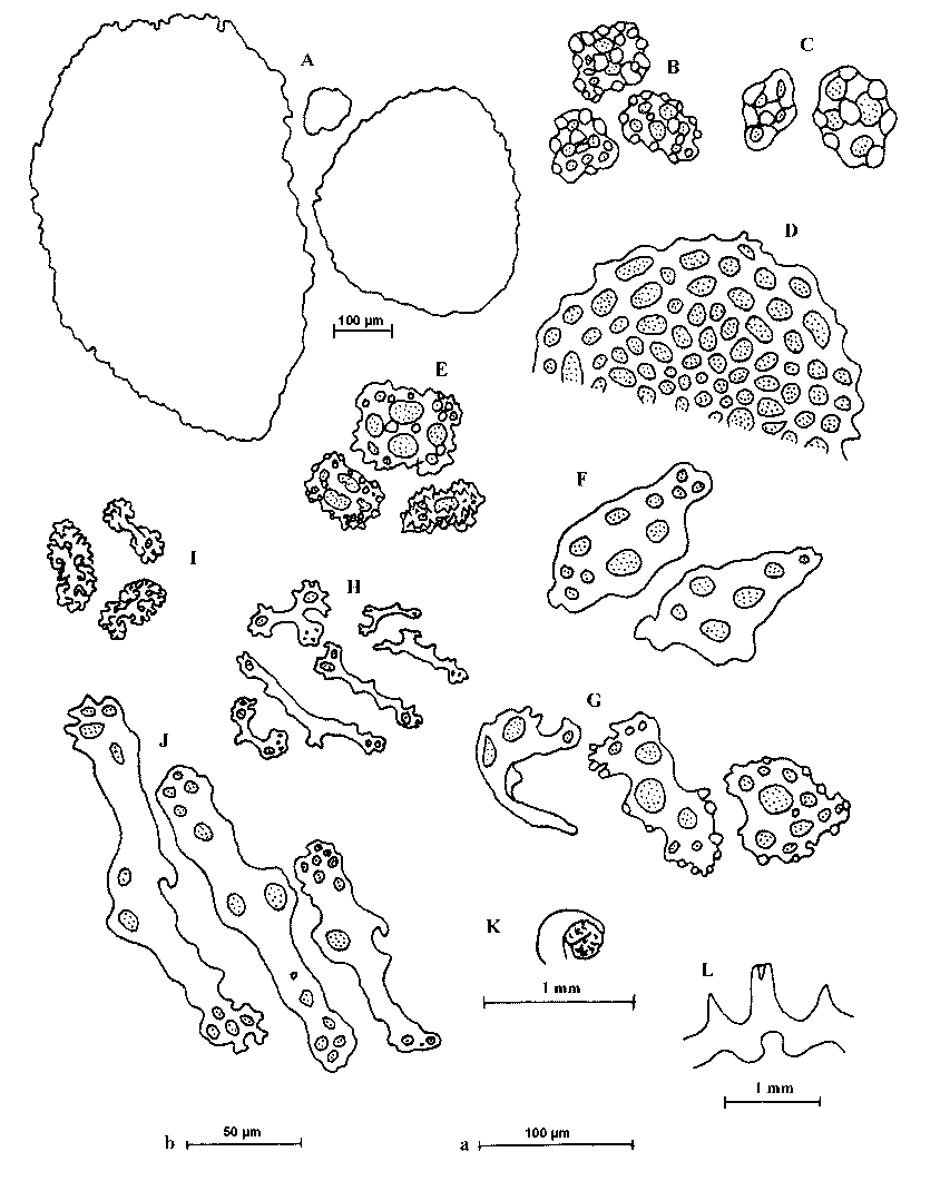
Calcareous ring ( Figure 9 View FIGURE 9 L) simple, well calcified, radial and interradial plates fused, radial plates with a bifid anterior prolongation for attachment of retractor muscle and a notched posterior surface; interradial plates shorter than radial plates, with a triangular anterior end and slightly indented posterior surface. Polian vesicle single, saclike; stone canal short, free; madreporite ( Figure 9 View FIGURE 9 K) spherical, grooved, lodged in the posterior notch of middorsal interrradial plate. Respiratory trees delicate, extremely transparent but well branched, right tree reaches about one third the body length from anterior end, left one shorter. Gonad not observed. Longitudinal muscles unpaired, thin, well developed anteriorly, fused to body wall for most of their length. Retractor muscles arise as single strands from the anterior third of the longitudinal muscles.
Dorsal body wall invested in imbricating scales and, besides these, include deposits of four types: multilayered buttons with large knobs; simple buttons with small knobs; usually complete baskets; and developing spicules of various shapes and sizes. Scales ( Figure 9 View FIGURE 9 A) rather complex, oval, 105–875 µm long; smallknobbed buttons of various shapes and sizes ( Figure 9 View FIGURE 9 B), 48–65 µm (mean 56 µm); largeknobbed buttons ( Figure 9 View FIGURE 9 C), 58–71 µm (mean 63 µm), with four or more holes; baskets characteristic, flat ( Figure 9 View FIGURE 9 E), 39–65 µm (mean 51 µm), with four large and four smaller alternating holes, surface spinose. Dorsal podia supported by perforated rods and plates ( Figure 9 View FIGURE 9 F), 84–165 µm (mean 94 µm), some with marginal tuberosities ( Figure 9 View FIGURE 9 G) and reduced endplates, 90–145 µm (mean 132 µm). Ventral podia with similar deposits but larger endplates, 170–240 µm (mean 202 µm) ( Figure 9 View FIGURE 9 D). Tentacles supported by elongated, perforated rods, 42–258 µm (mean 107 µm), smaller in the branches ( Figure 9 View FIGURE 9 H) and larger in the stalk ( Figure 9 View FIGURE 9 J), as well as rosettes ( Figure 9 View FIGURE 9 I), 16–32 µm (mean 24 µm).
Distribution
Tropical IndoWest Pacific, up to 20 m.
Remarks
The single specimen was at first thought to represent Psolidium ornatum ( E. Perrier, 1893) . However, Dr Rowe (pers. comm.) drew my attention to Plesiocolochirus dispar (Lampert) , a species which he thinks the specimen represents. I checked several descriptions of P. dispar and its synonyms and I concur with Dr Rowe without reservation. P. dispar has been recorded from the Red Sea and Somalia (see Clark & Rowe 1971) but this is its first record from southern Africa. However, the species was reported from Tuléar and Nosy Bay ( Madagascar) by Cherbonnier (1988) who also provides a detailed description of the species. The current specimen agrees well with that material.
| SAM |
South African Museum |
No known copyright restrictions apply. See Agosti, D., Egloff, W., 2009. Taxonomic information exchange and copyright: the Plazi approach. BMC Research Notes 2009, 2:53 for further explanation.
|
Kingdom |
|
|
Phylum |
|
|
Class |
|
|
Order |
|
|
Family |
|
|
Genus |
Plesiocolochirus dispar ( Lampert, 1889 )
| Thandar, Ahmed S. 2006 |
Pentacta gravieri
| Cherbonnier 1955: 162 |
Pentacta trimorpha
| Clark 1921: 171 |
Colochirus dispar
| Cherbonnier 1988: 164 |
| Clark 1971: 180 |
| Ekman 1918: 32 |
| Vaney 1905: 187 |
| Lampert 1889: 820 |