
Parapapio broomi Jones, 1937
|
publication ID |
https://doi.org/ 10.5252/geodiversitas2023v45a5 |
|
publication LSID |
lsid:zoobank.org:pub:CB66D102-090F-4AE7-89E8-D83E9024718B |
|
DOI |
https://doi.org/10.5281/zenodo.7815496 |
|
persistent identifier |
https://treatment.plazi.org/id/0277E376-FFE1-D471-FEE8-B5BD63C2F16F |
|
treatment provided by |
Felipe |
|
scientific name |
Parapapio broomi Jones, 1937 |
| status |
|
MATERIAL EXAMINED. — Cranium. BPB 1, partial skull with right P3/-M3/ ( Figs 5 View FIG , 6 View FIG ; Tables 3 View TABLE , 5 View TABLE ). — Mandibles. BPB 2 ( Fig. 11 View FIG ; Tables 5 View TABLE , 6 View TABLE ); BPB 3 ( Fig. 14 View FIG ; Tables 5 View TABLE , 6 View TABLE ); BPB 4 ( Fig. 9 View FIG ); BPB 13 ( Fig. 16 View FIG ; Table 6 View TABLE ); BPB 30 ( Fig. 17 View FIG ; Table 6 View TABLE ).
DESCRIPTION
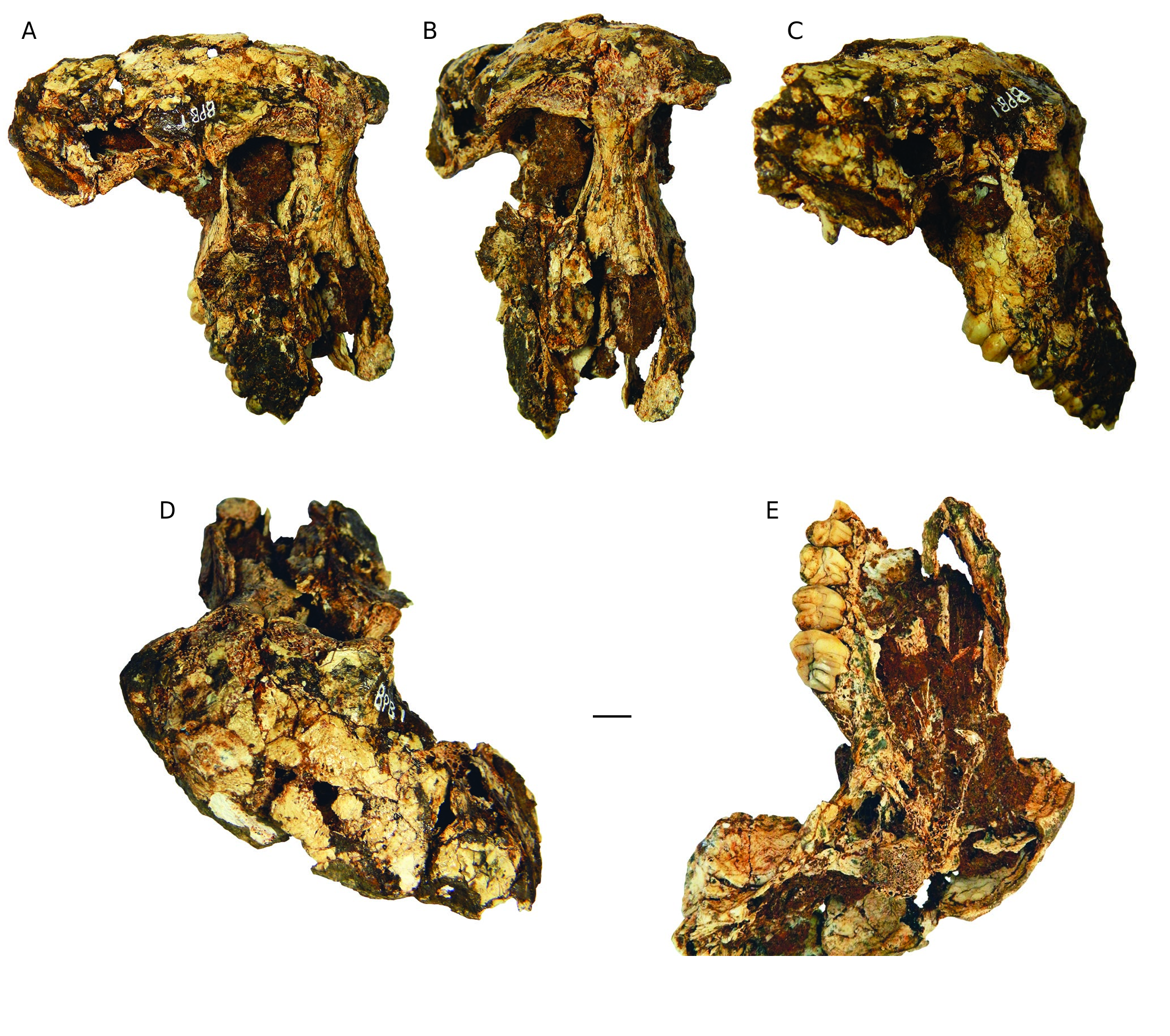
Cranium BPB 1 ( Figs 5 View FIG , 6 View FIG ; Tables 3 View TABLE , 5 View TABLE )
General description and preservation. This fossil is a highly distorted and crushed partial skull (right side of the skull and part of the left side of the face) ( Fig. 5 View FIG A-C). It lacks an important part of its left side probably due to erosion of the “ Parapapio spot”. At the time of discovery, the skull was partially exposed and the right side of the skull was protected in the breccia ( Fig. 4 View FIG ). Due to the fossilisation, the skull is almost flattened, the right part of the calvarium is practically in the same plane as the face, which complicates the anatomical study according to the classical anatomical plans. The fossil is 110.4 mm long antero-posteriorly (from the broken anterior part of the muzzle to the preserved posterior part of the skull). The face is 92.8 mm long (from the broken anterior part of the muzzle to the glabella) and the preserved width is approximately 53.6 mm (from the broken lateral edge of the left orbit to the lateral edge of the right orbit). The preserved part of the maxilla is approximately 50 mm long anteroposteriorly (from the fragment of the P3/ to the end of the bone of the dental arch posteriorly to the M3/).
The classical anatomical landmarks are not preserved here for the linear measurements and it is therefore impossible to make a further comparison with data from the literature or on more complete specimens. Much of the back of the skull is not preserved and originally the skull was longer, the muzzle is relatively short if we compare it with skulls of extinct and extant specimens of Papio , except for Papio izodi (see Gilbert et al. 2018). This skull belongs to a medium-sized to large papionine such as Parapapio . The skull also presents several other diagnostic features of Parapapio ( Freedman 1957, 1965; Szalay & Delson 1979, Jablonski & Frost 2010; Harrison 2011; Gilbert 2013) and differs from the smallest taxa of fossil Papio such as P. izodi and P. angusticeps ( Gilbert et al. 2015, 2018), namely, in lateral view ( Fig. 5C View FIG ), despite some small cracks, the interorbital and nasal regions indicate a slight concavity and lack of an ante-orbital drop. The maxillary ridge is weak in comparison with that of Papio and the maxillary fossa seems shallow. In anterior view ( Fig. 5B View FIG ), as well as in superior view ( Fig. 5D View FIG ), despite the lack of some superficial bone, the supraorbital torus is lightly built and not projected anteriorly, and the glabella is not prominent. In superior view, the ophryonic groove is absent. The anterior temporal line is weak. Only the right upper toothrow (P3/-M3/) is preserved but the P3/ is badly damaged ( Figs 5E View FIG ; 6A, B View FIG ).
Calvaria. It is represented mainly by the frontal and a part of its right side (sphenoid, anterior parts of temporal and parietal) ( Fig. 5C, D View FIG ). The frontal presents many cracks and is distorted. It lacks some fragments of bone. A hole located postero-laterally has a rounded aspect and perhaps could be related to a predation mark. Despite the distortion, the observations in the anterior and right lateral views suggest that the anterior part of the frontal was moderately rounded. Only the right inferior temporal line is preserved.This line is well defined (weak crest), short (seems to stop abruptly after the postorbital constriction). The frontal doesn’t exhibit an ophryonic groove, just a very shallow depression on the right side and probably on the opposite side but this part is badly damaged. The morphology of the temporal line and the curved aspect of BPB 1 are similar to BF 43 in which the temporal lines are pinched, as well in other skulls attributed to Pp. broomi ( Gilbert, 2013) . In antero-lateral and lateral views, the sphenoid is covered anteriorly by some breccia and broken posteriorly at the suture with the temporal. The remnant of temporal presents many cracks and is heavily distorted. In its inferior part, a groove marks the beginning of the zygomatic arch.
Basicranium. Only the anterior part of the right side of the basicranium is preserved ( Fig. 5E View FIG ). At the junction with the palate, the lateral pterygoid plate is well preserved, except for a lack of a central fragment, and as well the deep fossa pterygoidea. Laterally, the fossa mandibularis is preserved but is slightly distorted and flattened. Posteriorly, there is a wellpreserved robust posterior glenoid process which is easier to observe in the lateral view.
Orbital region. The face of BPB 1 is badly damaged and distorted but it is possible to estimate the original morphology ( Fig. 5 View FIG A-C). The face lacks some superficial bone but this doesn’t modify greatly the morphology but alters the aspect of the supraorbital torus which seems lightly built and not projected anteriorly. There is no important bulge between the two orbits which means that the glabella is not prominent ( Fig. 5C View FIG ). The inter-orbital pillar (between the superior median corners of each orbit) is slightly broad (11.6 mm) as in BF 43 (11.5 mm) and long. The right orbit is more complete (the median and the superior margins, as well the medio-inferior corner, are preserved) than the left where only the median margin and medio-superior corner remain. The preserved morphology of the right orbit (23.2 mm height) seems to suggest that the orbit is squarish with rounded corners as observed in BF 43. The median part of the orbit is mostly filled with matrix but not near the inter-orbital pillar which allows us to observe inferiorly a developed fossa for a well-preserved lacrimal duct. In the medio-superior corner of the margin of the left orbit, there is a small supra-orbital notch ( Incisura supraorbitalis ). It is impossible to see this anatomical structure in the right orbit because there is a crack in the same area. The right zygomatic process of the maxilla is damaged, but it is possible to observe its origin (or root). The malar height is short as in BF 43 (the malar height is the distance between the inferior orbital margin and the inferior border of the zygomatic process of the maxilla as defined in Gilbert et al. (2015)).
Muzzle and palate
The anterior part of the snout is badly damaged ( Fig. 5A, B View FIG ). The premaxilla is not preserved and neither is the nasal aperture. The nasal is preserved only posteriorly and has many cracks. It is slightly distorted but this doesn’t affect the morphology. It is convex transversely and slightly concave antero-posteriorly near the level of the inferior edge of the orbits as in BF 43. There is no ante-orbital drop. The root of the right zygomatic process is located above the middle of the M3/ as in BF 43 and suggests that the muzzle is relatively short. The right maxilla is preserved and some fragments of the left maxilla are preserved along the nasal from the orbit to the premaxilla. The right maxilla is distorted but there is no indication of a strong maxillary ridge and of a deep maxillary fossa.
Only the right side of the palate is preserved ( Fig. 5E View FIG ). The postcanine tooth row is slightly curved and the lingual wall of the dental arch is moderately tall which suggest that the palate was moderately deep. Below and lingually to the M3/, there is an antero-posteriorly oriented depression which corresponds to the groove of the posterior palatine foramen. The bone of the dental arch doesn’t end immediately posteriorly to the M3/ but presents the same aspect observed in BF 43, forming a robust bony bulge. This suggests that the hard palate ends behind the M/3.
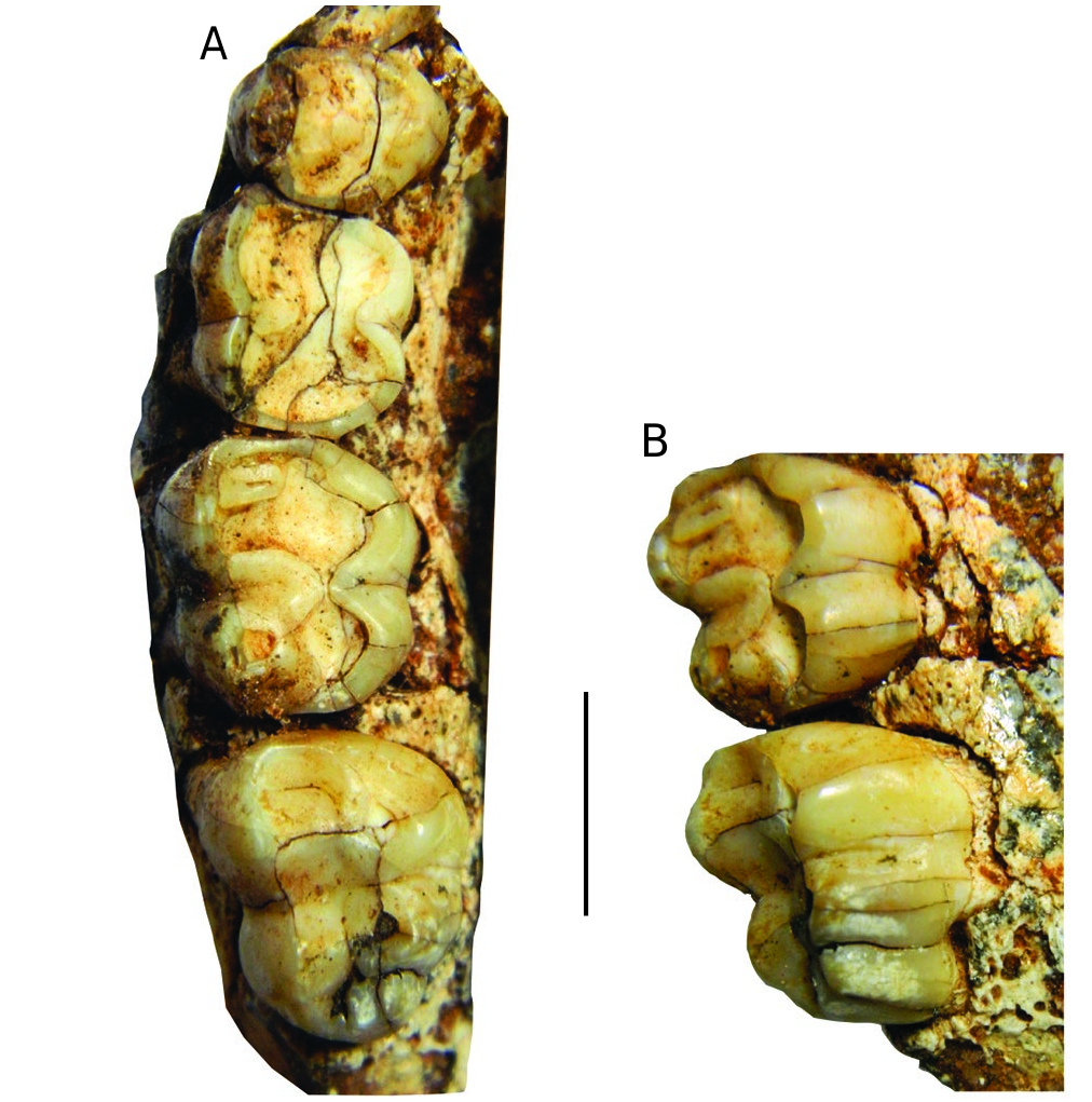
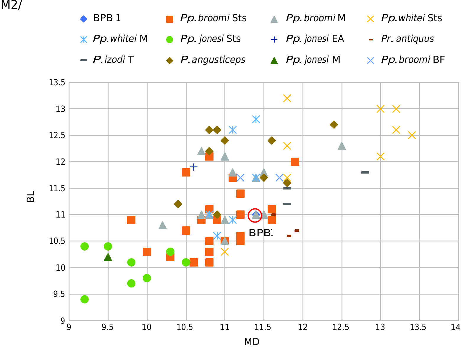
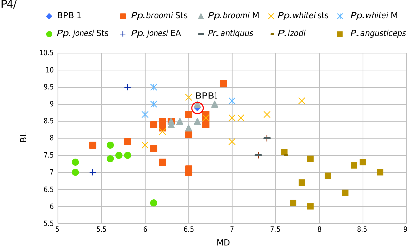
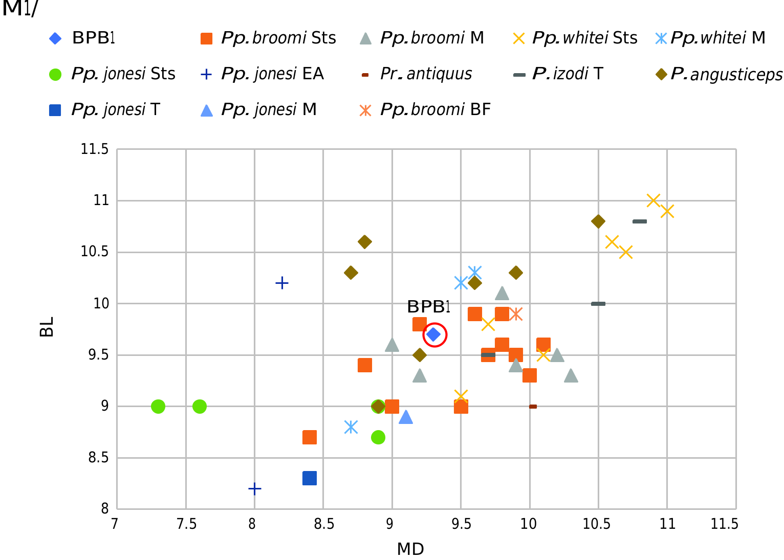
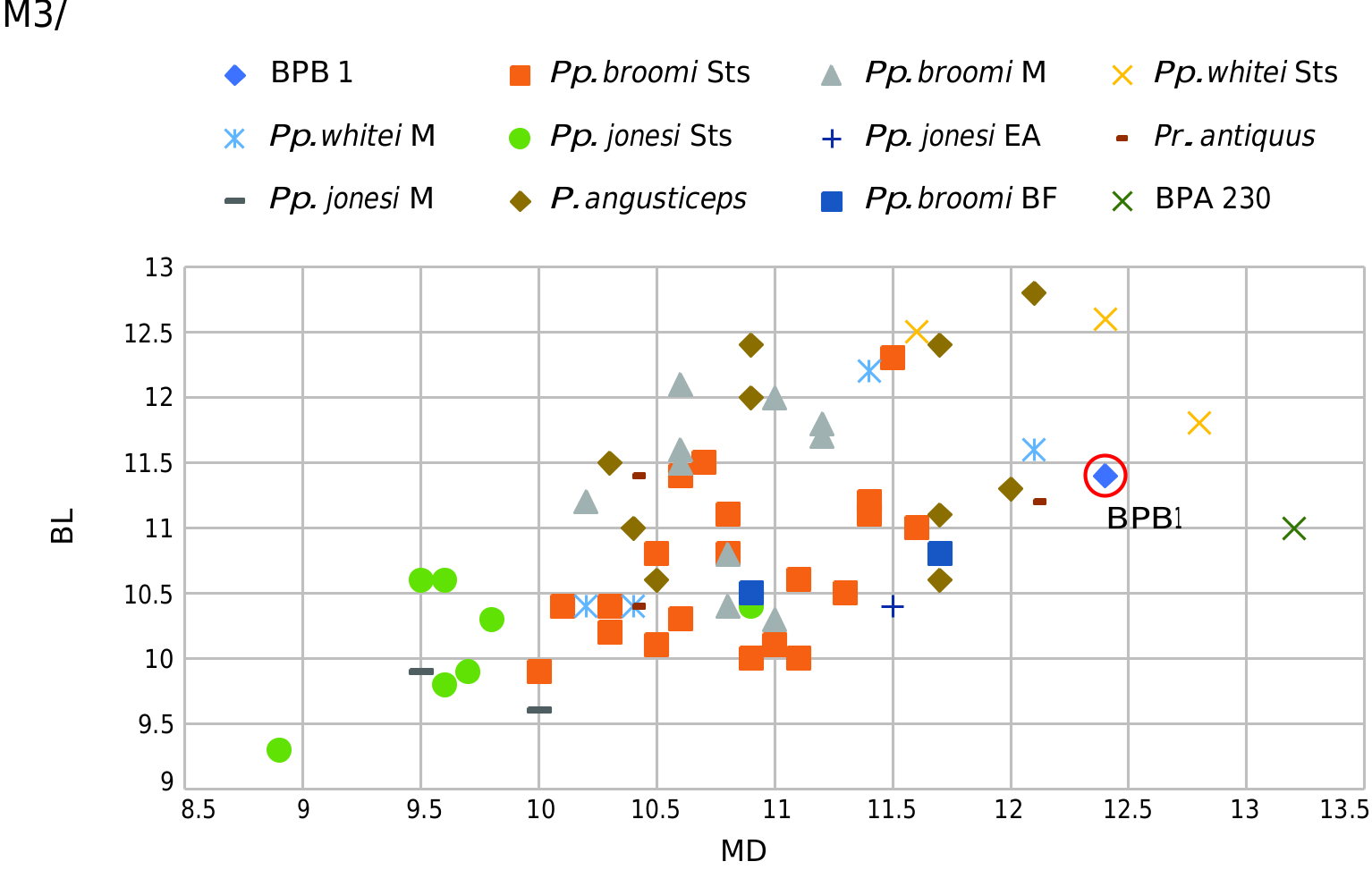
Dentition. The post-canine teeth are well preserved except for the P3/ which is just represented by a lingual fragment and the P4/ which has lost a fragment on the buccal edge of occlusal surface ( Fig. 6 A View FIG ; Tables 3-5 View TABLE View TABLE View TABLE ). The P4/ and M1/ are worn. The M2/ is less worn (more lingually than buccally). The M3/ is slightly worn at the apex of the protocone and the hypocone. The degree of wear of the preserved post-canine teeth is typical of a mature, but not very old, adult. Despite an important MPI (maxillary premolar index ( Fleagle & McGraw 1999, 2002) of 65.11 which correspond to the upper variation of Pp. broomi and perhaps the lower variation of Pr. antiquus ( Haughton, 1925) ( Gilbert 2007a) , the size of this premolar enters into the variation of Pp. broomi and Pp. whitei Broom, 1940 , from Sterkfontein and Makapansgat Limeworks ( Tables 3 View TABLE , 4 View TABLE ; Fig. 7 View FIG ). The molars increase in size from the M1/ to the M3/ ( Tables 3 View TABLE ; 4 View TABLE ). The range of variation in molar size overlaps with different taxa of fossil South African papionines ( Figs 8 View FIG ; 9 View FIG ) but differs from that of Papio jonesi . The size of the two molars of BPB1 plots within the range of variation of Pp. broomi , especially true for the M2/. The M3/s have a reduced distal loph as in BF 43. Heaton (2006) found this feature in Pp. broomi . The shape outline of the base of the crown is more trapezoidal than for the M2/. The well-preserved aspect of the occlusal surface of the M3/ permits observation of the intercuspid distance which is small and the molar reversed W-shaped in outline as described by McKee (1993). The M3/ presents an additional cusp linguodistally ( Fig. 6A, B View FIG ), a developed distoconulus ( Saheki 1966) as in an isolated M3/ from BPA (BPA 230 under study) and Sts 378A but less robust in this tooth than specimen BPB 1 ( Freedman 1957). This strongly increases the mesio-distal length of the M3/ of BPB 1 ( Fig. 10 View FIG ). In the oblique-lingual view ( Fig. 6B View FIG ), there is an additional strong root beneath the distoconulus for BPB 1 and the same applies to BPA 230. This distoconulus increases the length of the dental row P4/-M3/ or M1/-M3/ ( Table 3 View TABLE ). It seems that no distoconulus is observed in extant Papio ( Swindler 2002) .
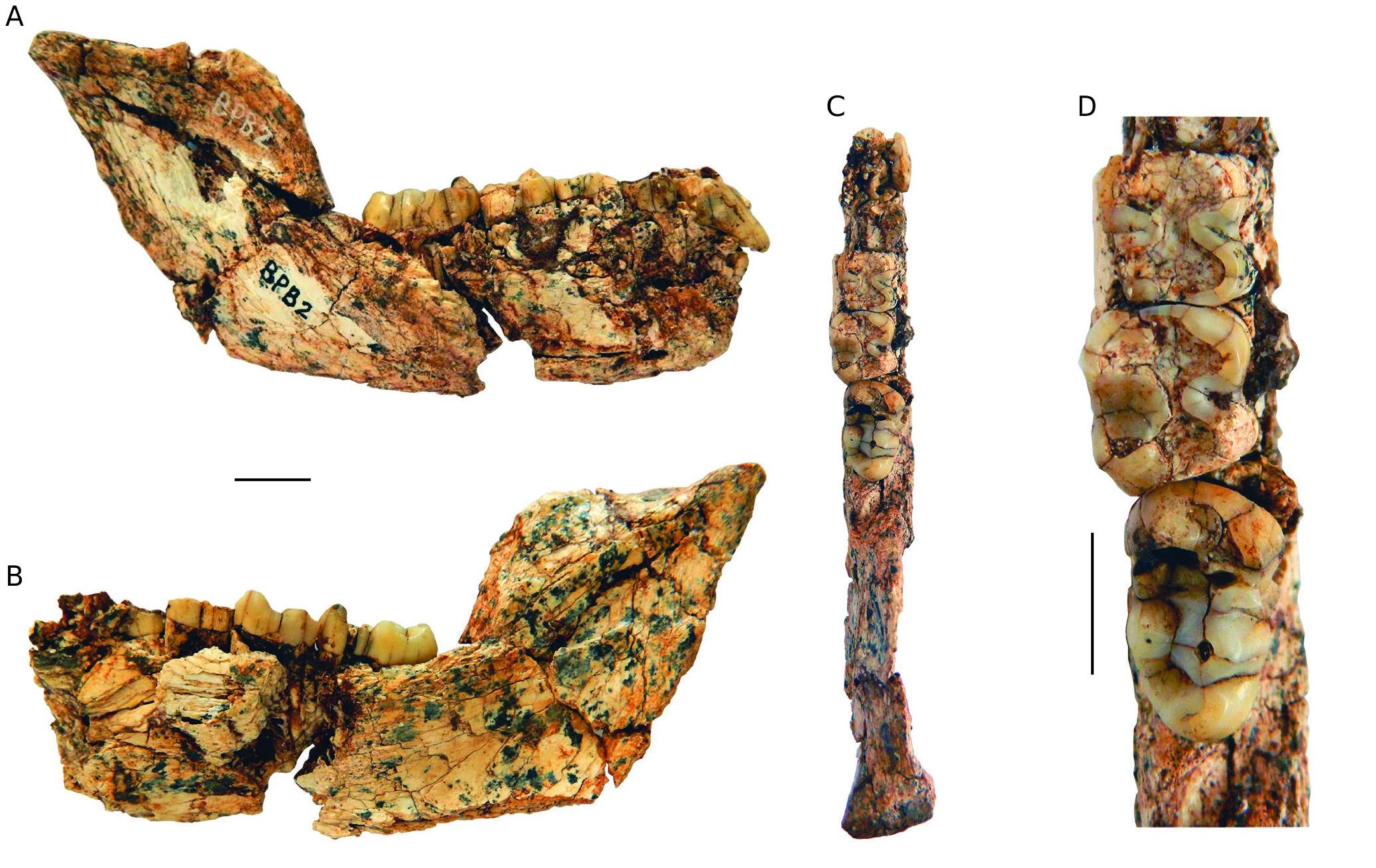
Mandible BPB 2 ( Fig. 11 View FIG ; Tables 5-7 View TABLE View TABLE View TABLE )
General description and preservation. BPB 2 is a partial right mandible broken mesially to the p/3 and the symphysis is absent (maximum length: 100.4 mm; breadth: 25.4 mm; height: 34.7 mm). The anterior part is badly damaged, and the two premolars are fragmentary. The corpus mandibularis is poorly preserved with some more or less important cracks and fragments of bone missing but it is better preserved on its posterior part but broken along the inferior edge. The ascending ramus is well preserved as is the mandibular condyle but the coronoid process is absent. The post-canine teeth are present but in various states of preservation.
Mandibular corpus and ramus. It is poorly preserved on the anterior portion which presents cracking, and some fragments of bone are missing. The mandibular corpus and the gonial region are broken along the inferior edge. Despite this damage, the mandibular corpus is moderately deep (preserved height between m/1-m/2 is 25.4 mm and between the m/2-m/3 is 24.9 mm). Buccally, there is no prominentia lateralis under the m/3 ( Fig. 11A View FIG ) unlike C. williamsi which presents a well-developed one. The buccal surface of the posterior part of the mandibular corpus is flat and vertical as in Parapapio . Inferiorly the lingual surface ( Fig. 11B View FIG ) presents an important submandibular fossa. The ascending ramus is almost in the alignment of the molar tooth row. In contrast, in BF 42B ( C. williamsi ), the ascending ramus is not so laterally located. The extramolar sulcus is narrow medio-laterally as well as mesio-distally. The ramus is low and 31.3 mm long antero-posteriorly. The distal edge of the ascending ramus tilts disto-superiorly. On the lingual surface, the mandibular foramen is low, located at the same level as the alveolar margin, and the groove of this foramen is deep and pronounced. The ascending ramus has lost the coronoid process. The mandibular condyle is well preserved and is more developed medially (maximum length: 8.5 mm; breadth: 13.7 mm).
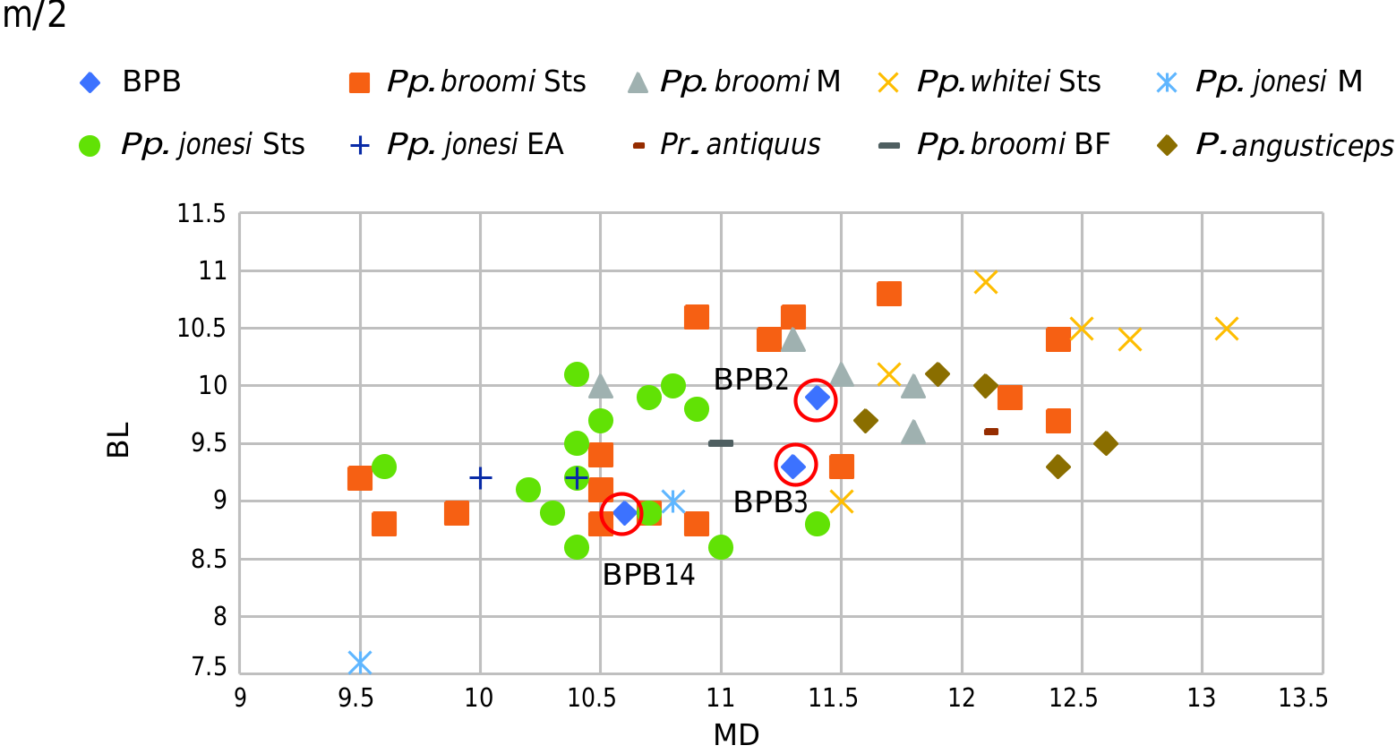
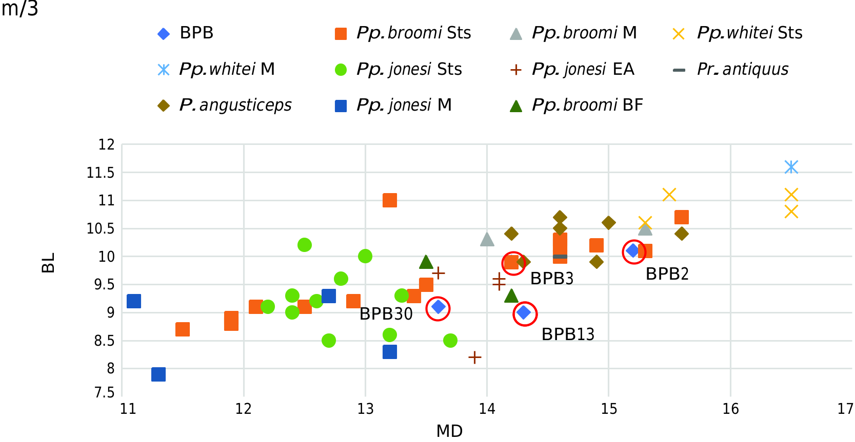
Dentition. The two premolars are very poorly preserved ( Fig. 11C View FIG ). The mesio-buccal flange on the p/3 ( Fig. 11A View FIG ) is the only well-preserved part and is short as in female individuals of Parapapio . The flange is fairly well developed and not honed by the upper canine which suggests that this specimen represents a female. The three molars have lost some fragments of enamel. The molars increase in size from m/1 to m/3 as is usual in papionines ( Tables 5-7 View TABLE View TABLE View TABLE ). The m/1 is the least well preserved and the most deeply worn of the three molars (the preservation doesn’t permit inclusion in the bivariate plots MD x BL). The molars possess relatively thick enamel and the less worn cusps are low ( Fig. 11D View FIG ). On the m/3, the prominent and salient hypoconulid is located buccally. This continues buccally by a developed distal marginal ridge and the distal fovea is deep, spacious and short mesio-distally as in many specimens of Pp. broomi (such as Sts 414A and Sts 565) and also but sometimes less pronounced in Pp. whitei (such as Sts 563). The dimensions of the two molars enter into the range of variation in molar size of different species of Parapapio but the m/2 doesn’t enter in the range of variation of Pp. jonesi ( Figs 12-13 View FIG View FIG ).
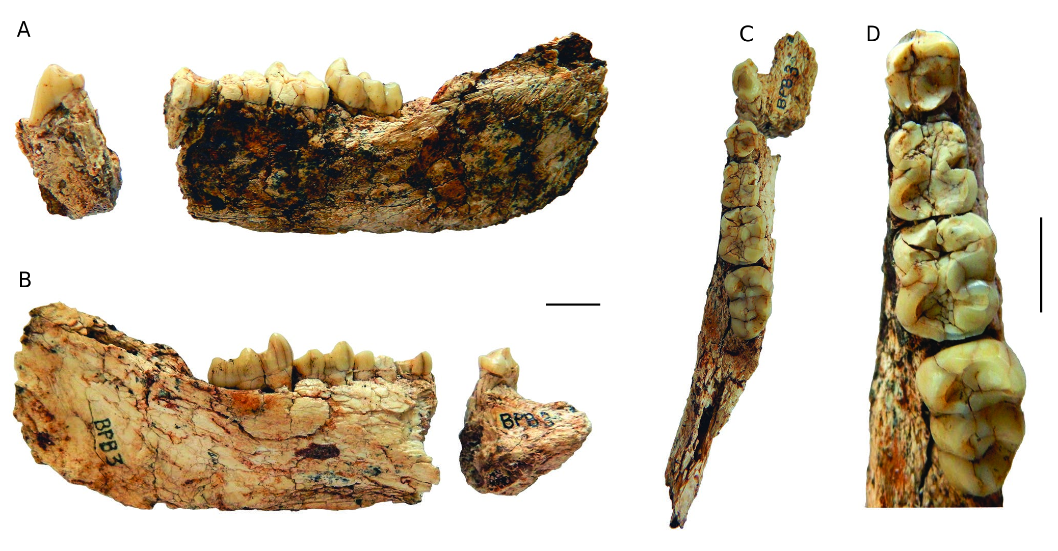
Mandible BPB 3 ( Fig. 14 View FIG ; Tables 5-7 View TABLE View TABLE View TABLE )
General description and preservation. BPB 3 represents a left mandible in two pieces, broken between the p/3 and p/4. The smaller fragment (21.9 mm long antero-posteriorly, 19.6 mm broad and 19.3 mm tall under the p/3) corresponds to the left part of the symphysis. The second fragment from the mesial side of the p/4 to the posterior of the ramus is 82.4 mm long antero-posteriorly and 40.1 mm high. The two fragments fit together well. The upper part of the ramus is absent and the gonial region is damaged. The bone presents some superficial cracking.
Symphysis. Only the left part of the symphysis is preserved. The inferior edge is broken. In buccal view ( Fig. 14A View FIG ), the symphysis extends at least as far posteriorly as the m/1 (a fragment is preserved at the inferior edge of the mandibular corpus below the contact p/4-m/1). In lingual view, the planum alveolare seems long and slopes moderately disto-inferiorly ( Fig. 14B View FIG ). This is moderately developed transversely and is moderately deep. The superior transverse torus is smooth. The digastric fossa seems small and shallow. The anterior surface is eroded.
Mandibular corpus. The mandibular corpus is better preserved than that of BPB2. It presents some superficial cracking, especially below the m/3. The inferior edge of the mandibular corpus is well preserved. It is moderately deep (height between m/1-m/2 is 25 mm and m/2-m/3 is 24.7 mm) and robust (mid-height width between m/1- m/2 is 11.5 mm and m/2-m/3 is 13 mm). The buccal surface ( Fig. 14A View FIG ) is slightly convex and vertical. Buccally, there is a mental foramen under the p/3 and no prominentia lateralis under the m/3. Distally the lingual surface ( Fig. 14B View FIG ) presents a shallow depression (submandibular fossa). The preserved part of the ascending ramus suggests that it is almost in the alignment of the molar tooth row. The extramolar sulcus is narrow medio-laterally as well as mesio-distally.
Dentition. The canine and incisors are absent ( Fig. 14C View FIG ). The premolars and molars are better preserved than in BPB 2 despite presenting some cracking (especially in p/4, m/1 and m/2) ( Fig. 14D View FIG ). The molars increase in size from m/1 to m/3 as for BPB 2 ( Tables 5-7 View TABLE View TABLE View TABLE ). The two premolars and the m/1 are deeply worn, m/2 is moderately worn and the m/3 has the two mesial cuspids worn (protoconid and paraconid) which suggest a mature adult individual. The degree of wear of the three molars corresponds to that observed in BPB 2. Probably, BPB 2 and BPB 3 specimens belong to the same individual. The mesio-buccal flange on the p/3 is fairly developed as is typical of females. The hypoconulid is well developed and located buccally as in BPB 2. The aspect of the distal fovea is very similar to that of BPB 2 as well. Concerning the dimensions of the m/2 and the m/3, they are the same as for BPB 2 ( Figs 12-13 View FIG View FIG ).
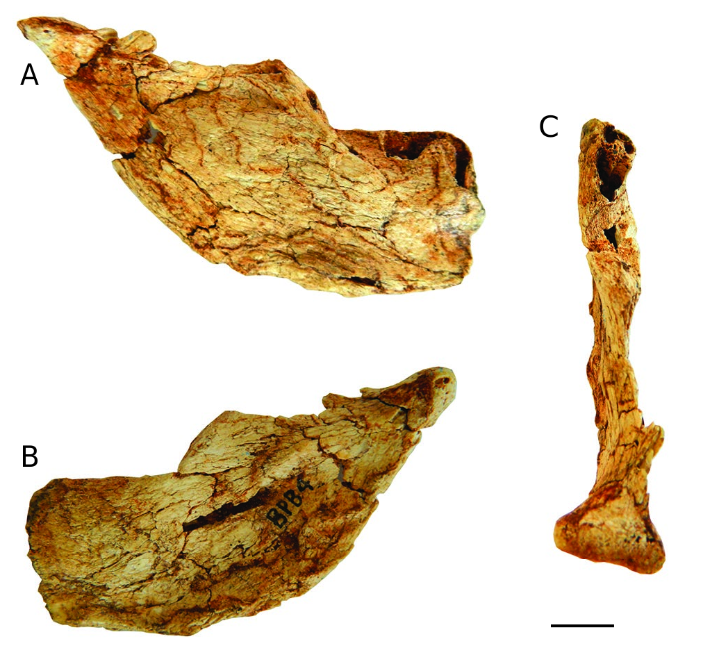
Mandible BPB 4 ( Fig. 15 View FIG )
General description and preservation. BPB 4 is the distal part of the right mandible (maximum length: 75.4 mm; height: 49.3 mm). The coronoid process is broken as is the mesial edge of the ascending ramus. The fossil presents some cracking, the most important are on the distal part of the ascending ramus. The mandibular condyle is well preserved but the coronoid process is absent. No tooth is preserved, only the sockets for the roots of the m/3.
Mandibular corpus and ramus. Only the portion below the m/3 is present and suggests that the mandibular corpus is vertical, high and moderately large (below the alveolar margin at m/3, height: 28.2 mm; breadth: 12.7 mm). The buccal surface ( Fig. 15A View FIG ) is slightly convex and there is no prominentia lateralis under the m/3. The extramolar sulcus is only preserved buccally. Inferiorly the lingual surface ( Fig. 15B View FIG ) presents a depression which corresponds to the submandibular fossa.
The gonial region is well preserved and only a small fragment of the inferior edge is broken. This region doesn’t expand distally or inferiorly but presents an open convex outline. In the lingual surface, at the junction of the gonial region with the distal edge of the ascending ramus, there are three smooth bulges which correspond to medial pterygoid tubercles. The ascending ramus is low and 36 mm long antero-posteriorly. The distal edge of the ascending ramus tilts disto-superiorly. On the buccal surface ( Fig. 15A View FIG ), the mandibular foramen is present and the surface bone covering the canal of this foramen is lacking, probably due to damage during fossilization. The mandibular condyle is well developed medially (maximum length: 7.1 mm; preserved breadth: 17.4 mm). The general morphology of the ascending ramus of BPB 4 is very similar to that of BPB 2.
Dentition. This fragment is edentulous, but the sockets for the roots of the m/3 are present, well preserved for the distal root and partially preserved for the mesial root ( Fig. 15A, C View FIG ).
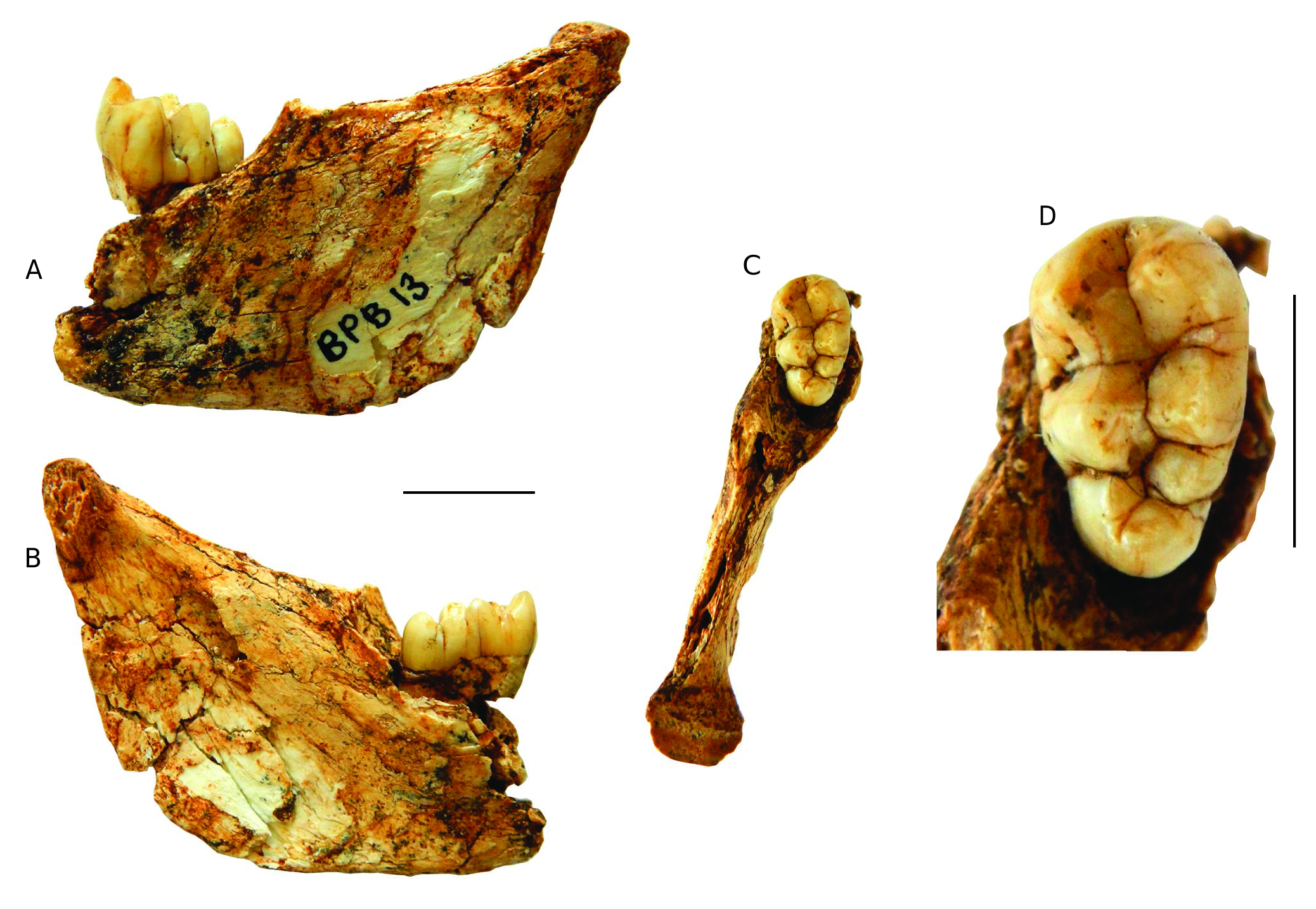
Mandible BPB 13 ( Fig. 16 View FIG ; Tables 6 View TABLE , 7 View TABLE )
General description and preservation. BPB 13 is the distal part of a left mandible with the m/3 (maximum length: 43.9 mm; height: 42.6 mm). The fossil presents some cracking. The coronoid process is broken and the mandibular condyle is damaged. The gonial region is preserved as is most of the posterior part of the ascending ramus. The tooth row is only represented by the well-preserved m/3.
Mandibular corpus. Just a part of the mandibular corpus is preserved below the m/3. The buccal surface ( Fig. 16A View FIG ) is almost flat and there is no prominentia lateralis under the m/3. The extramolar sulcus is short mesio-distally and narrow bucco-lingually as in BPB 3 ( Fig. 16C View FIG ).
The gonial region is well preserved, except for a few fragments of missing bone. The dorso-inferior outline of this region is convex. The lingual surface of the gonial region ( Fig. 16B View FIG ) is slightly concave. The mandibular foramen is well preserved and the mylohyoid notch is also well preserved. The ascending ramus is better preserved in its posterior part and the posterior edge of the ascending ramus tilts slightly dorsally. The anterior edge of this ramus is broken but the ramus is clearly low and long. The mandibular condyle is broken lingually and abraded (maximum preserved length: 5.9 mm and breadth: 9.9 mm).
Dentition. The m/3 is well preserved and just the apex of the two mesial cusps (protoconid and metaconid) are slightly worn ( Fig. 16D View FIG ). This fossil corresponds probably to a younger adult than BPB 2 and BPB 3. The cuspids are more centrally positioned than in Papio robinsoni (laterally positioned). This molar presents different extra-cusp and cusplet. A small Tuberculum intermedium is present between the metaconid and the entoconid. A low accessory cusp, near the base of the crown is also present between the protoconid and the hypoconid. The distal part of the molar is developed with a Tuberculum sextum. The hypoconulid is important and is located disto-lingually. Buccally, there is a clear distal marginal ridge and the distal fovea is deep. The size of the molar plots within the range of variation of Pp. broomi ( Tables 4-5 View TABLE View TABLE ; Fig. 13 View FIG ).
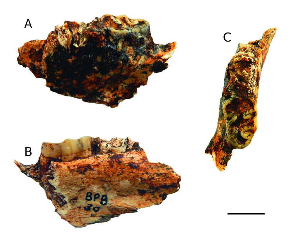
Mandible BPB 30 ( Fig. 17 View FIG ; Tables 6 View TABLE , 7 View TABLE )
General description and preservation. BPB 30 is part of a left mandible with fragmentary m/2 and worn m/3 (maximum length: 44.9 mm; breadth: between m/2-m/3: 10.9 mm; height: 29.3 mm).
Mandibular corpus. The mandibular corpus is only preserved below the m/2 and the m/3 and is not preserved over the entire height. It seems narrow inferiorly. On the lingual surface ( Fig. 17B View FIG ), a deep concave depression slopes from mesio-inferiorly to disto-superiorly and corresponds to the submandibular fossa.On the buccal surface ( Fig.17A View FIG ), there is a smooth concavity probably due to fossilization and it seems that the prominentia lateralis doesn’t exist. The mandibular corpus presents a slender aspect unlike Cercopithecoides . A short portion of the extramolar sulcus is preserved. This is reduced and located disto-buccally to the m/3 and not buccally as in Cercopithecoides .
The crown of the m/2 is represented only by a fragment of dentine ( Fig. 17C View FIG ). The m/3 is very worn suggesting that it represents an old adult and lingually it lacks some fragments of enamel at the metaconid and entoconid. The enamel is thick and the median buccal and lingual clefts are narrow as usual in Papionina . The hypoconulid is developed and located buccally. As for BPB 13, the size of the m/3 of BPB 30 enters into the range of variation of Pp. broomi ( Tables 6-7 View TABLE View TABLE ; Fig. 13 View FIG ).
REMARKS
In the literature, there is more information concerning the skulls than the mandibles to distinguish the different taxa of Papionini . So here, despite the preservation of BPB 1, this skull is most useful for the discussion. We must consider the biochronological age of the specimen and it is necessary to discuss older taxa (from East Africa) but also the Plio-Pleistocene Papionini . BPB 1 cannot belong to another Pliocene papionin taxon such as Pliopapio Frost, 2001 (5.2-4.2 Ma: Ethiopia) ( Frost 2001; Jablonski & Frost 2010) which has a clear ante-orbital drop and an ophryonic groove. It cannot be attributed to Procercocebus antiquus either ( Gilbert 2007a) which occurs at Taung ( South Africa), and is characterized by an extremely straight nasal profile, widely divergent anterior temporal lines, relatively larger premolars, more prominent maxillary ridges and relatively more extensive maxillary/suborbital fossae than Parapapio . As indicated in the general description of BPB 1, the morphology of the interorbital and nasal regions indicates a slight concavity and lack of an ante-orbital drop which exclude the possibility that this specimen represents an individual of the smaller taxa of fossil Papio such as P. izodi and P. angusticeps ( Gilbert et al. 2015, 2018).
Three species of Parapapio are recognized in South Africa ( Jablonski & Frost 2010; Harrison 2011; Gilbert 2013) during the Plio-Pleistocene: Pp. jonesi , Pp. broomi and Pp. whitei . Older fossils exist in the Early Pliocene at Langebaanweg ( Grine & Hendey 1981) and Waypoint 160 ( Gommery et al. 2008b, 2009, 2014) but they are too fragmentary or limited to assign to a specific taxon. In BPB 1, the maxillary ridge is more developed and the maxillary fossa is deeper than in Pp. jonesi . The premaxilla is not preserved in BPB 1 but it would not have extended much further anteriorly, as suggested by the anterior morphology of the maxilla, and as such the muzzle appears short and broad and its dorsum is flattened. All these features are typical of Pp. broomi . In Pp. whitei , the muzzle is long and narrow. The nasal bone and muzzle, especially in males, have a peaked aspect ( Gilbert 2013). The morphology of BPB 1 is similar to Pp. broomi specimens such as BF 43 from Pit 23 at Bolt’s Farm or M 202 from Makapansgat, so it can confidently be assigned to this species.
Pp. ado (Hopwood, 1936) , a smaller species, is known from Kenya ( Jablonski et al. 2008a) and Tanzania ( Harrison 2011) (4.1-3.49 Ma). Its frontal and maxilla have similar morphologies ( Harrison 2011) to Pp. broomi . These two species differ by the nasals which form a midline keel in Pp. broomi which is absent in Pp. ado . Despite the distortion of the nasals, the preserved transversal convexity in BPB 1 suggests that a midline keel was present. An older species exists, Pp. lothagamensis Leakey et al., 2003 ( Leakey et al. 2003; Jablonski & Frost 2010; Gilbert 2013) (7.4- c. 5 Ma: Kenya), but it is considered to be more primitive than the younger species ( Gilbert 2013).
The mandibles BPB 2 and BPB 3 probably belong to the same individual, which must have been a female. The teeth of these mandibles show a stage of wear corresponding to that of BPB 1. These two mandibles were found in a reduced sample of breccia taken from the “ Parapapio spot”. It is very likely that these three fossils belong to the same individual.
No known copyright restrictions apply. See Agosti, D., Egloff, W., 2009. Taxonomic information exchange and copyright: the Plazi approach. BMC Research Notes 2009, 2:53 for further explanation.