
Palaeomylus lii, MENG & WYSS & HU & WANG & BOWEN & KOCH, 2005
|
publication ID |
https://doi.org/ 10.1206/0003-0082(2005)473[0001:GMFTLP]2.0.CO;2 |
|
DOI |
https://doi.org/10.5281/zenodo.5634131 |
|
persistent identifier |
https://treatment.plazi.org/id/03B187E7-FFFE-FFF9-456B-F9A7A7F6FD0D |
|
treatment provided by |
Felipe |
|
scientific name |
Palaeomylus lii |
| status |
sp. nov. |
Palaeomylus lii , new species
HOLOTYPE: IVPP V 14128 View Materials , a fragmentary right maxilla with P4 and M1 ( fig. 3 View Fig ).
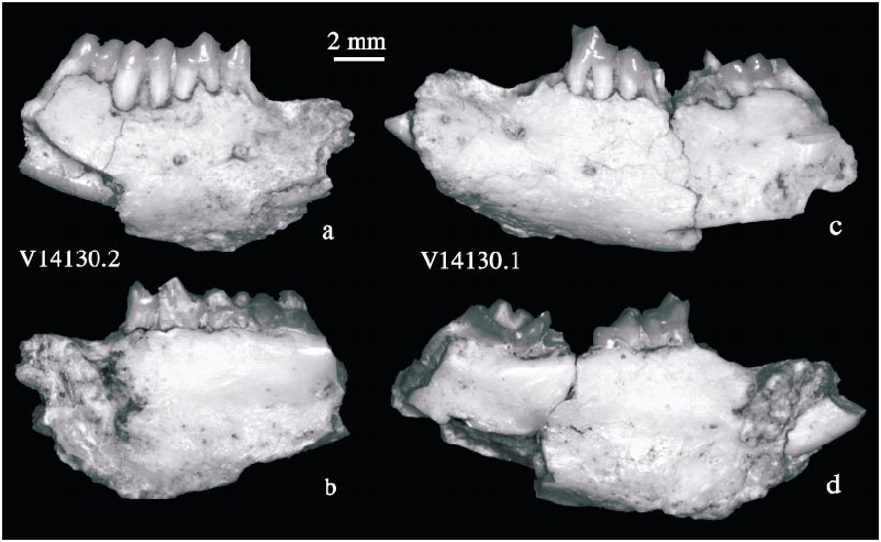
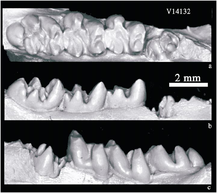
REFERRED SPECIMENS: V 14129.1, a fragmentary left mandible with m1–3; V 14129.2, a fragmentary left mandible with m1–3; V 14129.3, a fragmentary right mandible with m3; V 14129.4, a fragmentary left mandible with m2; V 14129.5, a fragmentary right mandible with m3; V 14129.6, a fragmentary left mandible with talonid of m2 (a young individual); V 14130.1, a fragmentary left mandible with p4, m3, and partial m1–2 (from the same individual of V 14130.2); V 14130.2, a fragmentary right mandible with p3–m2 (p4 and m1 broken); V 14130.3, a left partial femur and innominate (associated with V 14130.1–2); V 14131 View Materials , a fragmentary right mandible with m1–2; V 14132 View Materials , a left mandible with erupting p4 and m1–3. Numerous fragmentary mandibles with broken teeth are not numbered. See tables 1 View TABLE 1 and 2 View TABLE 2 for measurements.
LOCALITY AND AGE: Upper part of the Nomogen Formation at Bayan Ulan, Inner Mongolia; late Paleocene.
ETYMOLOGY: Palaeo (Greek), ancient; mylos (Greek), grinder or millstone, in analogy with Eurymylus , Eomylus , Rhombomylus , and Sinomylus . The trivial name honors Professor Chuankui Li for his landmark contributions to the study of Glires .
DESCRIPTION: The posterior edge of the anterior root of the zygoma lies lateral to the anterior half of M2. The tips of the P4–M1 roots are exposed in the floor of the orbit. The third upper premolar is not preserved; its alveolus indicates a doublerooted tooth that is narrower than P4. The nonmolariform P4 is also doublerooted and has an oval outline in occlusal view ( fig. 3 View Fig ). One cusp occurs at the anterolingual corner of the tooth; its anterior surface bears a flat wear facet. No contact facet for P3 occurs on the anterior surface of P4. Three ridges emanate from the lingual cusp. The anterior ridge (anteroloph) extends labially, forming the anterior edge of the tooth. The central one of the three ridges curves to meet the lingual base of the labial cusp. A small depression occurs between this ridge and the anteroloph. The third ridge (posteroloph) runs posteriorly from the lingual cusp, and bends labially at the posterolingual corner of the tooth, extending in a gentle curve to reach the labial margin of the tooth. The extended posterolingual corner suggests a hypocone. An elongate basin is formed between the posterior and the middle ridges. The labial cusp is conical and a short ridge projects from it posterolabially to join the posteroloph.
A large lingual root and two small labial ones occur on M1. The tooth’s lingual surface bears a shallow depression bordering the protocone and hypocone. The protocone forms a curved ridge that is continuous anteriorly with the preprotocrista and posteriorly with a crescentic hypocone ( fig. 3 View Fig ). The preprotocrista, made up of thick enamel, is low and curved; it ends labially at the small paraconule. Because the paraconule is positioned labially very near the paracone, the preprotocrista is unusally long. A short, curved postparaconule crista connects the paraconule and the base of the paracone. The preparaconule crista extends anterolabially to the anterolabial base of the paracone. The boundary between the protocone and hypocone is indistinct in occlusal view. The latter is slightly more lingually positioned. The metaconule is as large as the metacone and is situated much nearer the metacone than the protocone. The long postprotocrista is faint near the protocone but strengthens toward the metoconule. The metaconule’s posterior surface is rounded and covered by enamel. Light wear is reflected on the posterior surfaces of the metaconule and metacone. In contrast, the anterior surface of the metaconule is a heavily worn facet of bare dentine, which slopes into the large, transversely oriented trigon basin. A short premetaconule crista connects the metaconule and metacone. The paracone and metacone are roughly equal in size and are connected by a weak centrocrista. The paracone is the transversely wider of the two cusps and tapers lingually. A narrow cingulum is present at the labial base of the paracone. The metacone is roughly conical, but its tip is broken. There is no cingulum on the external side of the metacone. The postcingulum is low, long, and convex posteriorly. The hypocone shelf is sizable and concave. The thin enamel that covered the shelf has been eliminated by wear; the area adjacent to the metaconule still bears enamel. A contact facet for M2 is present on the posterior surface of M1.
Several fragmentary mandibles bear tooth marks. The best preserved mandibles are V14132 View Materials and V14130.1–2 ( figs. 4 View Fig , 5 View Fig ). V14132 View Materials is from a relatively young individual because its p4 is not erupted and the molars are little worn, whereas V14130.1–2 are from an old individual, of which the teeth are deeply worn. The symphysis of V14132 View Materials is partially preserved and is an inclined, simple articular surface. In V14130.1–2 the symphysis is much more extensive with uneven articular surface, the morphology being possibly age related. The lower diastema is 4.6 mm long in V14130.1 and 3.2 mm in V14132 View Materials ; the mandible is 6.7 mm deep at m1 and 3.48 mm thick at m 2 in V14130.1 and 4.8 and 3.0 at the same positions in V14132 View Materials . There are two mental foramina on the lateral surface of the mandible, a large one under p3 and a small one under p4. One or two very small foramina are also present lateral to the diastema. Ventrally the mandible bears numerous small fenestrations. The anterior edge of the masseteric fossa is even with the m3 trigonid and ends at a knob.
The lower dental formula is 1023. The incisor is slender, measuring 1.39 mm wide and 1.75 mm thick in V14130.1, and 1.1 by 1.36 mm in V14132 View Materials , again a difference due to age. It extends posteriorly to the talonid of m3 and is positioned ventromedial to the roots of the cheek teeth. Therefore, a protuberance is created on the lingual surface below the molars. In V14132 View Materials , the incisor is complete and is 15.7 mm long, of which the exposed tip is a small portion (2.2 mm). The entire lingual surface of this exposed tip bears a wear facet. The wear facet is oval and forms a step at its base. The tip of the incisor is at a position slightly lower than the occlusal surface of the cheek teeth ( fig. 4 View Fig ). The incisor enamel is thin. As in other Glires , it is distributed along the entire tooth longitudinally but mainly on the labial surface of the tooth. The incisor increases in width posteriorly.
The p3 is preserved only in the right mandible (V14130.2). It is small, nonmolariform, and doublerooted ( figs. 5 View Fig , 6 View Fig ). The trigonid consists of only one large cusp, of which the posterior surface bears a flat wear facet. A cingulid cusp lies at the anteromedial base of the main cusp. The talonid is a simple, low and transverse ridge.
The p4 is partially erupted in V14132 View Materials , and in occlusal view its crown pattern is visible ( fig. 7 View Fig ). The trigonid of the nonmolariform tooth consists of two cusps, with the lingual one (presumably the metaconid) being higher than the labial one (presumably the protoconid). Anterior and posterior ridges extend from the labial cusp to the anterior and posterior bases of the lingual cusp, respectively, defining a closed trigonid basin. The bases of the two cusps meet at the center of the trigonid basin. The talonid is narrower and lower than the trigonid. The lingual cusp (presumably the entoconid) on the talonid is conical and from it a narrow crest extends to a ridgelike cusp (presumably the hypoconid) at the labial side of the talonid. The trigonid and talonid are separated by a broad transverse valley in which there is no trace of the cristid obliqua. The p 4 in V14130.1 ( fig. 6 View Fig ) was worn. A crescentic trigonid basin develops between the trigonid cusps; the posterior wall of the trigonid forms a steeply sloping surface that sweeps posteriorly into the concave, structureless talonid basin.
The m1 and m2 are similar except that m2 is larger ( figs. 7–8 View Fig View Fig ). They differ from p 4 in that the talonid is wider and longer than the trigonid and has more differentiated cusps. The protoconid is much lower than the metaconid and has a wear facet on its tip that continues to its labial surface. The paracristid extends to the anterior side of the metaconid and is separated from the latter by a narrow groove. The paraconid is absent. The metaconid is pointed and extends anterolabially toward the paracristid. An extensive wear facet covers the anterolabial surface of the cusp. The protocristid is lower but thicker than the paracristid. Diverging from the condition seen in E. bayanulanensis , where the protocristid directly joins the metaconid, the protocristid curves around and reaches to the posterolingual side of the metaconid. Following moderate wear, the trigonid basin takes on the shape of a crescentic enamel lake between the metaconid and protocristid. The posterior wall of the trigonid is inclined anteriorly and bears a large wear facet. The trigonid becomes shaped by two major facets, a horizontal (occlusal) anterior one and a sloped posterior one, in later wear stages. The occlusal trigonid wear surface is level with the hypoconulid of the preceding tooth, thus forming a posterior functional extension of the talonid of the preceding tooth.
Specimen V14129.6 (not illustrated) is an unworn m2 talonid from a young individual. It shows that the cusps were originally pointed and the ridges sharp. Other specimens show various degrees of wear. The hypoconid is the largest cusp of the talonid and is labially extended. As with the protoconid, a wear facet marks the labial surface of the hypoconid. The cristid obliqua is short but broad, bears a mesoconid, and merges to the posterior wall of the trigonid at the midpoint. The entoconid is the highest cusp of the talonid, from which the welldeveloped hypolophid extends posterolabially to join the hypoconulid. It is eradicated early in wear and all cusps become confluent with the talonid basin. The hypoconulid is transversely stretched and becomes either completely obliterated or is recognizable only as a small notch separating it lingually from the entoconid. At this stage of wear the flat hypoconulid abuts the protoconid of the following tooth. The talonid basin is broad, concave, and smoothly polished after wear.
The m3, the longest lower molar, displays considerable variation ( figs. 6–8 View Fig View Fig View Fig ) between specimens. The metaconid remains a tall cusp even after the tooth is deeply worn, but is frequently broken. The talonid is slightly narrower but is much longer than on m2, owing to the presence of the third lobe formed of a large hypoconulid. In some specimens the m3 hypoconulid lobe consists of a single cusp, but in V14129.1 and V14132 View Materials it is bifurcated into two, relatively smaller cusps ( figs. 7 View Fig , 8 View Fig ). The entoconid is distinct and is aligned transversely with the hypoconid. It is separated from the hypoconulid by a broad, deep valley. Unlike in m1–2, the m3 hypolophid does not merge to the hypoconulid directly; instead, it joins the ridge that connects the hypoconid and hypoconulid. Following wear, the cusps and crests from flat surfaces with low, wide outer edges. Further wear erases all structures within the talonid, leaving only a broad, concave basin that is deepest lingually as shown in V14130.1 ( fig. 6 View Fig ).
A left partial femur and pelvis (V14131.3; fig. 9 View Fig ) were recovered from the same nodule containing V14130.1–2, indicating the likely association of these specimens. The anterior portion of the ilium is broken but a prominent posterior iliac spine, forming the anterior edge of a greater sciatic notch, is preserved. The medial surface preserves a partial, shallowly concave articular facet for the sacrum. A prominent femoral process lies anterior to the acetabulum that is deep and nearly hemispherical in shape and bears a welldefined rim except posteroventrally, where it is indented by the acetabular notch. The notch is deep and extends as a groove anterodorsally onto the upper surface of the acetabulum. The groove divides the lunate surface into two portions. The posterodorsal band of the lunate surface is narrow and bears a prominent ventral process that bounds the deep acetabular notch laterally. The rest of the lunate surface surrounds the nonarticular surface in the center of the acetabulum except at the acetabular notch. The medial surface of the innominate is a large, concave area. The sutures between ilium, ischium, and pubis are completely fused and no longer visible. The proximal portion of the left femur is preserved. The femoral head is well defined and has a globular articular surface. The greater trochanter is robust and is more proximally extended than the femoral head. The lateral surface of the greater trochanter is a low, curved ridge, which would extend distally to the third trochanter. The greater trochanter is marked posteriorly by a prominent trochanteric crest that overhangs a deep, pocketlike trochanteric fossa. The lesser trochanter forms a low triangular flange, projecting posteromedially from the shaft. A rounded ridge runs laterally from the lesser trochanter to meet the trochanteric crest of the greater trochanter.
COMPARISON: The collection from the late Paleocene Zhigden Member of the Naran Bulak Formation at Tsagan Khushu also includes articulated upper and lower dentitions of a new species of Sinomylus ( Kondrashov and Lopatin, 2003; Lopatin and Kondrashov, 2003). According to Kondrashov and Lopatin (2003), the lower jaws and teeth of the unnamed species are identical to those tentatively referred to Eomylus zhigdenensis by Dashzeveg and Russell (1988). Moreover, the mandible of the unnamed species of Sinomylus has an i3, leading Kondrashov and Lopatin (2003) to assign Sinomylus a dental formula intermediate between Mimotonidae and typical Eurymyloidea (since mimotonids have two pairs of incisors in both upper and lower jaws whereas eurymylids have single incisors in each jaw). Whether Sinomylus zhaii ( McKenna and Meng, 2001) also has an i3 is unknown.
Although the lower cheek teeth resemble those originally referred to E. zhigdenensis by Dashzeveg and Russell (1988), now allocated to a new species of Sinomylus ( Kondrashov and Lopatin, 2003) , the new taxon from Bayan Ulan certainly lacks i3, which distinguishes Palaeomylus from Sinomylus . In addition to absence of i3, cheek teeth of the new taxon also differ from those of ‘‘ E. zhigdenensis ’’ in several respects, as indicated in the diagnosis.


| IVPP |
Institute of Vertebrate Paleontology and Paleoanthropology |
| V |
Royal British Columbia Museum - Herbarium |
No known copyright restrictions apply. See Agosti, D., Egloff, W., 2009. Taxonomic information exchange and copyright: the Plazi approach. BMC Research Notes 2009, 2:53 for further explanation.
|
Kingdom |
|
|
Phylum |
|
|
Class |
|
|
Order |
|
|
Family |
|
|
Genus |