
Metamastigophorophyllon hamatum, Antić, Dragan Ž. & Makarov, Slobodan E., 2016
|
publication ID |
https://doi.org/ 10.11646/zootaxa.4211.1.1 |
|
publication LSID |
lsid:zoobank.org:pub:6B86C6BA-6AFE-4AAD-870D-04794C138D47 |
|
DOI |
https://doi.org/10.5281/zenodo.6074445 |
|
persistent identifier |
https://treatment.plazi.org/id/03B7878C-FFAE-CD41-FF4C-EC64B2FBED11 |
|
treatment provided by |
Plazi |
|
scientific name |
Metamastigophorophyllon hamatum |
| status |
sp. nov. |
Metamastigophorophyllon hamatum View in CoL sp. nov.
Figs 100–104 View FIGURE 100 View FIGURE 101 View FIGURE 102 View FIGURE 103 View FIGURE 104
Diagnosis. Differs from the other Metamastigophorophyllon species by the presence of a triangular, hairy, sternal lamella and a highly complicated inner branch of the lateral process. The inner branch consists of an anterior lamellar part clothed with setae, a tube-like structure with a clearly visible opening, and a characteristic hookshaped process with a visible opening on the anterior gonopods, or by male leg-pair 7 with a short basal process and, more apically, with a wide protrusion, both mesally orientated, on the prefemur.
Etymology. An adjective; to emphasize its unique, hook-shaped process on the inner branch of the lateral part of the anterior gonopods’ coxal process.
Material studied (total: 14 males, 18 females, 40 juveniles). Holotype. ABKHAZIA: male, Ochamchira District, village Otapi , near entrance to Cave “ Golova Otapa ” (“Otap’s Head”), Feb. 1983, S. Smirnov leg. ( ZMUM ρ3360).
Paratypes (total: 5 males, 24 juveniles). ABKHAZIA: 1 male, near Sukhumi, near Cave Kelassuri , litter, 11 Apr. 1983, S. Golovatch leg. ( ZMUM ρ3361).
GEORGIA: 2 males, 18 juveniles, 40 km W of Mestia, Kherkhvashi , E of Nakra (= Naki), 1250–1700 m asl, Quercus , Fagus , Carpinus , Picea , Abies , etc. forest, litter and bark, 21 Aug.–21 Sep. 1986, S. Golovatch leg. ( IZB) ; 2 males, 3 juveniles, 40 km W of Mestia, above Kherkhvashi , 1900–2200 m asl, timber-line ( Azalea , Picea , Abies ) and subalpine meadows, 21 Sep. 1986, S. Golovatch leg. ( ZMUM ρ3362) ; 3 juveniles, Mestia , 1500 m asl, Betula & Rhododendron on moraine, litter and under stones, 16 Sep. 1986, S. Golovatch leg. ( ZMUM ρ3363).
Other material (total: 8 males, 18 females, 16 juveniles). All from RUSSIA, Krasnodar Province : 3 males, 2 females, 1 juvenile, Tuapse District, 15 km SE of Novomikhaylovskiy, Psebe , deciduous forest, litter, under stones and in rotten logs, 29 Oct. 1981, S. Golovatch leg. ( ZMUM ρ3364) ; 1 male, Goryachiy Klyuch , 10 km S of village Fanagoriyskaya , mixed forest ( Fagus , Quercus , Picea , etc.) litter, under stones and in rotten logs, 30 Oct. 1981, S. Golovatch leg. ( ZMUM ρ3365) ; 2 females, 2 juveniles, Sochi, Dagomys , 250 m asl, Quercus , Carpinus , Fagus shrub, litter, logs, 18 May 1983, S. Golovatch leg. ( ZMUM ρ3366) ; 1 male, Goryachiy Klyuch, 12 km SW of Fanagoriyska, near Cave Fanagoriyskaya , Fagus & Acer forest, litter, logs, 19 May 1983, S. Golovatch leg. ( ZMUM ρ3367) ; 1 male, 7 females, 1 juvenile, Adygea, Pasture Abago near Guzeripl, Caucasian Nature Reserve , Abies & Fagus forest, Rhododendron thicket, 1100 m asl, litter, under bark and stones, 24–26 May 1985, S. Golovatch leg. ( ZMUM ρ3368) ; 1 male, 2 females, 10 juveniles, Severakaya District, Mt Derbiy , ca 15 km SW of Ubinskaya, 800–850 m, old Quercus , Fagus , Fraxinus , Alnus , etc. forest, litter, bark, 2 Jul. 1986, S. Golovatch leg. ( ZMUM ρ3369) ; 3 females, Adygea, Sakhray River , 20 May 2004, K. Voigtländer leg. ( SMNG) ; 1 male, 2 juveniles, same data, except 21 May 2004 ( SMNG), 2 females, Adygea, Poljana , 23 May 2004, K. Voigtländer leg. ( SMNG).
Type locality. ABKHAZIA: Ochamchira District, village Otapi, near entrance to Cave “ Golova Otapa ” (“Otap’s Head”).
Description. Body with 31 segments (including telson) in adults.
MEASUREMENTS. Males 10–10.5 mm long, vertical diameter of the largest pleurotergite 0.85–0.9 mm. Females 11.5–12 mm long, vertical diameter of the largest pleurotergite 1–1.05 mm.
COLORATION ( Fig. 100 View FIGURE 100 ). Dorsal and lateral sides of prozonites greyish, dorsolateral and ventrolateral sides with yellowish spots. Metazonites brownish or yellowish. Some specimens paler or darker.
HEAD. Slightly concave in males. Labrum with three medial teeth and 5+5 labral and 2+2 supralabral setae. Promentum triangular, without setae. Lingual plates with 6+5 setae. Stipites with ca 25+25 setae. Antennae 1.35 mm long in holotype. Length of antennomeres (in mm): I (0.08), II (0.2), III (0.3), IV (0.15), V (0.33), VI (0.16), VII (0.12) and VIII (0.01). Length/breadth ratios of antennomeres I–VII: I (1), II (2), III (3), IV (1.5), V (2.5), VI (1.5) and VII (1.4). Antennomeres II, IV, V, VI and VII with one, three, one, four and one sensillum, respectively. Number of ocelli 19–22, arranged in 5 rows in males; 20–23 in 5–6 rows in females.
COLLUM. Narrower than head, with six macrochaetae. Anterior edge semi-circular, posterior margin gently concave.
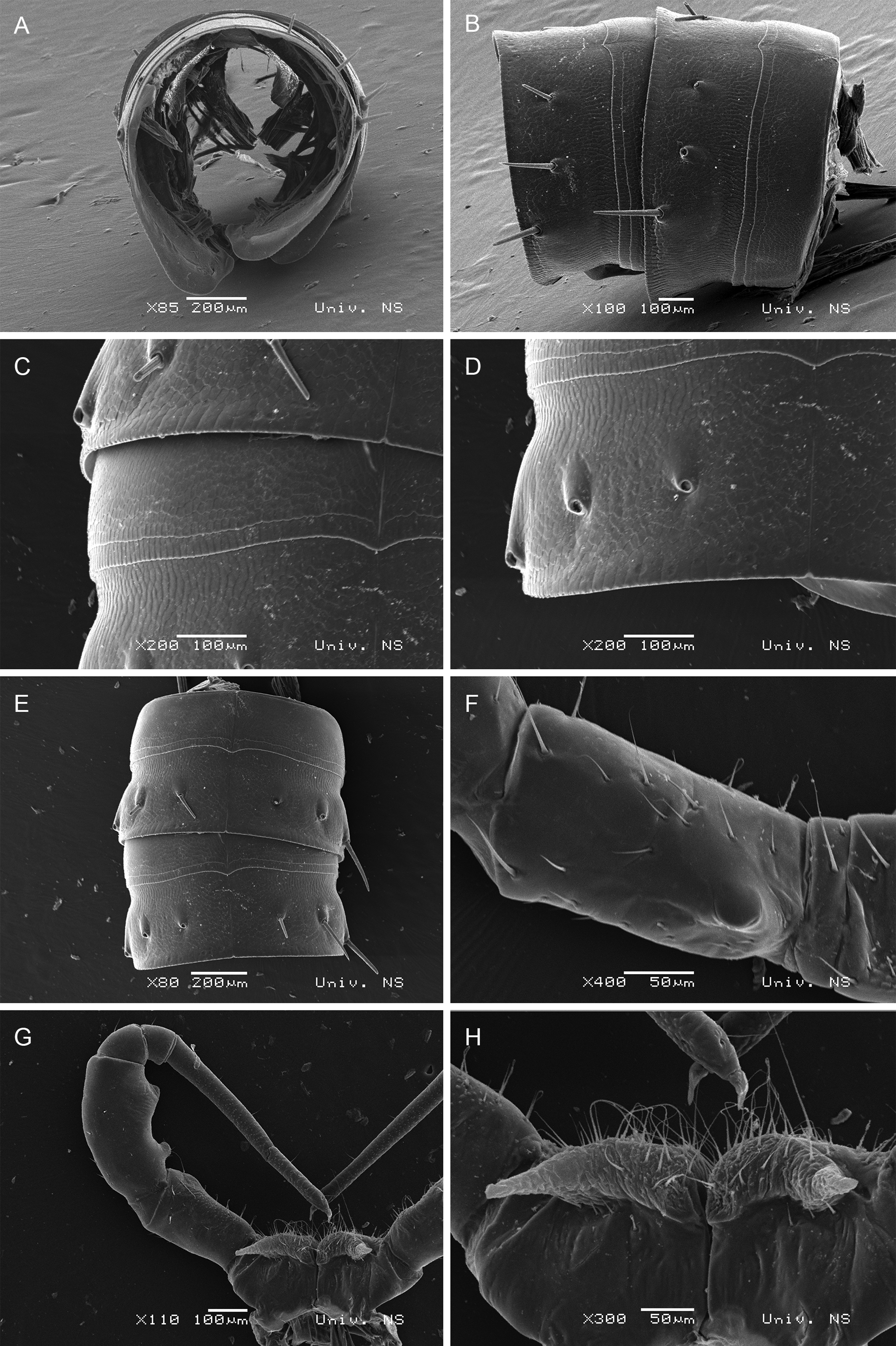
BODY SEGMENTS ( Figs 100 View FIGURE 100 , 101 View FIGURE 101 A–E). Lateral keels not developed; only barely visible lateral swellings present. Macrochaetae short, straight and trichoid, outer somewhat longer than inner and medial. CIX (pleurotergite 15) ~ 0.75; MIX (pleurotergite 15) ~ 1.1; PIX (pleurotergite 15) ~ 0.2; MA (pleurotergite 15) ~ 140˚. TELSON. Epiproct with a pair of spinnerets and 3+3 setae (1+1 paramedian, 2+2 marginal). Hypoproct with 1+1 apical setae. Paraprocts with 3+3 marginal setae.
WALKING LEGS. In both sexes, leg-pairs 1 and 2 with tarsal combs; prefemora with several long and robust setae; femora and postfemora with a group of several long and robust setae.
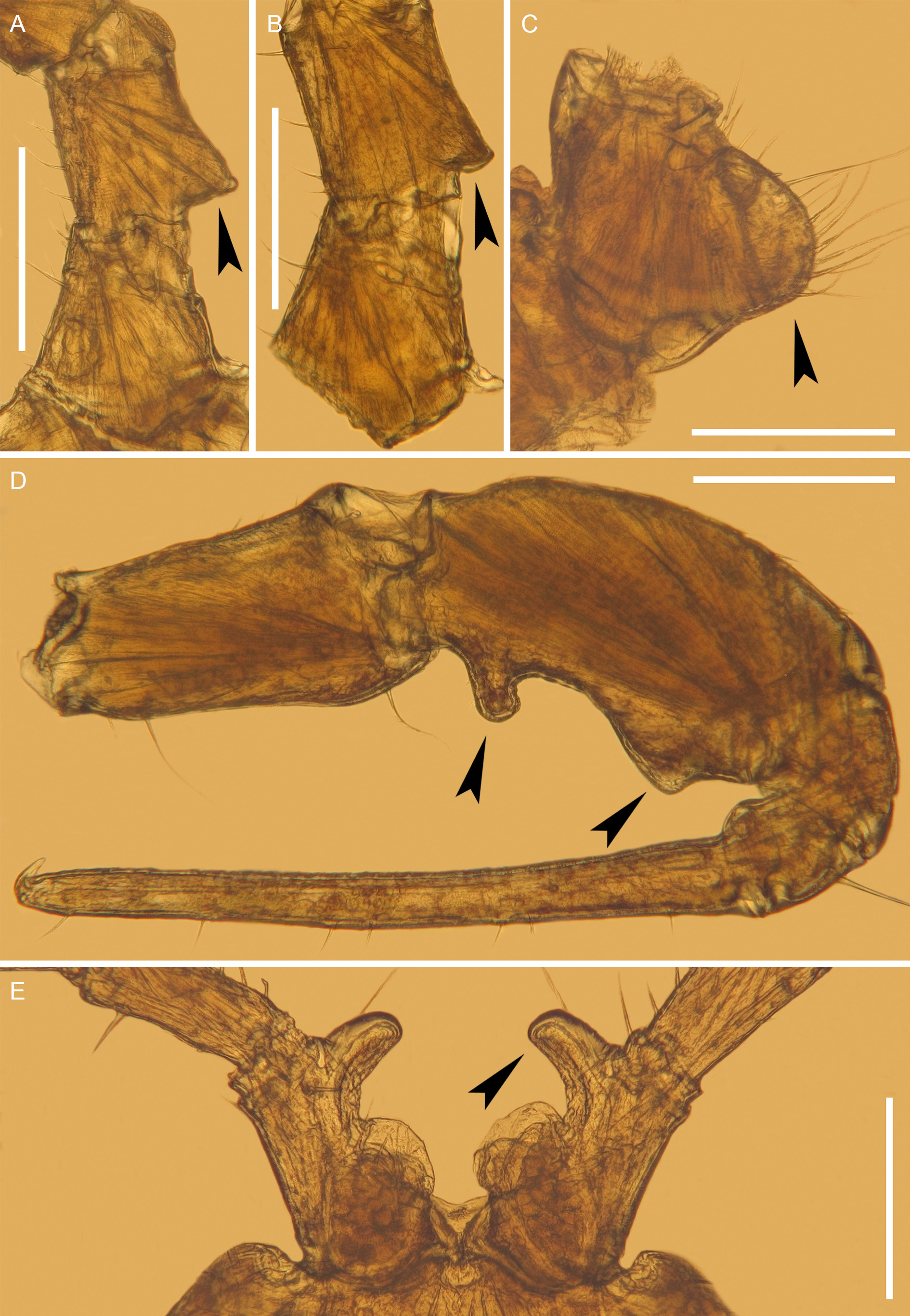
MALE SEXUAL CHARACTERS ( Figs 101 View FIGURE 101 F–H, 102). Leg-pairs 3–7 enlarged. Leg-pairs 3 and 4 each with all podomeres swollen; prefemur with a basal exterior protrusion. Leg-pair 5 with an extero-oral protrusion on prefemur. Leg-pair 6 without peculiarities. Leg-pair 7: coxae with a protrusion carrying numerous long setae, as well as one process; femora the most robust, with a short basal process and, more apically, with a wide protrusion, both orientated anteromesally; tarsus very long. Leg-pair 10 with coxal glands; with a well-developed coxal horn orientated caudad, these horns nicely abut between opening of coxal gland and remainder of coxae of leg-pair 11. Leg-pair 11 with coxal glands; without other peculiarities.
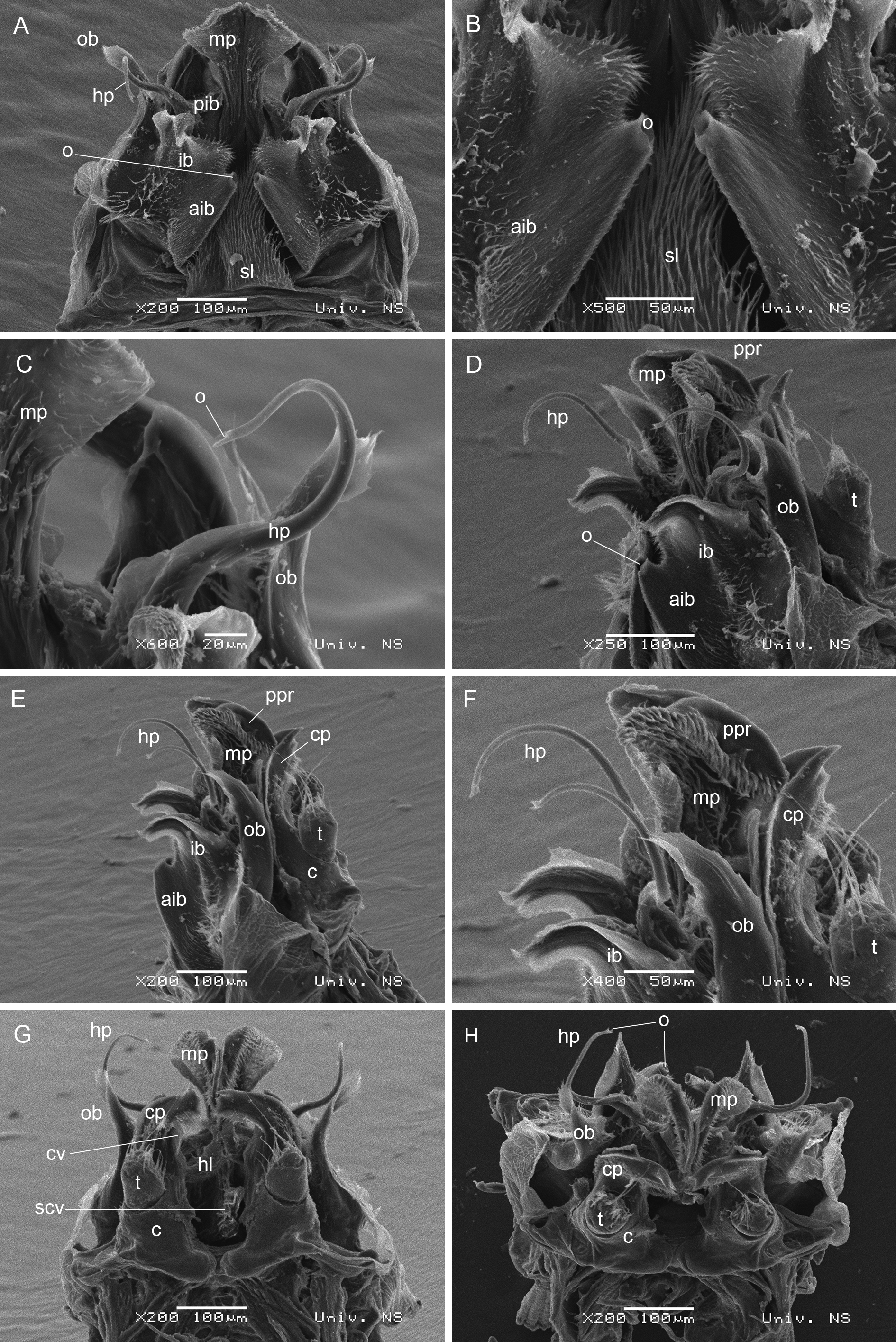
ANTERIOR GONOPODS ( Figs 103 View FIGURE 103 A–C, 104). Sternal plate anteriorly with a medial, triangular, hairy, posteriorly curved, sternal lamella (sl) [= S sensu Mauriès (1982): 387, figs 2 and 3]. Behind this lamella, at base, a small, nippled vesicle (wv) present. Coxal processes fused, only a small medial notch (n) present distally. Both coxal processes consisting of a medial part (mp) [= K sensu Mauriès (1982): 387, figs 1–3] and two lateral branches (inner and outer). Outer branch (ob) [= T sensu Mauriès (1982): 387, figs 1–3] simple, twisted, slightly curved laterally and anteriorly; apically, with several denticles; mesal edges with numerous setae. Inner branch (ib) [= T’ sensu Mauriès (1982): 387, figs 1–3] much more complex and consisting of an anterior part (aib) with a lamellar structure covered by setae; anteriorly, with a tube-like structure with a clearly visible opening (o). This lamellar structure connected to posterior part of inner branch (pib) which is in the form of characteristic hook-shaped process (hp) with a visible opening (o) and possibly with a function in sperm transfer. Medial part in the form of a wing from anterior and posterior views; laterally, covered by numerous setae; mesal edges folded inside and forming a projection (ppr) [= k sensu Mauriès (1982): 387, figs 1 and 3] orientated downwards. Immediately below this projection, a pair of hairy levers (hl) [= N sensu Mauriès (1982): 387, figs 1 and 3] present. Below these hairy structures, a vesicular structure (scv) can be seen (probably a syncoxal vesicle). The lateralmost, basal part of anterior gonopods carrying branched setae (bs).
POSTERIOR GONOPODS ( Figs 103 View FIGURE 103 D, 104D–H). Coxites (c) clearly divided. Telopodites (t) present on posterolateral side, clothed with setae. Distal part of coxal processes (cp) curved mesally; both with one tooth apically, while downward edges clothed with minute setae. Coxal processes orientated posteriorly. Coxal vesicles (cv) present on anterior side.
Distribution. Abkhazia, Georgia, Russia ( Fig. 166 View FIGURE 166 , red circle).


No known copyright restrictions apply. See Agosti, D., Egloff, W., 2009. Taxonomic information exchange and copyright: the Plazi approach. BMC Research Notes 2009, 2:53 for further explanation.
|
Kingdom |
|
|
Phylum |
|
|
Class |
|
|
Order |
|
|
Family |
|
|
Genus |