
Magelona picta, Mortimer & Kongsrud & Willassen, 2022
|
publication ID |
https://doi.org/ 10.1093/zoolinnean/zlab070 |
|
publication LSID |
lsid:zoobank.org:pub:278AA1B0-674E-414D-A47A-D87F43E2D6E4 |
|
DOI |
https://doi.org/10.5281/zenodo.6459428 |
|
persistent identifier |
https://treatment.plazi.org/id/039087DB-FFEE-FFF5-40E9-733DFED1FC2B |
|
treatment provided by |
Plazi |
|
scientific name |
Magelona picta |
| status |
sp. nov. |
MAGELONA PICTA View in CoL SP. NOV.
( FIGS 9 View Figure 9 , 10 View Figure 10 )
Zoobank registration: urn:lsid:zoobank.org:act:D51FC252-3FDE-4C25-AE0D-9FD07ACB0579 .
Type locality: Angola: 6.8526°S 12.2831°E, 50 m depth GoogleMaps .
Type material: Holotype, Angola: St. 7AN–03, a f i n 9 6%E t o h (Z M B N1 0 7 3 3 8, D N A -v o u ch e r) . Paratype, same sample as holotype, 1af in 96%Etoh ( ZMBN115737 View Materials , DNA-voucher) .
Etymology: The specific name is derived from the Latin word pictus, painted, referring to the pigmented body, in particular, the abdominal lateral lamellae.
Diagnosis: Prostomium marginally wider than long, no prostomial horns. Chaetigers 1–8 with slender foliaceous notopodial lamellae, long s u p e r i o r d o r s a l l o b e s a n d s l e n d e r, t r i a n g u l a r, ventral neuropodial lamellae. Lamellae of chaetiger 9 digitiform, postchaetal. All thoracic chaetae capillary. Abdominal lateral lamellae foliaceous and heavily pigmented. Abdominal hooded hooks tridentate, in two groups, vis-à-vis. No pouches observed, pygidium unknown.
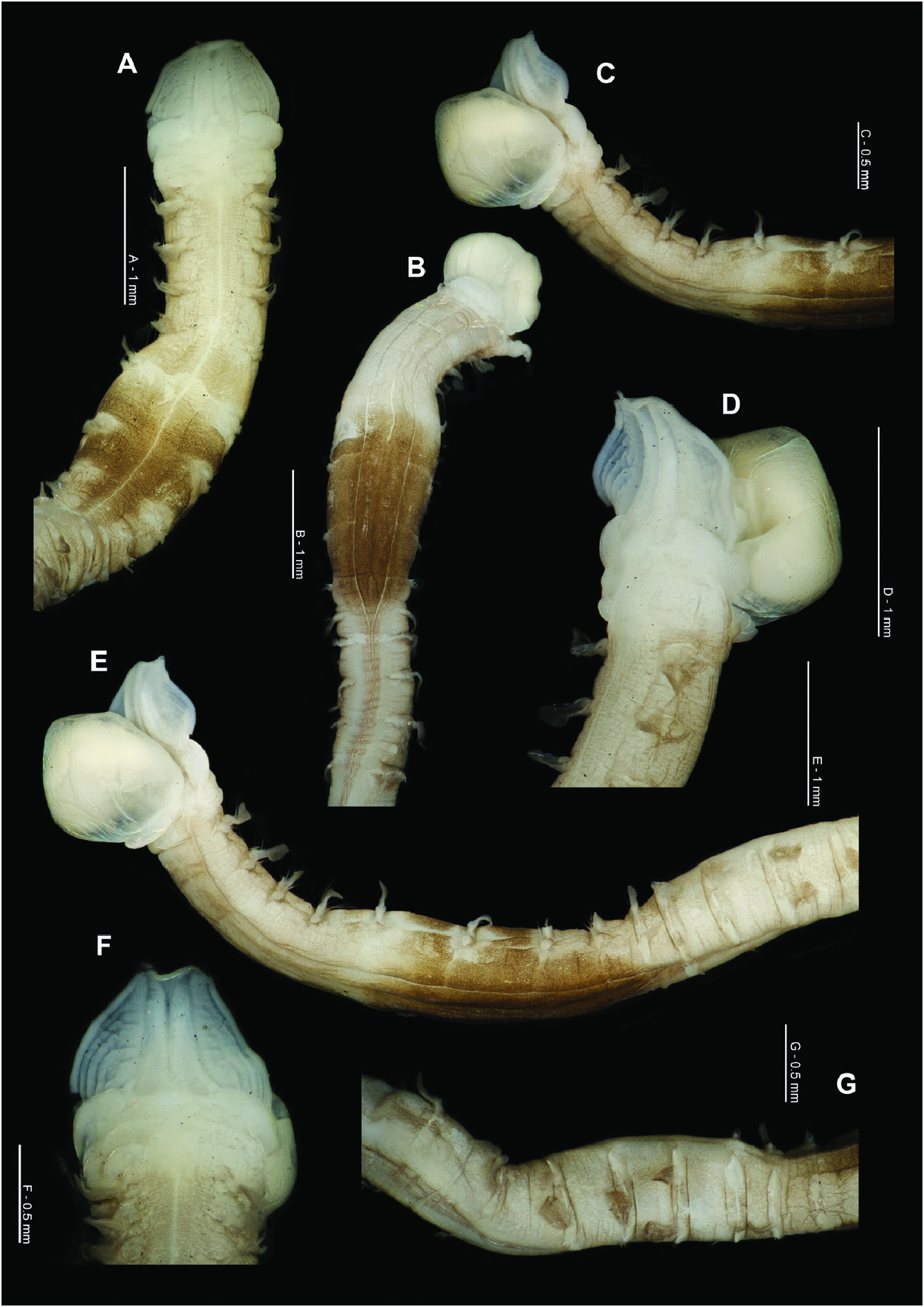
Description: A moderately stout species; junction between thorax and abdomen not hugely marked, but the dorsally flattened thorax is somewhat wider and thinner than the rounded abdomen ( Figs 9A View Figure 9 , 10B, E View Figure 10 ). Holotype, anterior fragment: prostomium 0.95 mm long, 1.0 mm wide; thorax 4.6 mm long (including prostomium), 1.0 mm wide (at widest point between chaetigers 5–6, only 0.75 mm in anterior thorax. NB specimen shows signs of being marginally dorsoventrally flattened); abdomen 0.6 mm wide; total length approximately 10.25 mm for 20 chaetigers (width measurements not including parapodia). Paratype, anterior fragment: 2.25 mm long for four chaetigers. Thoracic chaetigers of the mid-thorax bulbous and rounded when viewed dorsally ( Figs 9A View Figure 9 , 10A View Figure 10 ).
Prostomium rounded subtriangular ( Figs 9B View Figure 9 , 10F View Figure 10 ), marginally wider than long (L: W ratio 0.95). No prostomial horns, anterior margin straight, medially indented for holotype. Prostomium with one pair of thick longitudinal, dorsal muscular ridges, abutting for majority of length, diverging only at distal tips. Minute outer pair of ridges at their bases. Distinct prostomial markings either side of prostomial ridges, as arched lines and smaller more circular areas towards the centre. Burrowing organ of holotype almost fully everted, heart-shaped ( Figs 9B View Figure 9 , 10B, C, E View Figure 10 ), light longitudinal ridges inferiorly, appearing smooth superiorly. No palps retained, unknown.
Achaetous region behind the prostomium, approximately one and a half times the length of chaetiger 1 ( Figs 9A View Figure 9 , 10A View Figure 10 ). Chaetigers 1–8 similar ( Fig. 9C–I View Figure 9 ); parapodia biramous. Notopodia with low triangular prechaetal lamellae confluent with slender foliaceous postchaetal lamellae, slightly broader in anterior thorax, the upper edges of which are smooth. Single, long prechaetal superior dorsal lobe present on all thoracic chaetigers, except chaetiger 9. Neuropodia with low prechaetal lamellae confluent with long, slender, triangular lamellae with rounded tips, directly under chaetal bundle. Postchaetal lamellae slightly larger than prechaetal, particularly in anterior thorax ( Fig. 9C, D View Figure 9 ). Ventral lamellae decreasing greatly in size from chaetigers 1–4, and then of a similar size in the posterior thorax. Those of chaetigers 6–8 in a slightly postchaetal position.
Chaetiger 9: notopodial and neuropodial postchaetal lamellae similar in both rami, digitiform with rounded tips ( Fig. 9J View Figure 9 ). Chaetae emerging above notopodial, and below neuropodial lamellae. Chaetae of chaetigers 1–9 simple bilimbate winged capillaries ( Fig. 9L View Figure 9 ).
Parapodia of abdominal chaetigers ( Fig. 9K View Figure 9 ) sinuous, foliaceous lateral lamellae with slight basal constrictions. Lamellae extend postchaetally behind chaetal rows in anterior abdomen, triangular. Small, triangular processes observed at inner margins of chaetal rows (DML, VML). Abdominal chaetae tridentate hooded hooks ( Fig. 9M View Figure 9 ) all of a similar size, two superior fangs parallel, above main fang. Hooks in two approximately equal groups within each ramus, main fangs vis-à-vis ( Fig. 9K View Figure 9 ). Approximately eight hooks per ramus in the anterior abdomen. No abdominal pouches observed, but only 11 abdominal chaetigers examined. Pygidium unknown.
Colour: No living material observed. Preserved specimens cream with a distinct dark-brown pigment band present in the posterior thorax (chaetigers 5–8), comprising of many small dots ( Figs 9A View Figure 9 , 10 View Figure 10 ). Pigment band extends around the body from dorsal to ventral surface. However, some areas around the parapodia, particularly those of chaetigers 6 and 7 and along the mid-dorsal and mid-ventral lines, are clear of pigmentation ( Fig. 10A, E View Figure 10 ). Weaker pigmentation present over much of the body (except on prostomium and thoracic lamellae), similar in colour to pigment band, although much more diffuse. Pigmentation darker in furrows along the body. Abdominal lamellae heavily pigmented, comprising of dots over much of their surface. Staining with methyl green shows no distinct pattern, weakly stained over the entire body, except where heavily pigmented.
Distribution: Only collected from one station (off Angola) in the Gulf of Guinea Large Marine Ecosystem (LME), during West African surveys, 50 m ( Fig. 1 View Figure 1 ).
Remarks: This new species can be distinguished from other pigmented magelonid species within the MIWA region based on the following characters. In possessing a prostomium, which is wider than long with distinct prostomial markings either side of the prostomial ridges, M. picta differs from M. alleni , M. fasciata and M. mackiei . In addition to the distinct brown pigment band on chaetigers 5–8, M. picta possesses pigmentation over much of the body, including the abdominal lateral lamellae, not present in any of the other magelonid species in the MIWA region, apart from M. fasciata . However, in the latter species, this pigmentation occurs in a distinctly striped pattern, thus differing from M. picta . In possessing foliaceous thoracic notopodial lamellae, M. picta differs from M. alleni , M. guineensis , M. fasciata and M. mackiei . The presence of long thoracic superior dorsal lobes separates the new species from M. alleni , M. guineensis , M. fasciata and M. mackiei in which they are either short or absent. Magelona picta further differs from M. fasciata and M. mackiei in possessing tridentate hooded hooks in the abdomen, rather than bidentate. Of the MIWA magelonid species, M. picta shares the most similarities with M. nanseni , but differs in pigmentation patterns, with that of the latter species being restricted to the posterior thorax and light red in colour, whilst that of the former species is dark brown and over much of the body. Moreover, Magelona picta possesses foliaceous notopodial thoracic lamellae and abdominal lateral lamellae, which are not markedly constricted basally but with distinct postchaetal expansions behind chaetal rows, whilst M. nanseni has more slender notopodial thoracic lamellae and abdominal lamellae, which are basally constricted but without postchaetal expansions behind chaetal rows.
Of the other magelonid species known to carry posterior thoracic pigmentation, M. picta differs from M. cincta , M. equilamellae , M. japonica , M. variolamellata , M. symmetrica and M. polydentata in possessing thoracic superior dorsal lobes. It can be further distinguished from M. polydentata in possessing tridentate abdominal hooded hooks, rather than polydentate. Additionally, M. symmetrica differs in possessing lamellae that are triangular, both in the thorax and abdomen, and pigmentation that is limited to the posterior thorax.
Of all the previously described African species, M. picta shares some affinities with M. cepiceps from the Seychelles, but differs in the nature of chaetiger 9, e.g. absence of super dorsal lobes (present in M. cepiceps ) and in possessing elongate postchaetal neuropodial lamellae, without additional processes (broad, with additional prechaetal process in M. cepiceps ).


No known copyright restrictions apply. See Agosti, D., Egloff, W., 2009. Taxonomic information exchange and copyright: the Plazi approach. BMC Research Notes 2009, 2:53 for further explanation.