
Ilomantis ginsburgae Brannoch & Svenson
|
publication ID |
https://doi.org/ 10.1163/1876312X-47032141 |
|
DOI |
https://doi.org/10.5281/zenodo.6066869 |
|
persistent identifier |
https://treatment.plazi.org/id/4A09B002-3436-FFA7-FF88-FE83653BFA24 |
|
treatment provided by |
Plazi |
|
scientific name |
Ilomantis ginsburgae Brannoch & Svenson |
| status |
sp. nov. |
Ilomantis ginsburgae Brannoch & Svenson , sp.n.
Type species
Holotype ♀. Ihosy, 14–II–1967 (Muséum national d’Histoire naturelle, Paris, France).
Allotype ♂. Bekily, Rég. Sud de l’Ile, A. Seyrig (Muséum national d’Histoire naturelle, Paris, France). Paratype ♀. Ihosy, 14–II–1967 (Muséum national d’Histoire naturelle, Paris, France); Paratype ♂. Bekily, Rég. Sud de l’Ile, A. Seyrig (Muséum national d’Histoire naturelle, Paris, France).
Etymology
A noun in the genitive case, we named the species in honor of Ruth Joan Bader Ginsburg, Associate Justice of the Supreme Court of the United States, for her relentless fight for gender equality, as well as for her sartorial appreciation of the jabot, which is reminiscent of the postcervical plate of Ilomantis , a diagnostic character that embodies this judicial accessory.
Examined specimens
The list consists of 4 types and 13 additional specimens. Madagascar: Androy, 1♂, (Pays Androy (Nord)), Madagascar Sud, 1901/1926 (?)(this specimen has conflicting labels: one label indicates the specimen was collected in 1901, the other indicates the collection year was 1926), Alluaud, Genitalia No. 0 0 0 14 S. K. Brannoch ( MNHN); Ankiliarivo, 1♂, 26–III–1962, D. Wintrebert, Genitalia No. 0 0 0 12 S. K. Brannoch ( MNHN); Bekily, 1♀, Reg. Sud. De L’Ile, A. Seyrig ( MNHN); 1♂, Reg. Sud. De L’Ile, A. Seyrig ( MNHN); 1♀, Reg. Sud. De L’Ile, II–34, A. Seyrig, Genitalia No. 0 0 0 10 S. K. Brannoch ( MNHN); 1♂, Reg. Sud. De L’Ile, III–32, A. Seyrig, Genitalia R. Roy No. 4071 ( MNHN); Betioky (Sud), 1♀, Madagascar Sud-Ouest, 26–V–1968, D. Wintrebert Rec. ( MNHN); Clairière Erinkady (?), 1♀, 7–III–63, D. Wintrebert Reç. ( MNHN); Fort-Dauphin, 1♀, V–1937, A. Seyrig ( MNHN); Ifaty, 1♂, 20 km N Tulear 30 m, 10–27.XII.2003, S. Murzin & A. Shamaev leg, Genitalia No. 0 0 0 13 S. K. Brannoch ( MNHN); 1♂, 20 km N Tulear 30 m, 10–27.XII.2003, S. Murzin & A. Shamaev leg, Genitalia No. 0 0 0 15 S. K. Brannoch ( MNHN); Ihosy, 1♀, 14–II–1967, Genitalia No. 0 0 0 18 S. K. Brannoch ( MNHN); 1♀, 14–II–1967 ( MNHN); Lac Tsimanampetsotsa, 1♂, Madagascar Sud, Rés. Nat. Int. 10, 7/ 10–II–1969, P. Viette et P. Griveaud, Genitalia No. 0 0 0 11 S. K. Brannoch ( MNHN); Plateau Mahafaly, 1♂, I–1966, Paul Grivaud, GSMC 0001444 ( MNHN); Ranohira (W), 1♀ Madagascar Sud, 12-VI-63, Sité à Tapia, Q. Wintrebert Reç., Genitalia No. 0 0 0 16 S. K. Brannoch ( MNHN); Tuléar (20 km sud de Tuléar), 1♀ Madagascar S. S. Ouest, 30–III–1973, Descamps & Wintrebert Reç., Genitalia No. 0 0 0 17 S. K. Brannoch ( MNHN).
Natural history
Specimens examined were collected January through June in Southern Madagascar. No specific behavioral or ecological information is known.
Diagnosis
Pronotum with a medial keel that runs from the mid-prozone to the posterior margin of the metazone. Postcervical plate and exposed thoracic membrane of the cervix transverse. Females (ca. 15–20.6 mm) only marginally longer than males (ca. 14–20.4 mm) on average. Female genitalia: GA with reduced MO; GL with convergent AC. Male genitalia: pda small, adjacent to a moderately pronounced dextral lobe on the posterodextral margin; pva slightly arched along the anterior margin with a curved posterior margin. Distributed within the Northern region of the Republic of Madagascar. F =3DS/12AvS/4PvS; T =10–11AvS/9–13PvS.
Description
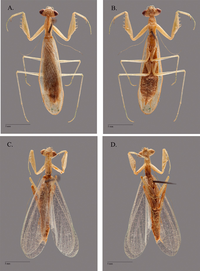
Female. Holotype ( Fig. 2 View Fig. 2 A,B). While male mantodean specimens have historically been assigned as holotypes, we have assigned a female specimen as the holotype for this new species. In so doing, we hope to underscore the point that when detailed character descriptions of both external and genitalic morphology are presented, a female holotype can be as informative as a male holotype. Traditionally, male holotypes were preferred because the male genitalia could be used to perform species-level determinations, while making species-level determinations in females was less certain due to ambiguous boundaries in external morphological characters. However, in this study we have demonstrated that female genitalia can be informative for species identifications, eliminating a potential problem with choosing females as the holotype for new species.
Body length 19.99–21.61; forewing length 14.48–15.48; hindwing length 14.64– 15.95; pronotum maximum length 3.83–4.29; prozone length 1.55–1.8; pronotum width 1.61–1.85; pronotum minimum width 1.34–1.44; head width 3.75–4.22; head vertex to clypeus 1–1.2; frons width 1.41–1.63; frons height 0.37–0.47; prothoracic femur length 4.92–5.23; mesothoracic femur length 4.26–4.93; mesothoracic tibia length 3.02–3.45; mesothoracic tarsus length 2.16–2.58; metathoracic femur length 5.02–5.63; metathoracic tibia length 5.22–5.85; metathoracic tarsus length 3.14–3.71.
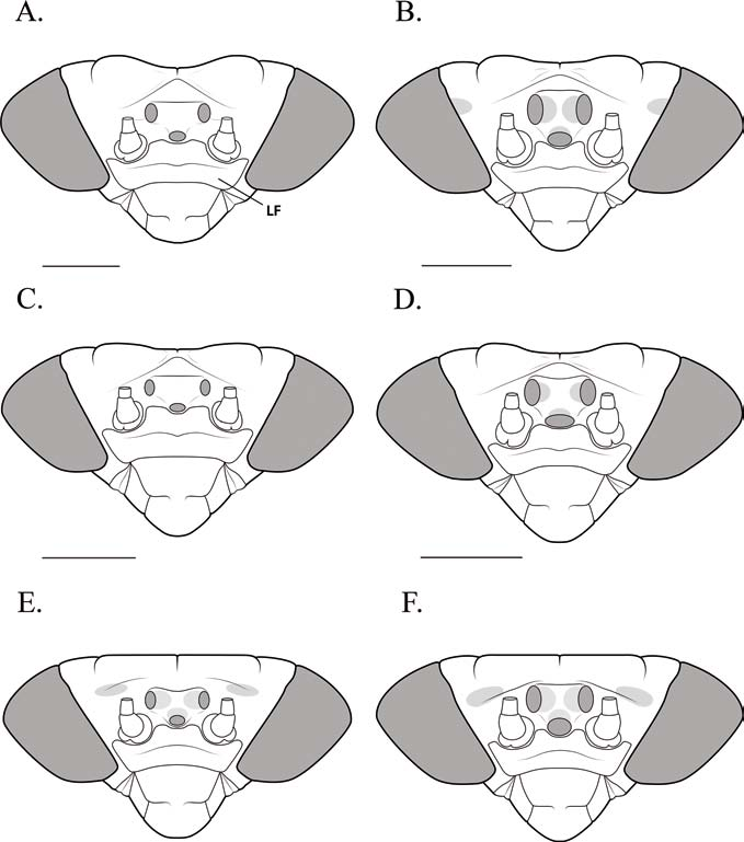
Head ( Fig. 4 View Fig. 4 C). Juxtaocular bulges present, slight. Vertex elevation yellow. Region posterior to the lateral ocelli slightly concave. Ocelli relatively small; lateral ocelli marginally larger than the median ocellus; area containing the ocelli slightly convex. Antennae extend to at least mid-body.
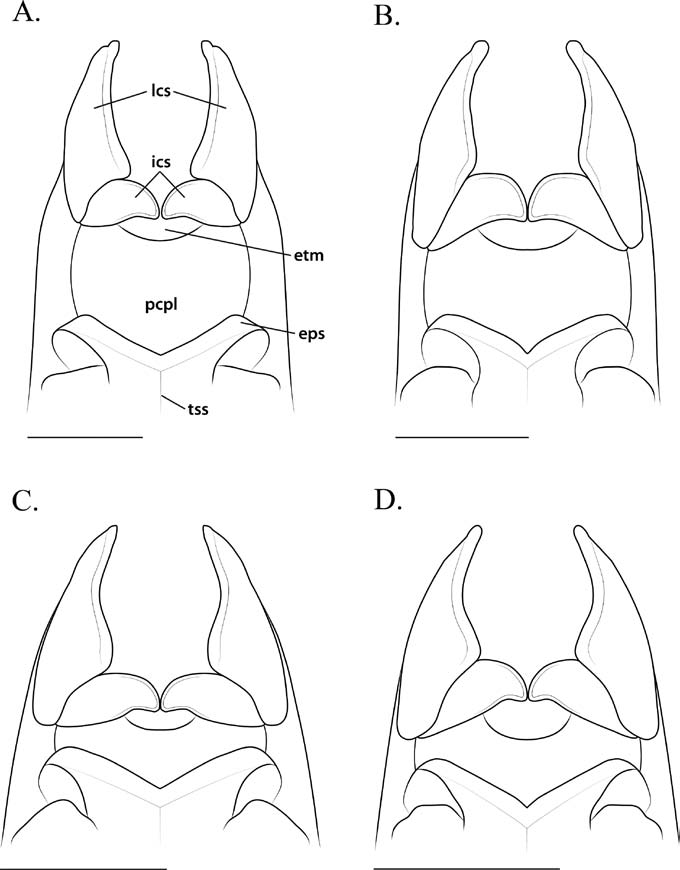
Thorax. Pronotal medial keel reaching mid-prozone ( Fig. 11 View Fig. 11 C). Postcervical plate and exposed thoracic membrane of the cervix transverse with an approximately hexagonal shape ( Fig. 13 View Fig. 13 C). Wings opaque with margins sparsely ciliated; anterior margin of forewing costal area with light green cells.
Prothoracic legs. Femur maximum width ca. 1.3 times the value of the male femur maximum width. F =3DS/12AvS/4PvS; T =11AvS/10–12PvS.
Meso- and metathoracic legs. Ciliated across surface. Mesothoracic basitarsus approximately 1.5 times the length of the remaining tarsal segments combined; metathoracic basitarsus approximately the same length as the remaining tarsal segments combined.
Abdomen. Supra-anal plate triangular, ciliated across surface, narrower than the subgenital plate. Cerci filiform, tapering.
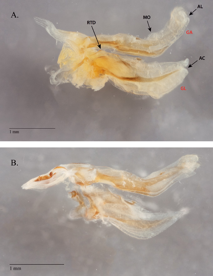
Genitalia ( Figs 3 View Fig. 3 B, 5C,D). Gonapophysis VIII (GA) slender, variably ciliated, and visibly curved from both the ventral (VH) and lateral (LH) perspectives; GA lightly surfaced with fossettes and sculpturing. From VH, GA angles inward; apical lobe (AL) ovoid, heavily ciliated. From LH, AL ranges from ovoid, to blunt, to thin and arched. AL has a membrane that projects from the internal dorsal margin of the structure, forming a shallow pocket as the membrane merges with the dorsal margin of GA, lending AL the appearance of being bilobed or mitten-shaped. From LH, GA is obtusely curved ventrad; GA ventral margin features a reduced medial outgrowth (MO), which projects posterior to the crest of the curve. GA dorsal margin is invaginated anterior to the curve towards the ventral habitus. Gonapophysis IX (GP) is relatively short, terminating in a weakly rounded apex; GP ventral margin features a subulate overhang, the medial tine (MT). Gonoplac (GL) is shorter than GA, broad, variably ciliated, and arched ventrad; from LH, GL base has a rectangular dilation on the anteroventral margin. GL narrows toward the apex; from LH, GL apex slants ventrad. GL features a significantly reduced apical cleft (AC), with the lobe of the dorsal margin appearing to converge with the ventral lobe, resulting in an approximately subulate apex. From LH, GL possesses a slight sclerotization that traverses the length of the structure, terminating just before AC. GL ventral and dorsal margins are relatively smooth.
Male. Allotype ( Fig. 2 View Fig. 2 C, D). Body length 19.34–20.44; forewing length 14.24–15.37; hindwing length 14.85–16.18; pronotum maximum length 3.46–3.84; prozone length 1.32–1.49; pronotum width 1.35–1.45; pronotum minimum width 1.12–1.3; head width 3.44–3.6; head vertex to clypeus 1–1.18; frons width 1.3–1.36; frons height 0.34–0.40; prothoracic femur length 4.19–4.49; mesothoracic femur length 3.78– 4.19; mesothoracic tibia length 2.48–2.94; mesothoracic tarsus length 1.71–2.17; metathoracic femur length 4.47–4.92; metathoracic tibia length 4.56–5.27; metathoracic tarsus length 2.74–3.66.
Head ( Fig. 8 View Fig. 8 D). Juxtaocular bulges present, highlighted by parietal sulci. Vertex elevation yellow. Region posterior to the lateral ocelli concave. Ocelli larger than females; lateral ocelli slightly larger than unpaired median ocellus. Ocelli sit within an elevated ocellar tubercle that has three brown markings on the cuticle between the ocelli. Antennae as in females.
Thorax. Pronotum as in females ( Fig. 11 View Fig. 11 D). Postcervical plate and exposed thoracic membrane of the cervix as in females ( Fig. 13 View Fig. 13 D). Wings hyaline and iridescent; fore- and hindwing margins and surface with light ciliation.
Prothoracic legs. Femur maximum width ca. 0.74 times the value of the male femur maximum width. F =3DS/12AvS/4PvS; T =10–11AvS/11–12PvS.
Meso- and metathoracic legs. Densely ciliated across surface. Mesothoracic basitarsus approximately 1.5 times the length of the remaining tarsal segments combined; metathoracic basitarsus approximately the same length as the remaining tarsal segments combined.
Abdomen. Supra-anal plate triangular, ciliated across surface; much narrower than the subgenital plate. Subgenital plate approximately rounded at the terminus, ciliated. Cerci long, compressed, tapering. Styli short.
Genitalia ( Fig. 10 View Fig. 10 ). Dorsal sclerotization of the left phallomeric complex is anteriorly narrow and elongate, broadening towards the posterior margin; anterior process (ap) ranges from narrow and elongate to compact, tapering anteriorly; ap anterior margin variably sclerotized; Apical process (paa) dilated on the anterior margin of its visible “base,” recurved distally, narrow; paa with a rounded apical margin. Lobo membranoso (loa) relatively short, apex moderately sclerotized. Ventral sclerotization of the left phallomere complex (i.e., the ventral phallomere) (L4A) longer than wide with an undulate, lightly sclerotized antero-sinistral margin; L4A anterior margin varies slightly in width; L4A posterior region lightly ciliated and bilobed; processo distale (pda) narrow, apically rounded, and well separated from a projecting dextral lobe that is broad and approximately rounded along the posterior margin. Anterior apodeme (an) anteriorly rounded with a moderately sclerotized sinistral margin; processo ventrale sclerificato (pva) obtusely curved, tumescent, with a heavily sclerotized posterior margin; piastra ventrale (pia) variably shaped, ranging from relatively straight to arcuate with a rugose surface. R1 posterior region lightly ciliated with a slightly tapered, rounded posterior margin; right arm (bm) varies in width, pointed apically.


| MNHN |
Museum National d'Histoire Naturelle |
No known copyright restrictions apply. See Agosti, D., Egloff, W., 2009. Taxonomic information exchange and copyright: the Plazi approach. BMC Research Notes 2009, 2:53 for further explanation.