
Icochilus extensus Ameghino, 1889
|
publication ID |
https://doi.org/ 10.11646/zootaxa.4543.2.2 |
|
publication LSID |
lsid:zoobank.org:pub:11F546CD-109F-4CBC-B87D-EA8D1AD0B96F |
|
DOI |
https://doi.org/10.5281/zenodo.5931281 |
|
persistent identifier |
https://treatment.plazi.org/id/B0798797-FFB2-2862-72CB-CC38FC2CFC22 |
|
treatment provided by |
Plazi |
|
scientific name |
Icochilus extensus Ameghino, 1889 |
| status |
|
Icochilus extensus Ameghino, 1889
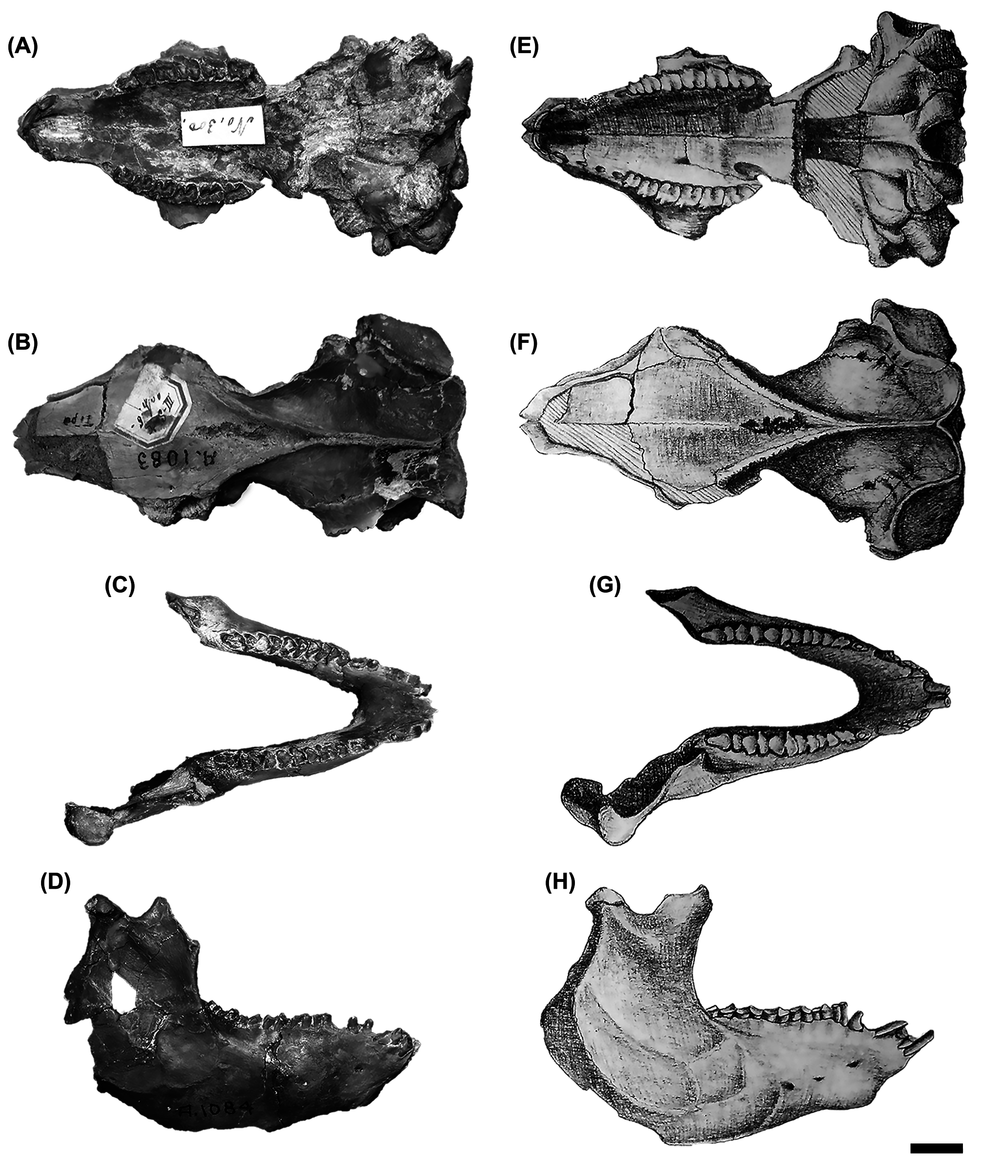
Lectotype (this contribution). MACN-A 1083 ( Figs. 2 View FIGURE 2 A–B) and MACN-A 1084 ( Figs. 2 View FIGURE 2 C–D), an almost complete skull and mandible with complete dentitions, both belonging to the same individual.
Paralectotypes (this contribution). Four isolated teeth, not located (see below).
Age and provenance of the type material. Santacrucian SALMA, early Miocene , Santa Cruz Province ( Argentina).
Original description ( Ameghino 1889: 471–472). “ Esta es la especie de mayor tamaño, cuya talla era un poco superior á la del conejo. Los premolares 2, 3 y 4 se distinguen fácilmente de los de las demás especies, por presentar sobre la parte anterior de la cara externa dos aristas estrechas separadas por una ranura angosta y profunda; de estas dos aristas, la anterior que forma el ángulo ántero-externo de cada diente es mas pequeña y mas baja, y la posterior mas ancha y considerablemente mas elevada tomando la forma de una columna perpendicular saliente. La parte posterior externa detrás de la columna mencionada es mas baja y plana, uniéndose á la cara posterior formando un ángulo redondeado. Los verdaderos molares superiores tienen las dos aristas de la parte anterior de la cara externa mas estrechas y de tamaño mas igual, sin que la segunda tome la forma de columna perpendicular saliente. Detrás de esta segunda árista viene una depresión perpendicular colocada sobre la mitad del ancho de la cara externa de la muela, de fondo cóncavo pero poco profunda. Cada uno de los verdaderos molares superiores presenta sobre el borde externo de la corona dos cúspides ó cerros puntiagudos, formados por el prolongamiento de las ondulaciones convexas perpendiculares de la cara externa. En la mandíbula inferior, los dos lóbulos que forman cada muela, son de forma mas distinta entre sí que en las otras especies; el lóbulo anterior tiene el lado interno mucho mas estrecho y en forma de arista perpendicular angosta y saliente dirijida hacia atrás, y el lado externo mas ancho y redondeado, con su eje mayor dirijido oblicuamente de afuera hacia adentro y de adelante hacia atrás, presentando la cara posterior de un ancho considerable; el lóbulo posterior es también igualmente muy estrecho sobre el borde interno y mas grueso sobre el externo, pero presenta su parte anterior que se une al lóbulo que lo precede considerablemente mas angosta que la posterior. La mandíbula inferior presenta tres agujeros mentonianos, el anterior mas grande, colocado debajo de la barra que separa el p. 1 del c. 1 en el punto en que la sínfisis se comprime transversalmente para tomar la forma de pico, el segundo mucho mas pequeño está colocado debajo del p. 2, y el tercero igualmente pequeño, se encuentra debajo de la parte anterior del p. 4. La cara externa de la rama horizontal debajo de los premolares es gruesa y convexa. La rama ascendente presenta el borde anterior con la base que sale del lado externo de la última muela levantándose hacia arriba formando una pequeña curva cóncava, pero su parte superior que constituía la apófisis coronoides se inclina hácia adelante ”.
English translation. This is the largest species, being a little larger in size than a rabbit. P2–4 are easily distinguishable from those of the other species, because they have two narrow edges separated from each other by a narrow and deep groove on the antero-external face; of these two edges, the anterior one, which forms the anteroexternal angle of each tooth, is smaller and lower, while the posterior is wider and considerably higher resembling a perpendicular projecting column. The postero-external region behind this column is lower and flat, joining the posterior face in a rounded angle. M1–3 also presents two antero-external edges but both are narrower and of equal size, and the posterior edge does not take the form of a perpendicular projecting column. Behind this second edge, there is a perpendicular depression, concave but shallow, placed about half the width of the external face of the molar. Each molar has two pointed cusps or hills on the external margin of the crown which are formed by the prolongation of the convex and perpendicular undulations of the external face. In the mandible, the two lobes that constitute each tooth, are more distinct from each other than in the other species; the inner side of the anterior lobe is much narrower than the external and similar to a narrow, perpendicular and protruding edge which directs backwards, whereas the external side is broader and rounded with its major axis obliquely directed, with the posterior face being considerable wide; the posterior lobe is also very narrow on the inner face and thicker on the external, but the anterior part that joins the preceding lobe is considerably narrower than the posterior lobe. The mandible has three mental foramina, the anterior is the largest and it is placed below the diastema that separates p1 from c at the point where the symphysis is transversely compressed acquiring the shape of a beak, the second is much smaller in size and it is placed below p2, and the third is equally smaller and is placed below the anterior margin of p4. Below the lower premolars, the external face of the horizontal mandibular ramus is thick and convex. The ascending ramus of the mandible presents its anterior border with the base that emerges from the external side of m3 rising upwards forming a small concave curve, but its upper part, which constitutes the coronoid process, slopes forwards.
Comments. Ameghino (1889: 471–472, plate 15: figs. 4–5) described and illustrated this species based on a partial skull and mandible of the same individual, along with four isolated teeth—I1, right upper premolar, right upper molar and left lower molariform—( Ameghino 1889, plate 15: figs. 6–9).
According to Ameghino’s catalogue and Mones (1986), the skull MACN-A 1083 and the mandible MACN-A 1084 are the type specimens of Ic. extensus . Following Ameghino’s catalogue, both are part of the same animal, which is confirmed by us because they articulate to each other. MACN-A 1083 and MACN-A 1084 correspond to figures 4 and 5, respectively, of the Atlas ( Ameghino 1889: plate 15), and are herein reproduced ( Figs. 2 View FIGURE 2 E–H). The isolated teeth ( Ameghino 1889: plate 15, figs. 6–9), which were not even mentioned in the written catalogue, were not located in the collection and, due to the fact that MACN-A 1083 and MACN-A 1084 already exhibit these teeth, we conclude that they do not belong to the same individual as the skull and mandible; nevertheless, these teeth would be part of the original syntypes. As the original diagnosis was based on the characteristics of the skull and mandible, we designate MACN-A 1083 plus MACN-A 1084 as the lectotype of Ic. extensus . Consequently, the remaining syntypes become paralectotypes ( ICZN 2000: Art. 73.2.2).
No known copyright restrictions apply. See Agosti, D., Egloff, W., 2009. Taxonomic information exchange and copyright: the Plazi approach. BMC Research Notes 2009, 2:53 for further explanation.