
Herculina polysagittae
|
publication ID |
https://doi.org/ 10.11646/zootaxa.4211.1.1 |
|
publication LSID |
lsid:zoobank.org:pub:6B86C6BA-6AFE-4AAD-870D-04794C138D47 |
|
DOI |
https://doi.org/10.5281/zenodo.6074501 |
|
persistent identifier |
https://treatment.plazi.org/id/03B7878C-FF6C-CD80-FF4C-EA32B54FE809 |
|
treatment provided by |
Plazi |
|
scientific name |
Herculina polysagittae |
| status |
|
Herculina polysagittae View in CoL gen. et sp. nov.
Figs 157–159 View FIGURE 157 View FIGURE 158 View FIGURE 159
Diagnosis. Differs from the only other known congener, H. oligosagittae gen. et sp. nov., by the shape of the coxal processes of the anterior gonopods, and by the presence of more numerous arrow-shaped filaments on the coxites of the posterior gonopods.
Etymology. To emphasize the presence of numerous arrow-shaped filaments on the coxites of the posterior gonopods.
Material studied (total: 1 male, 1 female). Holotype. GEORGIA: male, S of Bakuriani, Pinus & Fagus forest, 1750 m asl, litter, logs, 13 May 1983, S. Golovatch leg. ( ZMUM ρ3437).
Paratype (total: 1 female). GEORGIA: 1 female, same data as holotype ( ZMUM ρ3438).
Type locality. GEORGIA: S of Bakuriani , Pinus & Fagus forest, 1750 m asl.
Description. Body with 31 segments (including telson) in adults.
MEASUREMENTS. Holotype male 9 mm long, vertical diameter of the largest pleurotergite 0.7 mm. Paratype female 9.5 mm long, vertical diameter of the largest pleurotergite 0.7 mm.
COLORATION ( Fig. 157 View FIGURE 157 ). Pale yellowish, with a darker head.
HEAD. Flattened in male. Labrum with three medial teeth and 4+4 labral and 2+2 supralabral setae. Promentum triangular, without setae. Lingual plates with 4+4 setae, on each plate arranged in one row. Stipites with ca 17+17 setae. Antennae 1.4 mm long in holotype. Length of antennomeres (in mm): I (0.08), II (0.16), III (0.36), IV (0.19), V (0.32), VI (0.16), VII (0.11) and VIII (0.02). Length/breadth ratios of antennomeres I–VII: I (1), II (2), III (4), IV (2), V (3), VI (1.6) and VII (1.2). Antennomeres II, IV, V, VI and VII with one, three, one, four and one sensillum, respectively. Number of ocelli 9–11, arranged in 4 rows in male; 11–13 in 4 rows in female.
COLLUM. Narrower than head, with six macrochaetae. Anterior edge semi-circular, posterior margin gently concave.
BODY SEGMENTS ( Fig. 157 View FIGURE 157 ). Lateral keels like lateral swellings. Macrochaetae long and trichoid. CIX (pleurotergite 15) ~ 0.85; MIX (pleurotergite 15) ~ 2; PIX (pleurotergite 15) ~ 0.5; MA (pleurotergite 15) ~ 110˚.
TELSON. Epiproct with a pair of spinnerets and 3+3 setae (1+1 paramedian, 2+2 marginal). Hypoproct with 1+1 apical setae. Paraprocts with 3+3 marginal setae.
WALKING LEGS. In both sexes, leg-pairs 1 and 2 with tarsal combs; prefemora with several long and robust setae; femora and postfemora with a group of several long and robust setae. Leg-pairs 12–15 each with a small, inner, subtriangular, coxal protrusion in males (female not examined).
MALE SEXUAL CHARACTERS ( Fig. 158 View FIGURE 158 ). Leg-pairs 3–7 enlarged, especially so leg-pairs 5 and 6. Leg-pairs 3 and 4 each with a basal exterior protrusion on prefemur. Leg-pairs 6 the most robust, bizarre, femora extremly enlarged, tarsi with a basal inner protrusion. Leg-pair 5 similar to leg-pair 6, but with smaller podomeres. Leg-pair 7 with a posterior, hook-shaped, coxal horn. Leg-pair 10 with coxal glands and a short subtriangular coxal protrusion. Leg-pair 11 with coxal glands, and an exterior and an interior, rounded, coxal protrusion.
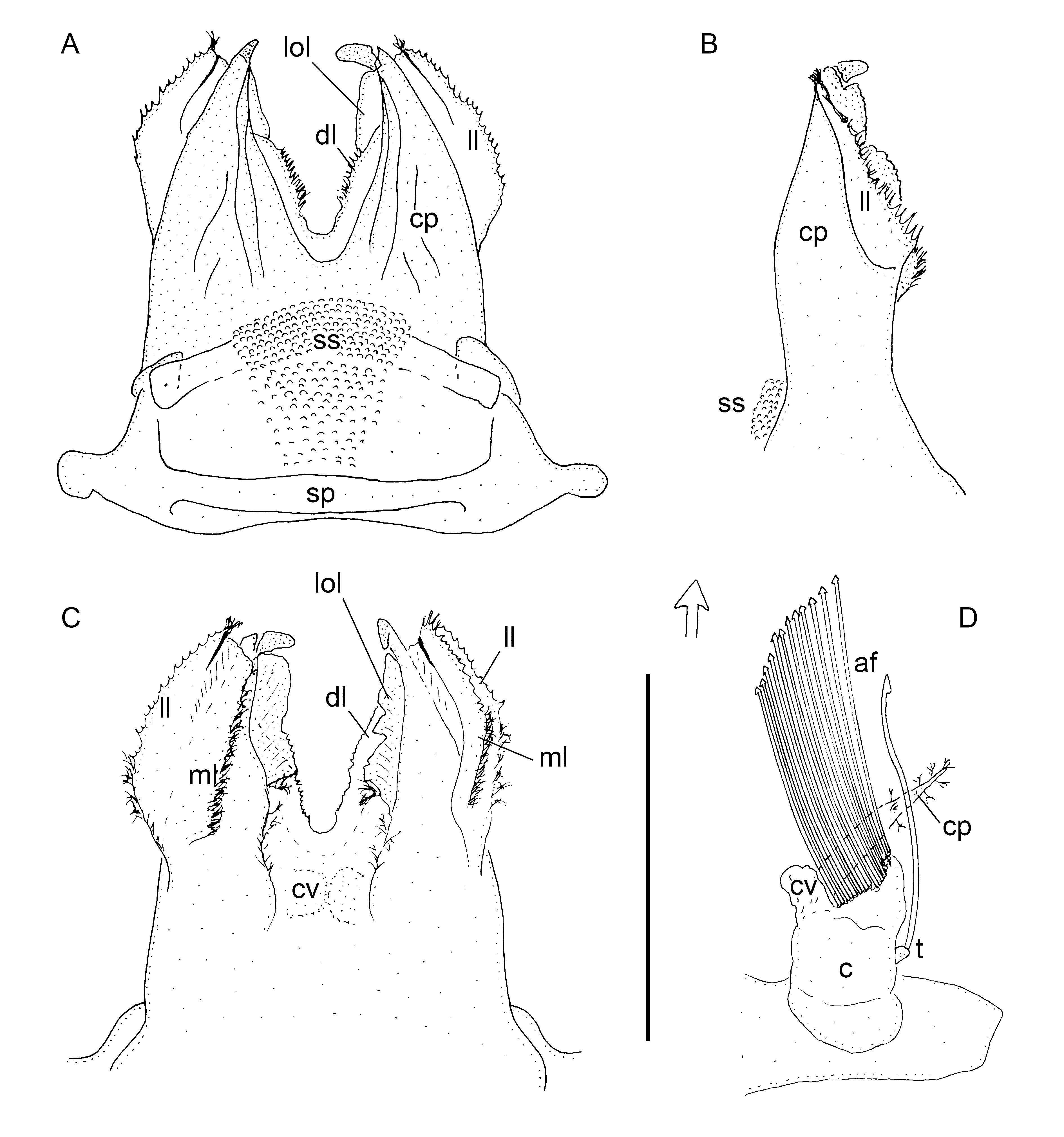
ANTERIOR GONOPODS ( Fig. 159 View FIGURE 159 A–C). Sternal plate (sp) anteriorly with a nippled sternal sac (ss). Coxal processes (cp) fused in basal third; distal two-thirds divided, but connected by a denticulate lamella (dl). Posteriorly, lateral edges of coxal processes carrying a very wide lamella divided into a mesal (ml) and a lateral (ll) part, both parts with setae and denticles. One more longitudinal lamella (lol) present on both mesal sides of coxal processes in posterior view. The latter lamella can only be partly seen in anterior view. Coxal vesicles (cv) barely visible in posterior view.
POSTERIOR GONOPODS ( Fig. 159 View FIGURE 159 D). Coxites (c) divided. Laterally, with a small telopodite (t) with a single, very long, arrow-shaped filament (af). The most characteristic parts of coxites are numerous arrow-shaped filaments (af). Posteriorly, coxites with lamellar coxal processes (cp) with setae. Coxal vesicles (cv) located on anteromesal side.
Distribution. Georgia (known only from type locality) ( Fig. 170 View FIGURE 170 , red triangle).


| ZMUM |
Zoological Museum, University of Amoy |
No known copyright restrictions apply. See Agosti, D., Egloff, W., 2009. Taxonomic information exchange and copyright: the Plazi approach. BMC Research Notes 2009, 2:53 for further explanation.