
Fridericia roembkei, Schmelz, Rüdiger M. & Collado, Rut, 2013
|
publication ID |
https://doi.org/ 10.11646/zootaxa.3647.2.4 |
|
publication LSID |
lsid:zoobank.org:pub:33866E2B-6B0F-4124-A6A6-2B057E642149 |
|
DOI |
https://doi.org/10.5281/zenodo.5612042 |
|
persistent identifier |
https://treatment.plazi.org/id/301187BC-2C0B-FFF1-88B0-FF314C1BFCF1 |
|
treatment provided by |
Plazi |
|
scientific name |
Fridericia roembkei |
| status |
sp. nov. |
Fridericia roembkei View in CoL sp. nov.
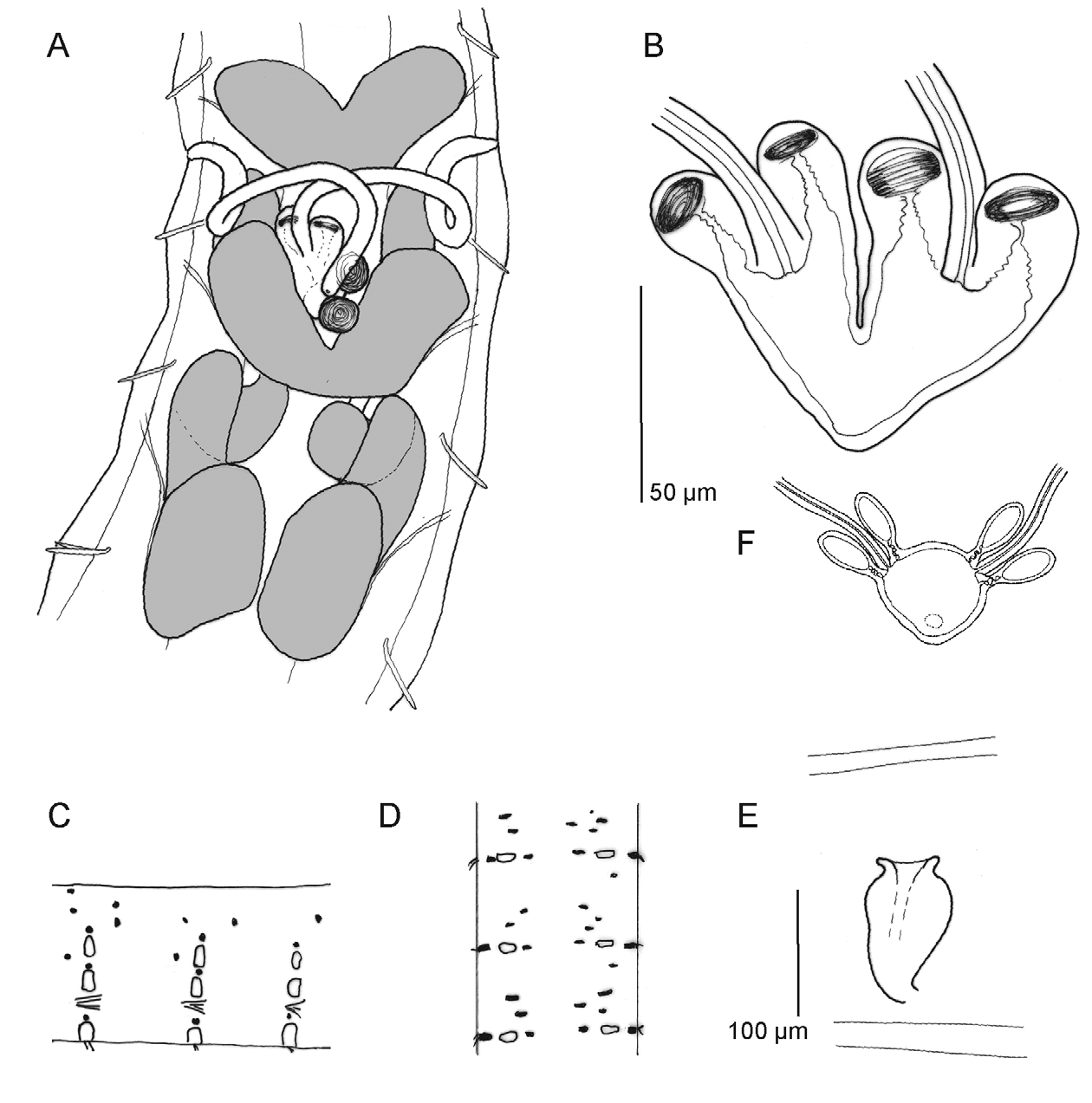
( Figs 2 View FIGURE 2 A–E, 5B, Table 3 View TABLE 3 )
Holotype. MNHML MB29-0 0 0 302, adult spcm, stained whole mount. Portugal, Coimbra, in soil from the experimental field area of the Coimbra Higher School of Agriculture (ESAC), meadow site ( Table 2 View TABLE 2 ); II 2012.
Paratypes. 60 spms. MNHML MB29-000303-311, 9 adult spms, stained whole mounts. ZMH OL 14520, stained whole mounts: 10 spms, 9 adults, 1 subadults. ZMH OL 14521, fixed in Bouin's fluid, preserved in 70% ethanol: 8 spms. ZMH OL 14522, fixed in 70% ethanol, preserved in 100% ethanol: 10 spms.
Other material. 134 spms investigated in vivo, preserved in collective sample vials, in the authors' collection.
Etymology. Named in honour of Jörg Römbke, enchytraeidologist, soil ecologist and ecotoxicologist at ECT Oekotoxikologie GmbH Flörsheim, research director of the TME experiments, in grateful recognition of his varied initiatives to support taxonomic work with enchytraeids.
Diagnosis. Less than 40 segments, max. 4 chaetae per bundle, clitellum girdle-shaped, cells absent between bursal slits, coelomo-mucocytes without refractile vesicles, nephridia present at 10/11, chylus cells post-clitellar, seminal vesicle absent, sperm funnel small, spermathecae joint entally, without ectal gland, ectal duct longer than body diameter, two stalked diverticula with ciliated subchamber and ciliar movement.
Description. Small Fridericia species. Length 8–10 mm when relaxed, 4–5 mm when contracted (viv), 6–7.5 mm (fix), diameter 0.2–0.25 mm (viv), 0.2–0.26 at V, 0.28–0.36 mm at XII–XIII, 0.22–0.29 mm in postclitellar segments (fix). Segment number (29)–34–38 (N = 40), mostly (75%) 36–38. Chaetae max. 4 per bundle, formula 2,3,4 – 4,3,2: (2,3,)4 – 4,3,2. Posterior 12–20 segments with only 2 chaetae per bundle; often all lateral postclitellar bundles with 2 chaetae. Chaetae in caudal segments largest, 65–70: 5.5 μm, size of largest anterior chaetae c. 52–55: 5.5 μm. Inner chaetae in bundles of 4 always smaller than outer, c. 2/3 the size of outer chaetae. Epidermal gland cells ( Fig. 2 View FIGURE 2 C,D) in 1–3 rows per segment, mid-row at chaetal level, cells pale or yellowish, in regular, alternating pattern, pale cells with clear outline, quasi-rectangular, yellow cells with indistinct outline, smaller; gland cells indistinct or seemingly absent in some specimens. Body wall comparatively thin, 10–15 μm thick, longitudinal muscle layer not or only slightly thicker than layer of ring muscles plus epidermis; cuticle very thin (<1 μm), barely visible at 400x magnification. Septa thin throughout.
Brain 100–110: 60–63 μm, posteriorly truncate, anteriorly slightly convex, sides slightly merging anteriad, almost parallel. Pharyngeal glands connected dorsally in IV, connected or separate in V, separate in VI. Dorsal lobes more or less of same size, ventral lobes strongly increasing in size from IV over V to VI. Oesophageal appendages with few short terminal branches. Chylus cells in 1/ 2XII –XV, 2–2.5 segment lengths; cells in conspicuous longitudinal rows, canals not widened into lacunae. Dorsal blood vessel from XVI–XVII. Midgut pars tumida not distinguished, possibly circumferal. In two consecutive posterior segments, intestinal epithelium with a network of fine canals. Preclitellar nephridia 5 pairs, 6/7–10/11, length c. 125 μm (fix), length ratio anteseptale: postseptale 2: 5, no constriction at septum, medial rise of efferent duct; postclitellar nephridia from 13/14. Coelomo-mucocytes pale with blurred vesicles, lenticytes minute, very numerous, optically dominating.
Clitellum girdle-shaped, absent between bursal slits, prominent, distinctly higher (c. 5– 7 x) than non-clitellar epidermis, cells in dense to indefinite rows, hyalocytes and granulocytes alternating, laterally of bursal slits only granulocytes; average diameter of hyalocytes 15 μm, of granulocytes 10 μm, cell height 15–20–(25) μm (fix). Seminal vesicle absent, developing sperm anteriorly in XI. Spermatozoa not numerous on sperm funnel, sperm heads not measurable in vivo, at least 60 μm (fix), probably longer. Sperm funnel small, c. 2x as long as wide (e.g. 100: 50 μm, or 120: 60 μm, fix), tapering distad, collar about half as wide as funnel body. Vas deferens in wide loops parallel to ventro-lateral body wall, therefore difficult to see, 8 μm wide. Male copulatory organ: bursa longitudinal, male gland c. 80 μm long, 50 μm wide, 40 μm high (fix), kidney-shaped, with thick-walled bursa in concavity. Subneural glands and other accessory glands absent. Spermatheca: no ectal gland; ectal duct c. 250 μm long, longer than body diameter, c. 4x as long as ampulla, coiled in contracted specimens, diameter 12 μm, with distal swelling up to 20 μm; proximal endpiece (ental bulb) not widened; ampulla with two stalked diverticula oriented ectad, diverticula with ciliated subchamber and rotating spermatozoa in peripheral chamber; ampullae joint entally and with one common attachment to oesophagus dorsally or dorso-laterally. Aspect of joint ampullae plus diverticula varied: common lumen inflated, or collapsed, inner surface smooth or wavy, organs contorted with left ampulla to the right and vice versa, or ampullae compressed in dorso-ventral position with diverticula approached and in staggered position ( Fig. 2 View FIGURE 2 A,B). One mature oocyte at a time, occupying up to 3 segment lengths.
Remarks. Fridericia roembkei sp. nov. belongs to a group of Fridericia species characterized by two spermathecal diverticula with a ciliated subchamber: F. perrieri (Vejdovský, 1878) , F. o m e r i Stephenson, 1932, F. rendsinata Dózsa-Farkas, 1972 , F. u l r i k a e Rota & Healy, 1999, F. h e a l y a e Schmelz, 2003, F. dozsae Schmelz, 2003 (Schmelz 2003: 296, 341 f.), and partly also F. galba (Hoffmeister, 1843) . It differs, together with F. m a rg i na ta described below, from all species of this group in the proximal fusion of the spermathecal ampullae. Using the tabular comparison of Fridericia species with two spermathecal diverticula in Dózsa-Farkas (2009), F. roembkei sp. nov. belongs to a group of 10 species with proximally fused spermathecae. It differs from all species of this group in the absence of spermathecal ectal glands, in an exceptionally long spermathecal ectal duct, and in the ciliation of the diverticula. Differences to F. marginata sp. nov., a remarkably similar species, are dealt with in the remarks section of that species, see also Figure 5 View FIGURE 5 and Table 2 View TABLE 2 .
The aspect of the spermathecae in F. roembkei varies considerably. The ampullae are often compressed, twisted, and in upright position, as shown in Fig. 2 View FIGURE 2 A, or they are inflated into a spherical chamber—much more than shown in Fig. 2 View FIGURE 2 B—mimicking the aspect in F. gamotheca ( Fig. 2 View FIGURE 2 F). Both aspects can be found at different times in the same living specimen. These unusual variations may be related to the thin and apparently soft ampullar walls.
In the key to species of Fridericia in Schmelz (2003), F. roembkei would key out together with F. g a m o t h e c a Issel, 1905. F. gamotheca as originally described (Issel 1905) and redescribed (Rota 1995) from several Italian localities is distinguishable from F. roembkei by the more oval spermathecal diverticula, a shorter spermathecal ectal duct (1.5x times as long as the ampulla), a larger sperm funnel, and higher segment number (40–44). Furthermore, diverticula are not ciliated (Schmelz pers. obs.) and the inflated aspect of the joint ampullae is a constant feature, not a transitory state as in F. roembkei . Some further possible differences are not straightforward because of variations noted in Rota (1995) regarding shape of diverticula, seminal vesicles and spermathecal ectal glands. Another variant of F. gamotheca from Morocco of uncertain taxonomic status (Schmelz 2003: 193f.) has spherical diverticula, only 28–33 segments (Dózsa-Farkas 1989), and a ventral clitellum as described for F. roembkei : girdle-shaped, absent between bursal slits (Schmelz 2003). It differs from F. roembkei in a short and thick ectal duct and in the presence of spermathecal ectal glands. The morphological variability documented in the redescriptions of Dózsa-Farkas (1989) and Rota (1995) have led Collado et al. (2012) to assume that F. g a m o t h e c a is a species complex.
Remarkable in F. roembkei is further the presence of both pale and yellow epidermal gland cells ( Fig. 2 View FIGURE 2 C,D), distinct only in living specimens, and clearly representing two different cell types. The pale cells are quasirectangular, filled with pale vesicles and mostly arranged in one row at chaetal level, while the yellow cells are smaller spots of irregular outline, not arranged in one row but nonetheless distributed with some regularity.
No known copyright restrictions apply. See Agosti, D., Egloff, W., 2009. Taxonomic information exchange and copyright: the Plazi approach. BMC Research Notes 2009, 2:53 for further explanation.