
Dentocypria smithi, Savatenalinton, Sukonthip, 2017
|
publication ID |
https://doi.org/ 10.11646/zootaxa.4243.2.4 |
|
publication LSID |
lsid:zoobank.org:pub:90BB82BA-4C3D-47B5-B6B1-67FD161956A9 |
|
DOI |
https://doi.org/10.5281/zenodo.5684840 |
|
persistent identifier |
https://treatment.plazi.org/id/039C87E2-FF86-FFAF-FF0F-FEE0DB778D02 |
|
treatment provided by |
Plazi |
|
scientific name |
Dentocypria smithi |
| status |
gen. nov. |
Dentocypria smithi n. gen. n. sp.
( Figs 11–14 View FIGURE 11 View FIGURE 12 View FIGURE 13 View FIGURE 14 )
2016 Physocypria sp. 2—Savatenalinton & Suttajit: 11, Table 2.
Holotype. Male, soft parts dissected in glycerine on a sealed slide, valves stored dry in a micropalaeontological slide (MSU-ZOC.168).
Allotype. Female, stored like the holotype (MSU-ZOC.169).
Paratypes. Three dissected males (MSU-ZOC.170–172) stored like the holotype, four undissected males (MSU-ZOC.173–176) stored dry in micropalaeontological slides, two dissected females (MSU-ZOC.177–178) stored like the holotype, two undissected females (MSU-ZOC.179–180) stored dry in a micropalaeontological slide and c. 25 males and 30 females in 70% ethanol.
Repository. All specimens are deposited in the MSU-ZOC.
Type locality. Bueng Kalo (swamp), Uttaradit Province. Material collected on 26 Sep. 2005, coordinates: 17° 36΄ 38˝ N and 100° 08΄ 35˝ E. Accompanying ostracod fauna: Bradleystrandesia weberi ( Moniez, 1892) , Strandesia sp. (juveniles).
Other localities. 1) Roadside canal, Phichit Province and 2) Wangwa canal , Phichit Province. For locality details see description of D. chantaranothaii n. gen. n. sp.
Etymology. The species is named after Dr. Robin James Smith (Lake Biwa Museum, Japan) in recognition of his outstanding contributions to ostracod research in general and Asian ostracodology in particular.
Diagnosis. Carapace in lateral view subovate, dorsal margin strongly arched, valve surface reticulated, CpD elliptical. Male A2 with transformed t2 and t3 setae, t2 longer than t3 and reaching tip of terminal segment. CR: Ga and Gp claws subequal, Sa short (length c. 1/3 of Ga), Sp very long (reaching beyond tip of ramus). Right prehensile palp of male T1 with large distal protrusion on first segment. Hemipenis with a-lobe markedly longer than b-lobe, a-lobe elongated, b-lobe subtriangular with pointed and distinctively dorsally curved end.
Differential diagnosis. Dentocypria smithi n. gen. n. sp. is characterized by the reticulate ornamentation, the short Sa of the CR and the very long a-lobe of the hemipenis. It cannot be confused with any other species of Dentocypria n. gen.
Measurements (mean, in µm). Female, LV (n = 2), L = 511, H = 336; RV (n =2), L = 483, H = 343; Carapace (n = 2), L = 449, H = 322, W = 257. Male, LV (n = 2), L = 440, H = 273; RV (n = 2), L = 417, H = 285; Carapace (n = 3), L = 403, H = 275, W = 205.
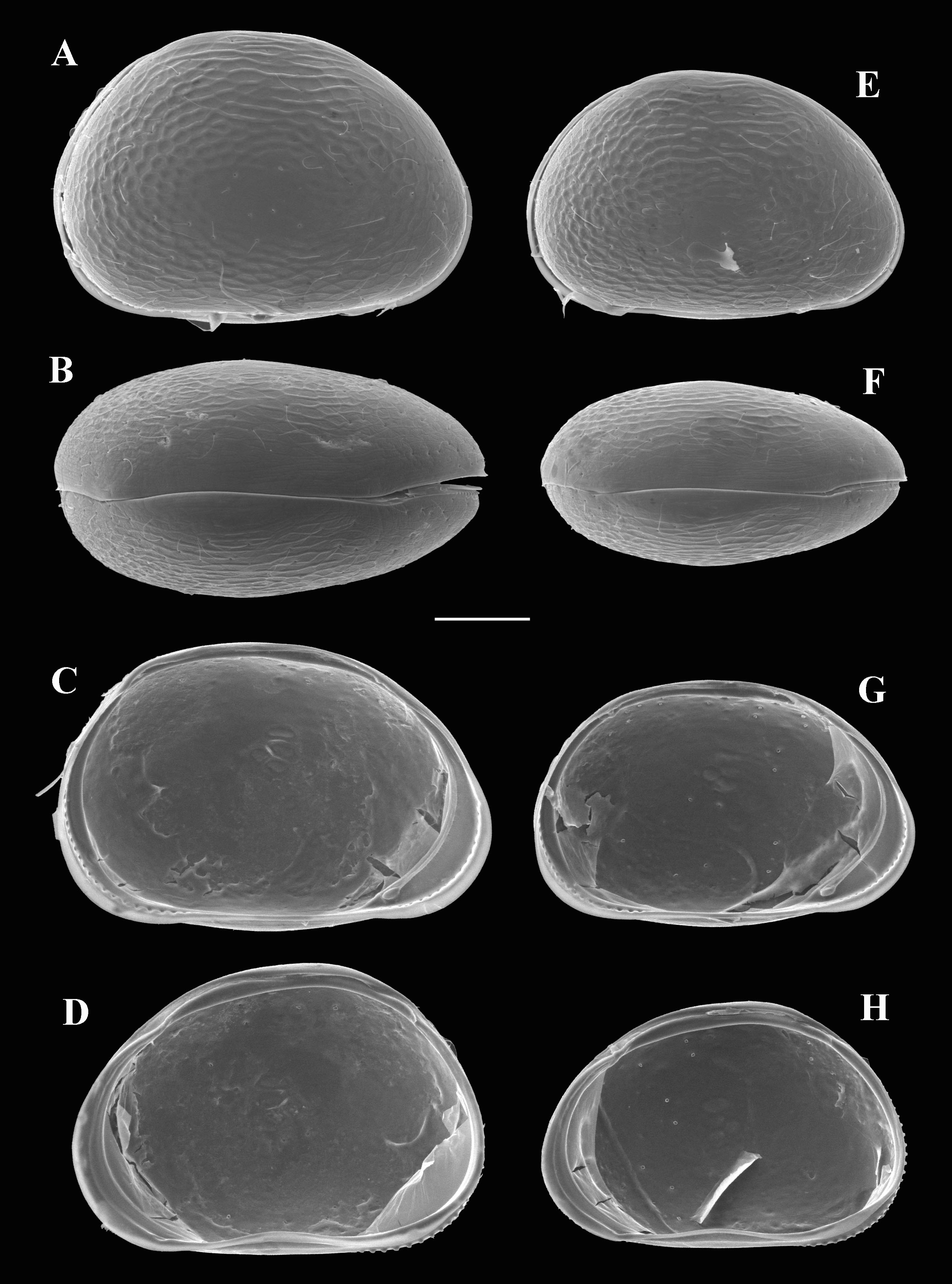
Description of female. Carapace in lateral view ( Fig. 11 View FIGURE 11 A) subovate, anterior margin rounded, slightly narrower rounded than posterior margin, LV overlapping RV anteriorly ventrally and posteriorly, dorsal margin strongly arched, greatest high situated at mid-length, valve surface reticulated.
CpD ( Fig. 11 View FIGURE 11 B) elliptical, with greatest width situated slightly behind mid-length.
LVi ( Fig. 11 View FIGURE 11 C) with selvage slightly inwardly displaced along anterior margin, internal tooth on antero-ventral part and tubercle-sockets anteriorly and posteriorly, calcified inner lamella anteriorly wider than posteriorly, with one inner list anteriorly.
RVi ( Fig. 11 View FIGURE 11 D) with submarginal selvage anteriorly, complementary pit of internal tooth on antero-ventral part, valve margin tuberculated anteriorly and posteriorly, posterior tubercles more prominent than anterior ones, calcified inner lamella anteriorly wider than posteriorly, with one inner list anteriorly.
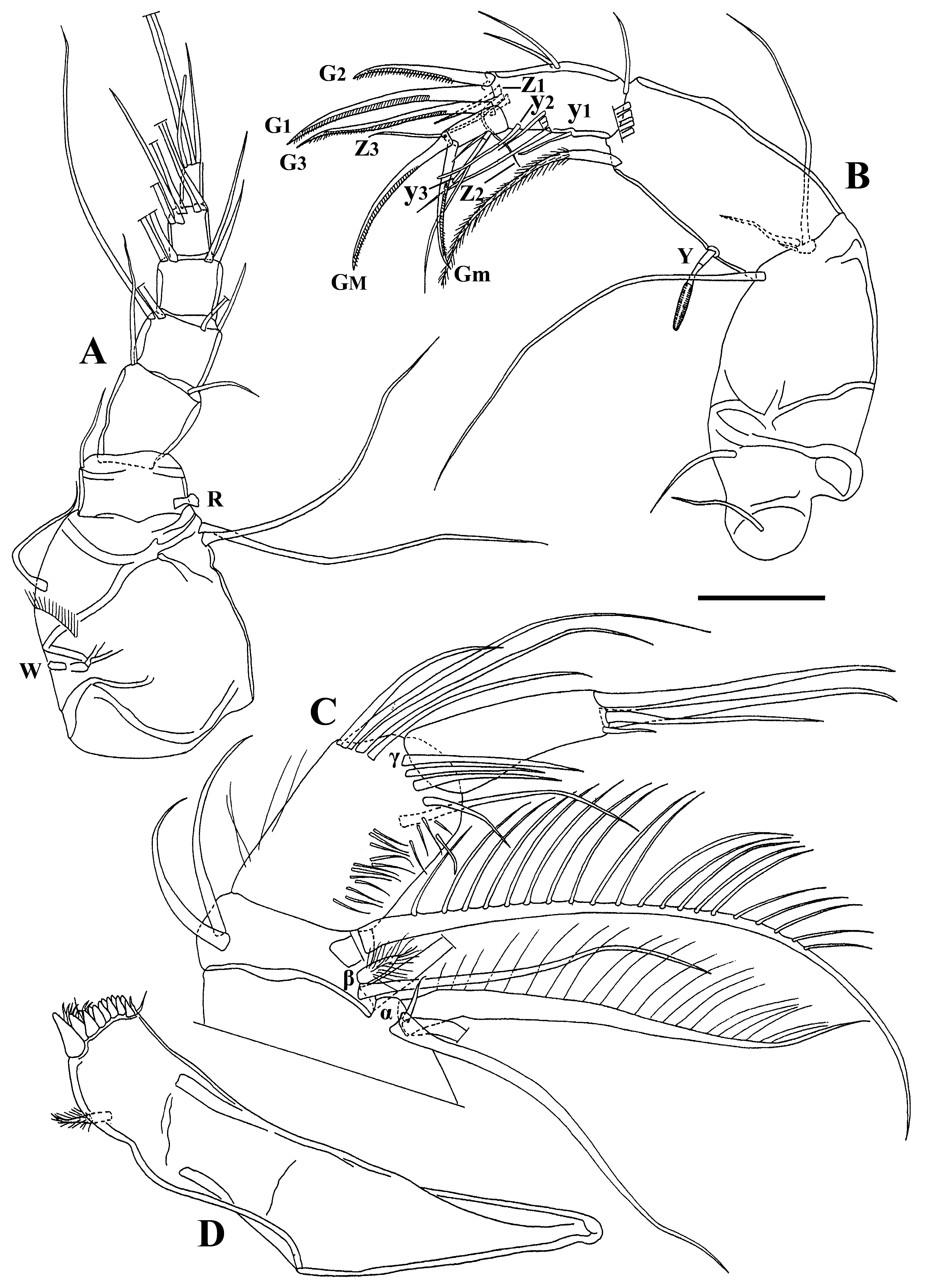
A1 ( Fig. 12 View FIGURE 12 A): first segment with small proximal Wouters organ, one long dorso-subapical seta (reaching beyond tip of segment) and two long ventro-apical setae. Second segment slightly wider than long, with one long dorso-apical seta (reaching beyond mid-length of third segment) and Rome organ. Third segment bearing two (dorso-apical and ventro-apical) setae, shorter seta reaching tip of fourth segment. Fourth segment with two long dorsal setae and two subequal ventral setae, short one reaching tip of fifth segment. Fifth segment dorsally with two long setae, ventrally with two (one long, one shorter) setae, short one reaching tip of terminal segment. Penultimate segment with four long apical setae and one shorter apical seta. Terminal segment with three (two long, one short) apical setae and aesthetasc y a, length of the latter c. 3/4 of that of short apical seta.
A2 ( Fig. 12 View FIGURE 12 B): exopodite with three (one long, two short) setae, long one reaching beyond tip of first endopodal segment. First endopodal segment with five long (reaching tip of terminal claws) and one short natatory setae, shortest seta slightly longer than half of lenght of penultimate segment, aesthetasc Y long, ventro-apical seta long, reaching beyond tip of terminal segment. Penultimate segment undivided, distally with three serrated claws (G1, G2, G3), G2 short (length c. 2/3 of that of G1), aesthetasc y2 very long (reaching beyond tip of terminal segment), z1 seta short, z2–z3 setae long; this segment medially with two unequally long dorsal setae (both setae reaching tip of segment), four ventral setae (t1–t4) of unequal length (shortest seta t1 reaching beyond tip of segment), and ventral aesthetasc y1, the latter long (reaching tip of segment). Terminal segment distally with two serrated claws (GM and Gm), length of Gm c. 3/4 of that of GM; medially with ventral aesthetasc y3, length of aesthetasc y3 less than half of that of accompanying seta.
Md-palp ( Fig. 12 View FIGURE 12 C) as in D. mesquitai n. gen. n. sp. with few differences: apical γ-seta long (not reaching tip of terminal segment), short seta on ventral side of penultimate segment not reaching tip of segment, terminal segment very elongated (length c. 4 times of that of width) bearing two claws, one long seta and one short seta.
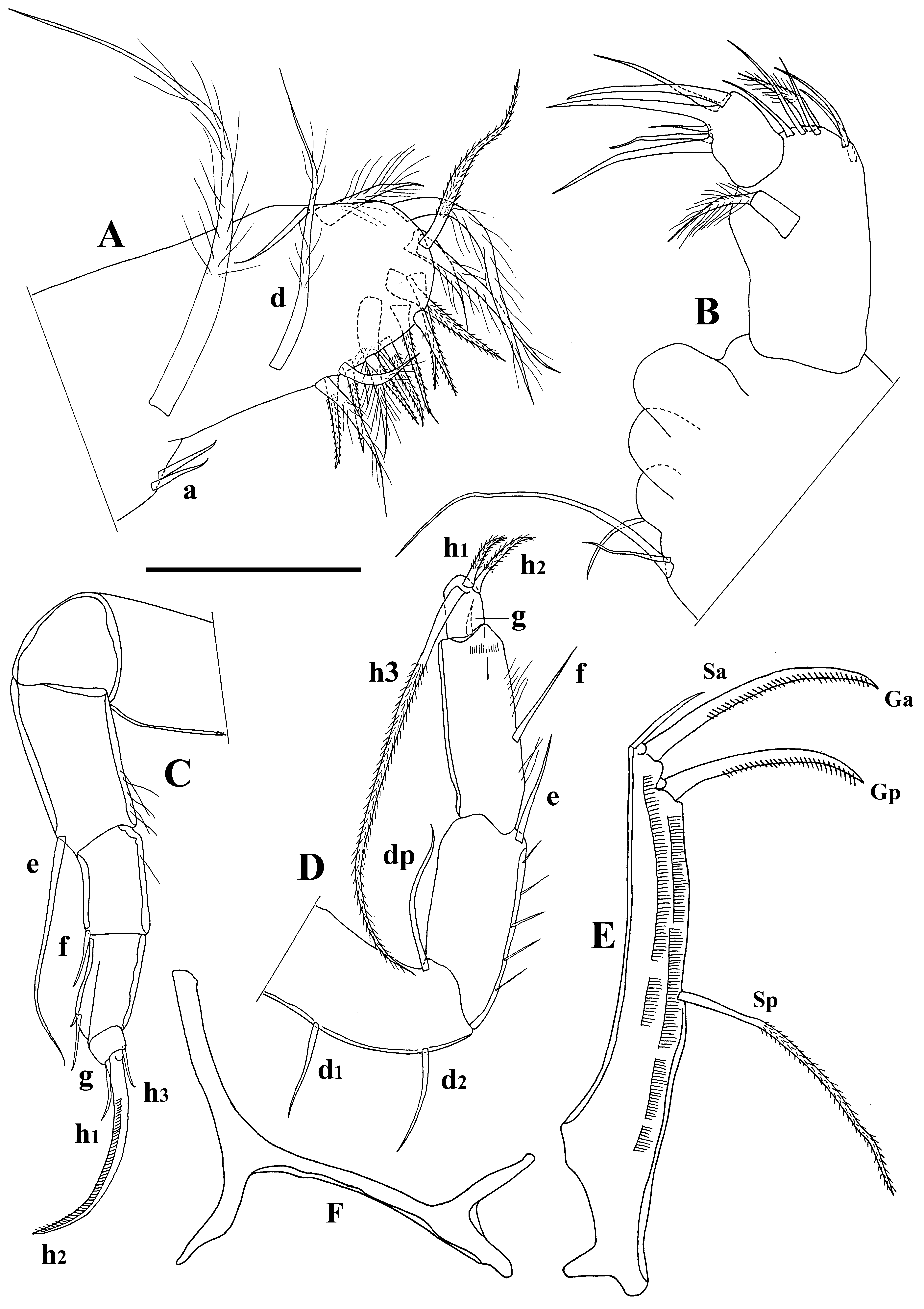
Mx1 ( Fig. 13 View FIGURE 13 B) as in D. mesquitai n. gen. n. sp. with few differences, e.g. sideways-directed bristles on first endite unequally long, length of long one c. 3 times of that of short one.
T1 ( Fig. 13 View FIGURE 13 A) as in D. mesquitai n. gen. n. sp. with different setae lengths.
T2 ( Fig. 13 View FIGURE 13 C) as in D. mesquitai n. gen. n. sp. with few differences, e.g. h3 seta of terminal segment long (not spine-like) and length of serrated claw (h2) almost identical with that of penultimate segment.
T3 ( Fig. 13 View FIGURE 13 D) as in D. mesquitai n. gen. n. sp. with different setae lengths.
CR ( Fig. 13 View FIGURE 13 E) as in D. mesquitai n. gen. n. sp. with few differences, e.g. length of Ga and Gp subequal, Sa short (length c. 1/3 of that of Ga), Sp very long (reaching beyond tip of ramus).
CR attachment ( Fig. 13 View FIGURE 13 F) as in D. mesquitai n. gen. n. sp.
Description of male. Carapace and valves ( Fig. 11 View FIGURE 11 E–H) as in female, but somewhat smaller. All limbs as in female, except for terminal two segments of A2 ( Fig. 14 View FIGURE 14 A) and T1 ( Fig. 14 View FIGURE 14 B–C).
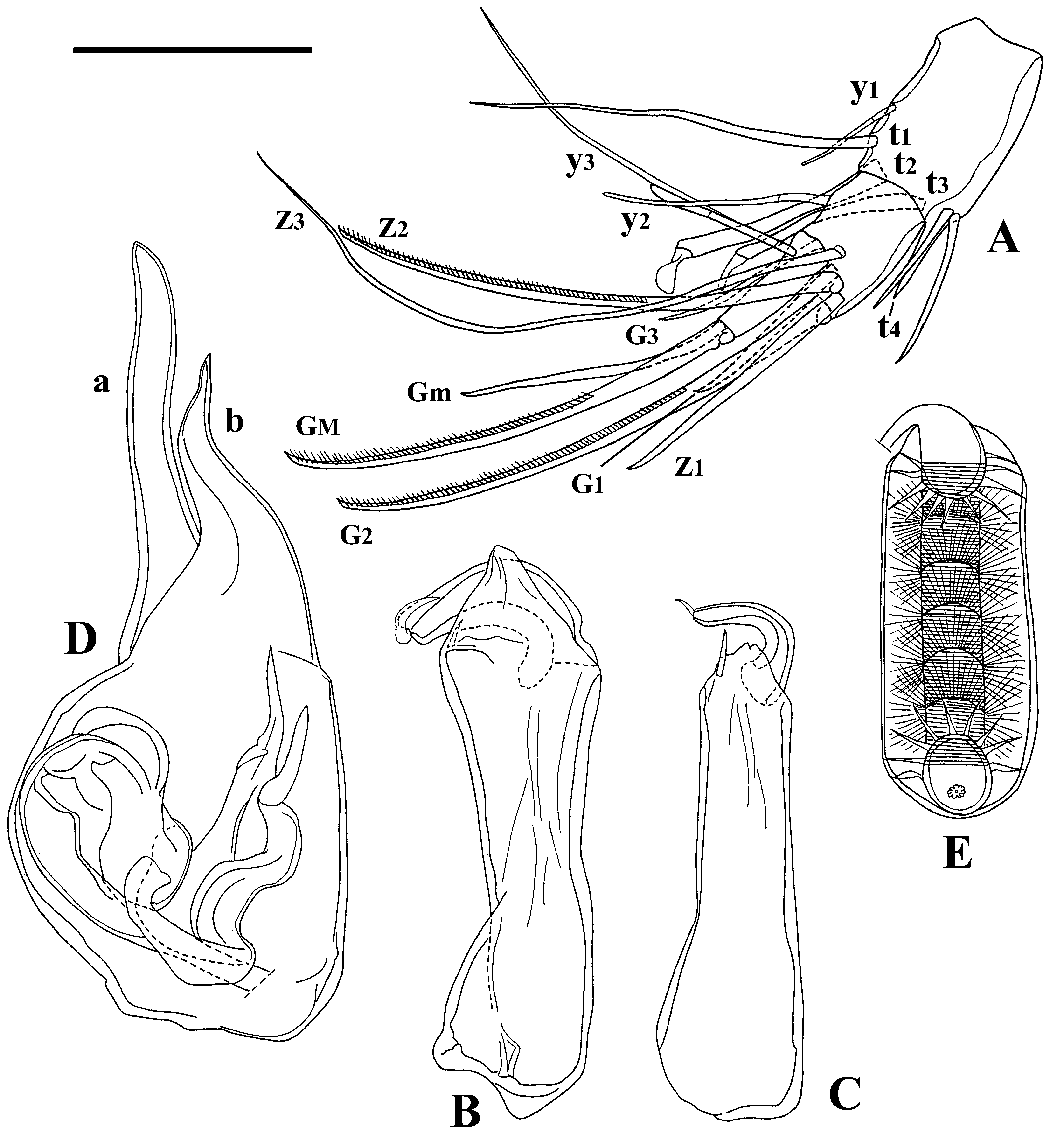
A2 with divided penultimate segment, transformed t2 and t3 setae, t2 longer than t3 and reaching tip of terminal segment. Setae z1 and z2 of penultimate segment transformed into claws; claw G1 reduced; claw G3 reduced to seta (reaching far beyond tip of terminal segment); Gm on terminal segment reduced (length c. 2/3 of that of GM).
T1 with asymmetrical prehensile palps (endopodites). First segment of right prehensile palp ( Fig. 14 View FIGURE 14 B) with large distal subtriangular protrusion and bearing one short apical spine; second segment large, subtriangular. Left prehensile palp ( Fig. 14 View FIGURE 14 C) elongated with first segment bearing one long apical spine; second segment narrow, curved and pointed.
Hemipenis ( Fig. 14 View FIGURE 14 D): a-lobe elongated, b-lobe subtriangular, with pointed end, b-lobe somewhat shorter than a-lobe, a-lobe elongated, b-lobe subtriangular with pointed and distinctively dorsally curved end.
Zenker organ ( Fig. 14 View FIGURE 14 E) elongated, length c. 2.7 times width, set with seven chitinous spiny whorls.
Remarks. Dentocypria smithi n. gen. n. sp. is superficially similar to Physocypria biwaensis ( Okubo, 1990) . It can be distinguished by the more tumid carapace in dorsal view, more numerous and prominent tubercles on the right valve margin and the reticulated valve ornamentation.
No known copyright restrictions apply. See Agosti, D., Egloff, W., 2009. Taxonomic information exchange and copyright: the Plazi approach. BMC Research Notes 2009, 2:53 for further explanation.