
Caririemys violetae
|
publication ID |
https://doi.org/ 10.5281/zenodo.175708 |
|
DOI |
https://doi.org/10.5281/zenodo.5659848 |
|
persistent identifier |
https://treatment.plazi.org/id/D3125809-FFD5-FFFA-FF3F-6522FABCE228 |
|
treatment provided by |
Plazi |
|
scientific name |
Caririemys violetae |
| status |
|
Caririemys violetae gen. et sp. nov.
Figures 2–7 View FIGURE 2 View FIGURE 3 View FIGURE 4 View FIGURE 5 View FIGURE 6 View FIGURE 7
Etymology: The specific name, violetae , honours Violeta Arraes de Alencar Gervaiseau former rector of the Universidade Regional do Cariri (URCA), who fosters paleontological studies in this region.
Holotype: partial skeleton consisting of an incomplete carapace, part of the last cervical vertebra, dorsal and caudal vertebrae, right pelvis and femur. The specimen is housed at the Museu Nacional (MN) of the Federal University of Rio de Janeiro ( UFRJ) under the number MN 6919–V.
Type Locality and horizon: the specimen was preserved in a calcareous nodule, typical from the Romualdo Member (Santana Formation), possible found in the region of Santana do Cariri, where most fossils from this deposit came from.
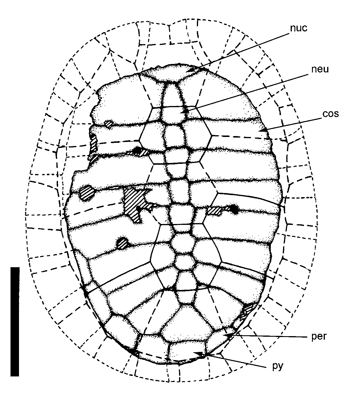
Diagnosis: Carapace oval and moderately domed (as in Cearachelys ); complete neural series reaching the suprapygal (as in Araripemys and Cearachelys ); neural plate 8 shows an extensive contact with costal 7 and 8.
Description.
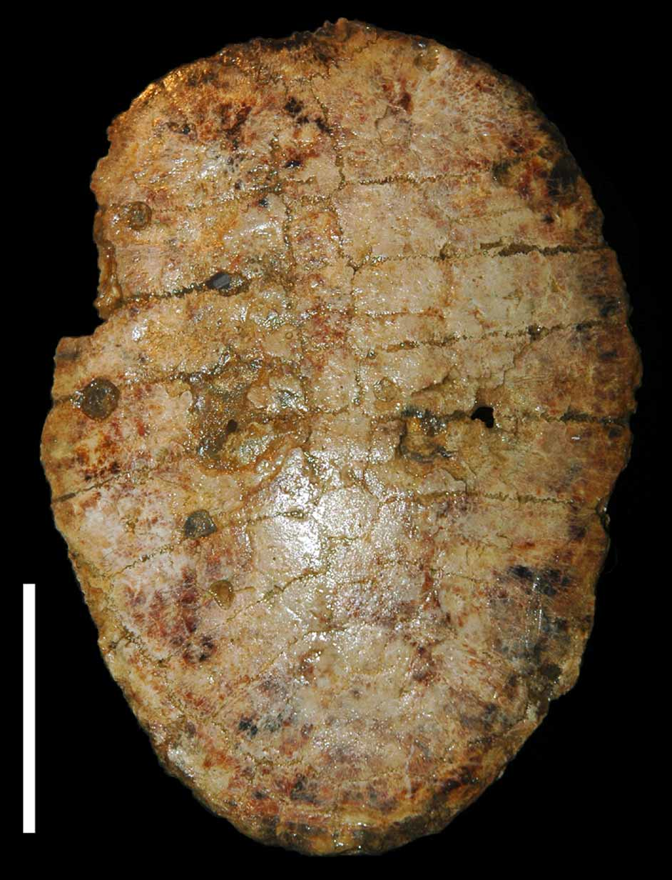
Carapace: The carapace of Caririemys violetae is moderately domed, oval, and almost complete ( Fig. 2– 3 View FIGURE 2 View FIGURE 3 ). It is composed of an incomplete nuchal plate, eight neurals, and eight pairs of costals. Peripherals 8, 9, 10 and 11 of the left side are preserved while only part of the right peripherals 10 and 11 were found. The suprapygal is well preserved but the pygal plate is incomplete. There are no fontanelles, and all the bones tightly sutured as in the most pleurodires ( Gaffney et al. 2001).
Only the caudal part of the nuchal plate is preserved. It is a broad element that contacts the neural 1. No evidence of a cervical scale is observed indicating that this element was absent in this taxon.
Caririemys violetae has a complete neural series, which is not typical of Pelomedusoides. The first neural is similar to the one of Cearachelys placidoi Gaffney, Campos & Hirayama, 2001 , by being six sided and contacting the neural 2, the first costal, and by having short and paired contacts with the second costal. The second neural has four sides, none of which contacts the costal 1 and 2. The third neural is six sided and has a short contact with costal 2, and a wide contact with costal 3. The fourth, fifth and sixth neurals are six sided and contact costal 3–4, 4–5, and 5–6 respectively. The seventh neural is the smallest of the neural series and shows a wide contact with costal 6 and 7. The eighth and last neural is six sided and has a wide contact with costal 7 and 8.
The suprapygal has a triangular shape and contacts the last neural, the peripheral 11 and the pygal.
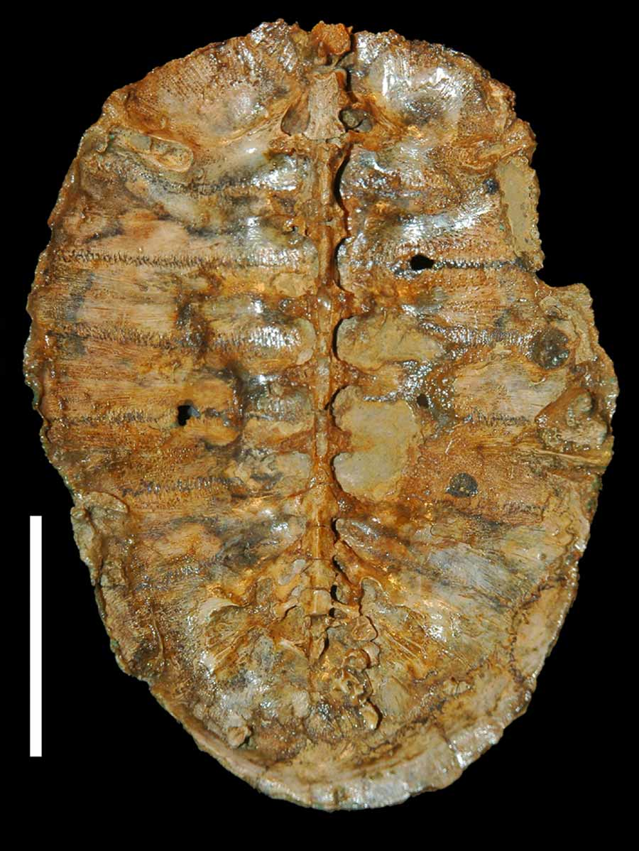
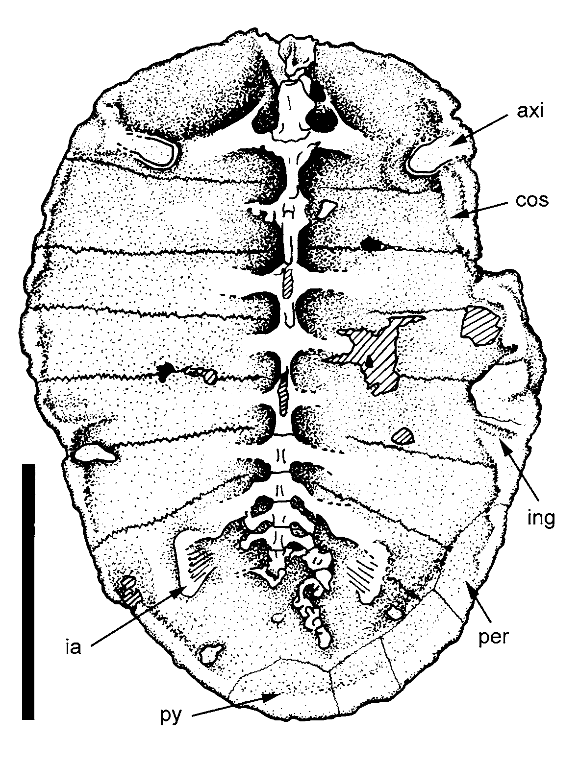
Since the plastron in not preserved, the internal part of the carapace could be exposed ( Fig. 4–5 View FIGURE 4 View FIGURE 5 ). The last cervical vertebrae (probably the eighth), nine dorsals, three sacrals and some disarticulated caudals (eleven) are preserved.
The axillary buttresses are well marked and contact the first costal. The ingunal buttresses is placed on the fifth costal. Both iliac articulations are preserved and they are positioned at costals 7 and 8. This articulation surface is triradiated in dorsal aspect, with cranial, caudal, and caudolateral projections. This feature differs from the condition of non-Pelomedusoides pleurodires (sensu Lapparent de Broin 2000), that lacks well developed projections ( Meylan 1996; Lapparent de Broin & Murelaga 1999; De La Fuente & Iturralde-Vinent 2001).
Caririemys violetae presents well discernible second and fourth vertebral scutes and the first and fourth pleural scutes. Caudally, vertebral scutes are wider than long and cover the entire neural series, and medial portions of the costal plates. The preserved sulci between vertebral scutes 1–2, 2–3, 3–4 and 4–5 respectively divide neural plates 1, 3, 5 and 8.
All dorsal vertebrae are well preserved and show no signs of deformation. The last dorsal is sacralized, a common feature in pleurodires, and has a broad transverse process that contacts the iliac articulation. There are three sacrals, with the last one incomplete. The end of the transverse process is laminar and forms the medial margin of the iliac articulation. The caudal vertebrae are displaced from their natural position. Some lack the neural arch that was unfused with the centrum, suggesting that MN 6919–V represents a young individual.
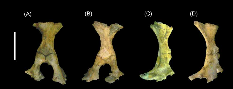
Pelvis: The pelvic girdle of Caririemys is well preserved, but only represented by the right elements ( Fig. 6 View FIGURE 6 a–d). It has mainly a vertical orientation, a typical feature of eupleurodires (Lapparent de Broin 2000; De La Fuente 2003). The thyroid fenestra forms a large aperture, as observed in some other podocnemidoids (Lapparent de Broin & Werner 1998; De La Fuente 2003). The pelvic bones are gracile and firmly attached to each other. In the acetabulum the contact surface of those elements is smooth and well defined while medially their contact form a serrated suture. The ilium forms a central waist, with ventral and dorsal expansions. This bone articulates dorsally with costal plates 7 and 8. Ventrally the ilium forms the dorsal surface of the acetabulum and connects the pubis cranioventrally and to the ischium craniocauldally. The pubis has the dorsal and ventral portions expanded. The dorsal segment of the pubis forms the cranioventral portion of acetabulum. The isquium is also dorsally and ventrally expanded forming the caudoventral part of the acetabulum. The acetabulum is oval, contrasting with the more rounded condition observed in Araripemys .
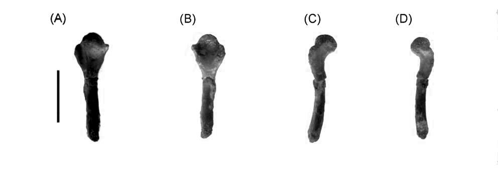
Femur: Only right femur is preserved in MN 6919–V, and lacks the distal portion ( Fig. 7 View FIGURE 7 a–d). The shaft is cylindrical and slightly arched, while the head projects dorsally. The trochanters are of equal size, with the minor trochanter extended cranioproximally, and the major trochanter extended caudoproximally.
No known copyright restrictions apply. See Agosti, D., Egloff, W., 2009. Taxonomic information exchange and copyright: the Plazi approach. BMC Research Notes 2009, 2:53 for further explanation.
|
Kingdom |
|
|
Phylum |
|
|
Class |
|
|
Order |
|
|
Family |
|
|
Genus |
Caririemys violetae
| Oliveira, Gustavo Ribeiro De & Kellner, Alexander Wilhelm Armin 2007 |
Cearachelys placidoi
| Gaffney, Campos & Hirayama 2001 |