
Bohra planei, Prideaux & Warburton, 2023
|
publication ID |
https://doi.org/ 10.11646/zootaxa.5299.1.1 |
|
publication LSID |
lsid:zoobank.org:pub:9CA85AEC-7128-4118-A50D-FCD16502F5E0 |
|
DOI |
https://doi.org/10.5281/zenodo.8017945 |
|
persistent identifier |
https://treatment.plazi.org/id/47F6EBDE-2594-4710-B440-D1D940951248 |
|
taxon LSID |
lsid:zoobank.org:act:47F6EBDE-2594-4710-B440-D1D940951248 |
|
treatment provided by |
Plazi |
|
scientific name |
Bohra planei |
| status |
sp. nov. |
Bohra planei sp. nov.
http://zoobank.org/ urn:lsid:zoobank.org:act:47F6EBDE-2594-4710-B440-D1D940951248
cf. Dorcopsis , large form: Plane (1967): pp. 50–55, figures 11–13. Hoch & Holm (1986), p. 188.
cf. Dendrolagus sp. : Woodburne (1967), p. 82.
Dorcopsis View in CoL large species: Flannery et al. (1983b), p. 77.
cf. Dorcopsis sp. : Flannery et al. (1989), pp. 151–152.
“ Dorcopsis View in CoL ”: Menzies & Ballard (1994), p. 131.
Silvaroo sp. indet.: Dawson (2004b), p. 284, table 1.
Silvaroo sp. indet. 2: Dawson (2004b), pp. 286–287.
? Dorcopsis sp. : Dawson (2004b), p. 288.
Dorcopsis View in CoL sp. nov.: Black et al. (2012), p. 1050.
Holotype. UCMP 70128 View Materials , partial right adult hindlimb, including femur, distal half of tibia, distal end of fibula, talus and calcaneus. The fibula fragment could not be located in the UCMP collection in August 2007.
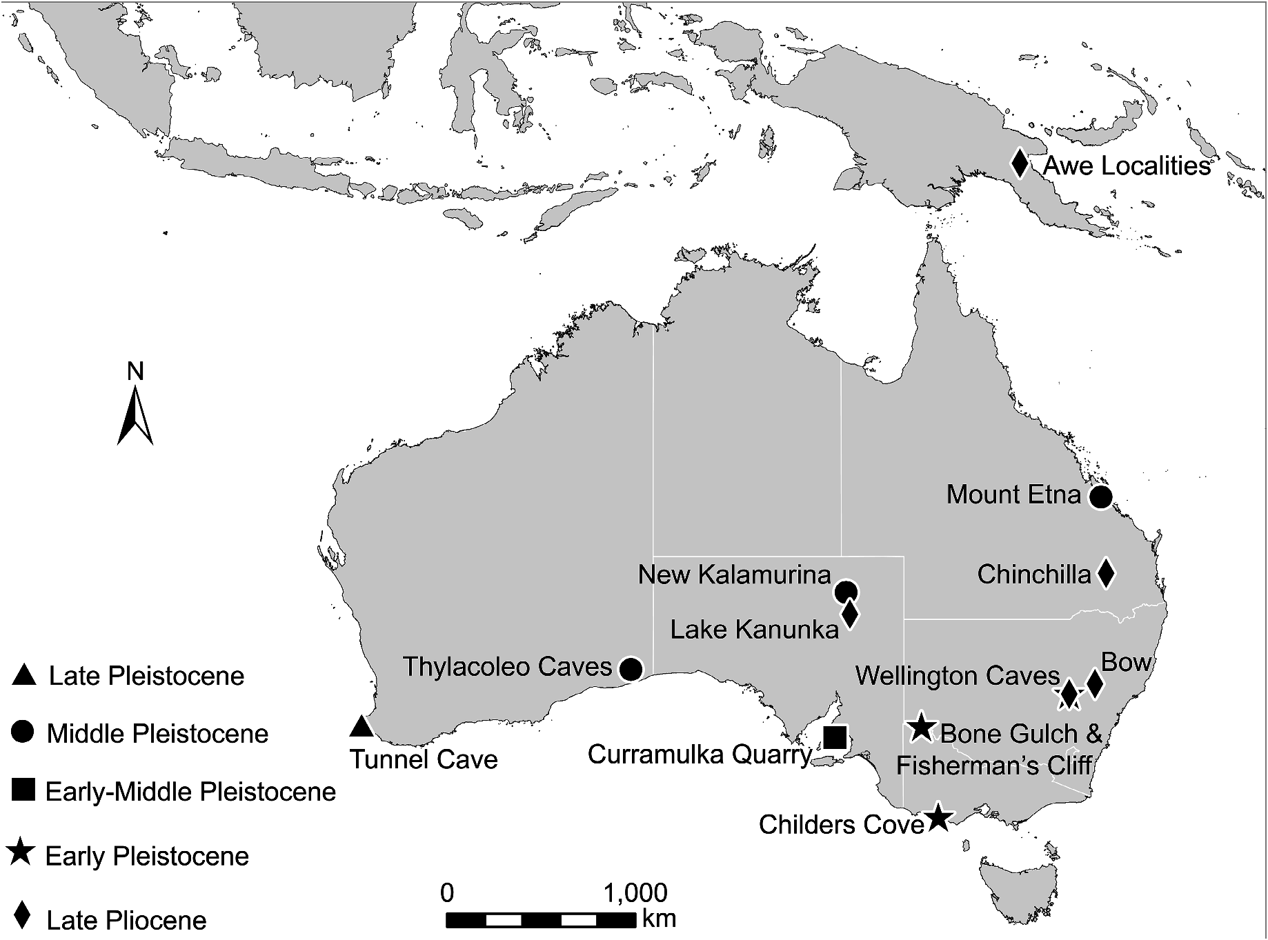
Type locality. Watut 3 ( UCMP V6356 ), located on the eastern bank of the Upper Watut River , downstream from Awe Fauna Type Locality (= Watut 1, V6234 ), Morobe Province, Papua New Guinea. Judging from the location map (figure 15 in Plane 1967), V6356 lies perhaps 500 m northeast of Awe Fauna Type Locality. UCMP 70128 View Materials was collected by Michael D. Plane in 1963 and is the only specimen recorded from Watut 3. The specimen was collected from a blue-grey claystone stratigraphically lower than Awe Fauna Type Locality .
Referred specimens. Charlie Lawrence Locality ( V 6172), Wiganda Creek, Upper Watut Valley, Morobe Province, Papua New Guinea. AM F49468, right adult maxilla (preserving M1–4). This specimen was collected in 1961 and presented to the Australian Museum by T. Brown. Before becoming submerged by a pond, the site was located “on the lower stretch of Wiganda Creek where the Gold and Power Company were working alluvial gold” ( Plane 1967: 63). No stratigraphic details were recorded.
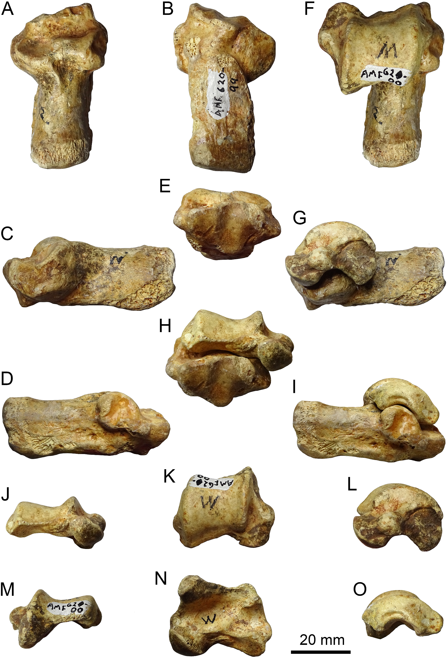
Widubosh goldmine, Upper Watut Valley. NHMD 193283 (formerly GMK 55709 ), partial right calcaneus. This specimen was collected by Ella Hoch on 29 August 1983 .
Etymology. This species is named in honour of Michael D. Plane (1933–2017), who collected the holotype and first described the Awe LF and Otibanda Formation.
Diagnosis. Bohra planei most closely resembles B. nullarbora and B. bila in upper molar size and morphology. Upper molars differ from those of B. nullarbora by having a cusp C region of the stylar crest on M1–3, incurved postpara- and premeta-cristae, and a broader, more open posterior metaloph face on M4. Upper molars differ from those of B. bila by having the bifurcation between the postparacrista and stylar cusp C region further from the paracone apex and a lingually thinner precingulum.
Femur is transversely broad and craniocaudally compressed proximally with very broad greater trochanter, broad, shallow trochlea and rugose adductor scars running along the length of the caudomedial aspect of the shaft. Distal epiphysis and talar articular facet of the tibia are relatively narrow in comparison to other Bohra and Dendrolagus . Calcaneus is transversely broad and dorsoventrally flattened, with distinct plantar epiphyseal ridge and medial crest. Medial and lateral talar facets are mesially constricted and more confluent with each other than in all other species of Bohra . The distal extremity is narrow relative to the talar and fibular facets, and the calcaneocuboid facets relatively deep and narrow and with a more acutely stepped articulation than in other species of Bohra . Talus transversely broad, dorsoventrally flattened, with broad, shallow trochlea, medially extended malleolus, broad and shallow malleolar fossa, short talar neck and smoothly contiguous calcaneal facets.
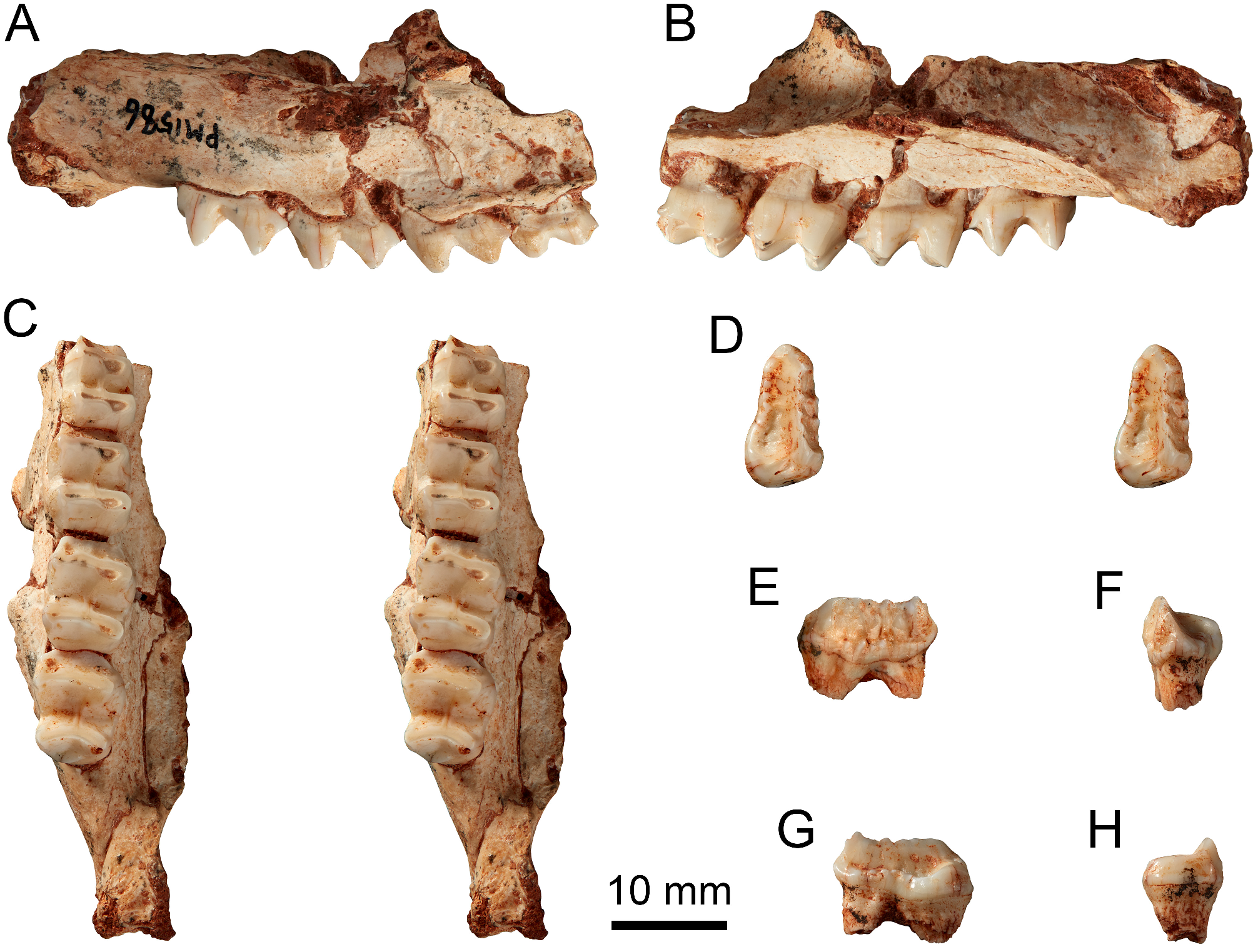
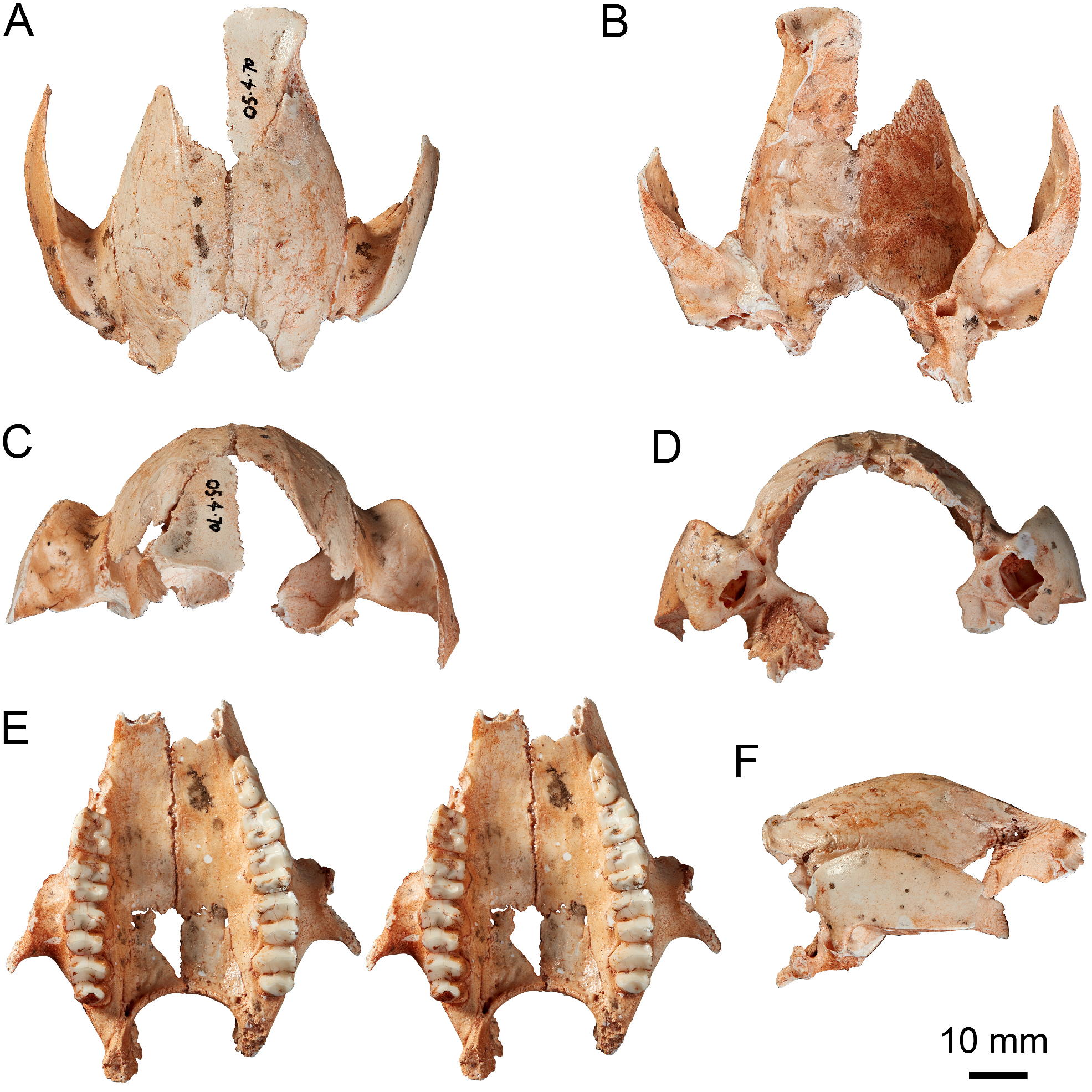
Description and comparisons. Maxilla and palatine. Very little is preserved of the maxilla of AM F49468, but there is sufficient to indicate that the base of the masseteric process was positioned above the anterior root of M 3 in lateral view ( Figure 30A, C View FIGURE 30 ). The shape of the fracture through the palate that represents the mesial limit of this specimen is consistent with a line of breakage along the maxilla–palatine suture ( Figure 30C View FIGURE 30 ). This is orientated anteromesially from adjacent to the M4 protoloph. What remains of the palatal portion of the maxilla is broadest adjacent to the anterior root of M3.
The position of the masseteric process relative to M 3 in AM F49468 matches that observed in adult specimens of B. nullarbora , B. bila and B. bandharr . The anteromesial orientation of the inferred maxilla–palatine suture is a close match for that of B. nullarbora ( Figure 27E View FIGURE 27 ), including the point at which it inflects anterolingually, as also is the relative width of the maxilla mesial to M3. By contrast, in B. bandharr , the maxilla–palatine suture runs anteroposteriorly for more of its length before inflecting anterolingually adjacent to M3 ( Figure 14C View FIGURE 14 ), while it runs anteroposteriorly up to a position mesial to the M2 protoloph in B. bila ( Figure 21B View FIGURE 21 ).
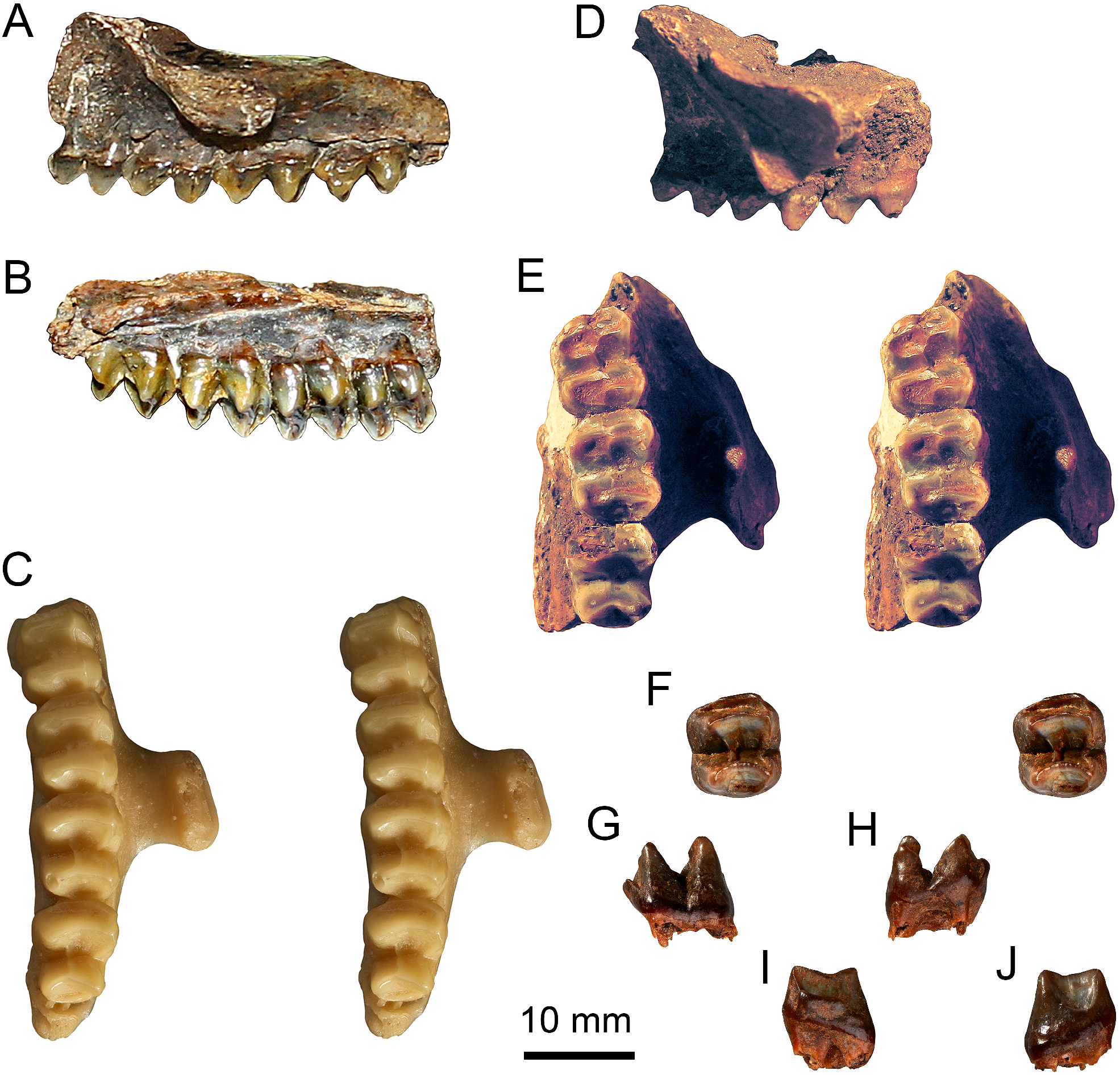
Upper dentition. The single known maxillary specimen (AM F49468) preserves M1–4. The protoloph of M1 is marginally narrower than the metaloph, but from M2 to M4 the protoloph becomes increasingly wider than the metaloph ( Figure 30C View FIGURE 30 ; Table 7 View TABLE 7 ). The molars are moderately worn, with dentine breached along the lengths of the lophs of M1–2, on the protoloph and metaconule of M3, and on the protocone of M4. Despite this, they were clearly low crowned, and it is evident from the less worn molars that the postparacrista, postprotocrista, premetacrista and postmetacrista are very fine and low ( Figure 30 View FIGURE 30 ). The postparacrista and premetacrista are distinctly incurved on all molars. On M1–3, the cusp C region of the stylar crest is evident, diverging from the postparacrista about halfway up the paracone face, such that it forms an inverted Y shape.The stylar crest remnant is projected buccally at 50° on M1 to the longitudinal axis of the crown, at 40° on M2 and at 30° on M3 ( Figure 30C View FIGURE 30 ). The postmetaconulecrista forms a small but distinct shelf on the posterior face of M4.
The upper molars of B. planei are smaller than in B. bandharr and B. illuminata , and closest in size and morphology to those of B. nullarbora and B. bila ( Tables 1 View TABLE 1 , 4–6 View TABLE 4 View TABLE 5 View TABLE 6 ). By comparison, B. nullarbora lacks any sign of a cusp C region of the stylar crest on any molars, and has anteroposteriorly orientated postpara- and premeta-cristae, a less lingually tapered precingulum, and a narrower, deeper, more ‘pocketed’ M4 posterior metaloph face. In B. bila , a cusp C region of the stylar crest is present on M1–2, but not M3, and differs by diverging from the postparacrista closer to the paracone apex. As in B. bila , the postpara- and premeta-cristae are incurved, but not as well developed as in B. wilkinsonorum . The configuration of these crests and the stylar crest on M1–3 is very like that of the species of Dorcopsulus , but in the much larger B. planei there is no obvious cusp D region of the stylar crest. It should be noted, however, that there is a small wear facet on the metacone anterior face buccal to the premetacrista on M1–2, which, in a minimally worn specimen, may reveal that this portion of the stylar crest was indeed retained in B. planei . The upper molars of B. planei share very weakly development of the anteroposterior crests with B. nullarbora and B. bandharr .
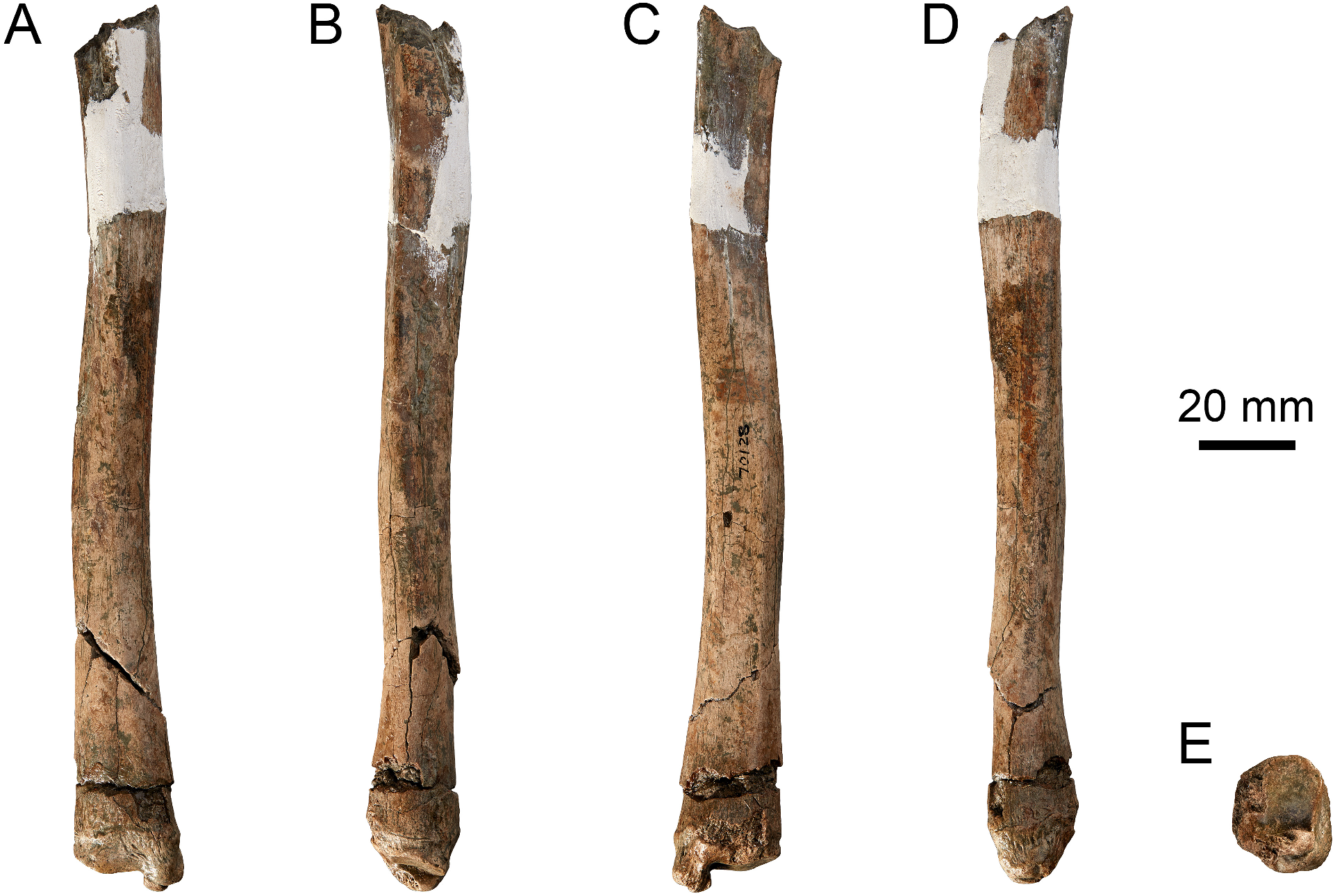
Femur. Well preserved and largely complete, the holotype femur is missing only the lateral condyle of the distal epiphysis ( Figure 31 View FIGURE 31 ). The femoral head is large and roughly hemispheric. The proximal epiphyseal margin is more distally placed than the caudal margin of the head. The greater trochanter is very rugose at its apex, particular in cranial view, and strongly laterally flared at its base ( Figure 31A View FIGURE 31 ). The lesser trochanter is robust and long, with a distally extended ridge and distinct sulcus on its craniodistal aspect ( Figure 31C View FIGURE 31 ). The femoral shaft is slightly compressed in the craniocaudal plane, and is cranially convex. The caudal surface of the diaphysis is marked by a rugose adductor muscle scar, which runs obliquely from below the lesser trochanter to a depression marking the insertion for the m. quadratus femoris midway along the length of the bone ( Figure 31B–C View FIGURE 31 ). A second scar marking the more distal portions of the mm. adductores femoris then continues along the posterior midline to the popliteal surface. The insertion of the m. quadratus femoris is rugose and concave ( Figure 31B View FIGURE 31 ). The caudomedial fossa for the m. gastrocnemius is shallow and the caudolateral fossa for the m. flexor digitorum superficialis is very shallow. The distal femoral condyles are relatively low and the trochlea groove relatively shallow ( Figure 31A–B, F View FIGURE 31 ). The medial condyle is transversely quite narrow.
The femur of the B. planei holotype is approximately 33% larger in size than in D. lumholtzi and 20% larger than in B. illuminata . The greater trochanter is taller than in species of Dendrolagus , and its rugose apex is distinct. The base of the greater trochanter bears a distinct fossa on its cranial surface for the m. vastus lateralis, which is of similar depth in B. illuminata and B. nullarbora , but noticeably wider. The laterally flared distal extremity is most like Thylogale billardierii ( Desmarest, 1822) . The thickened lateral margin or crest upon which the m. gluteus medius inserts is proportionally shorter than in most macropodines, terminating proximally to the distal margin of the lesser trochanter, as in B. illuminata and species of Dendrolagus ( Figure 7A–D View FIGURE 7 ). The lesser trochanter is more flared proximally than in species of Dendrolagus , Setonix and Thylogale , and may have been similar in B. illuminata and B. nullarbora , though the area is abraded in the only known specimens of each species. The shaft is similar in relative robustness to B. illuminata , and less robust than in B. nullarbora and species of Dendrolagus ( Figure 7 View FIGURE 7 ). The adductor muscle scars are more marked than in B. nullarbora , while in B. illuminata there are narrow but relatively deep longitudinal scars for the mm. adductores femoris along the medial aspect of the femoral shaft as in Dendrolagus ( Figure 7C View FIGURE 7 ; Warburton et al. 2012). The concave insertion of the m. quadratus femoris is unusual, in contrast to the typically raised tubercle in most macropodines. In this attribute, B. planei resembles T. billardierii . The diaphysis flares toward its distal end of shaft more than in species of Dendrolagus and terrestrial species but similar to B. illuminata and B. nullarbora .
The shallow fossae for m. gastrocnemius and m. flexor digitorum superficialis contrast markedly with the deep fossae in ground-dwelling species of Macropus Shaw, 1790 , Osphranter Gould, 1842 and Thylogale , but are similar to the morphology in B. illuminata and species of Dendrolagus . The craniocaudally compressed distal epiphysis is reminiscent of species of Dendrolagus ( Figure 7F View FIGURE 7 ). The transversely narrow medial condyle is similar in morphology to B. illuminata .
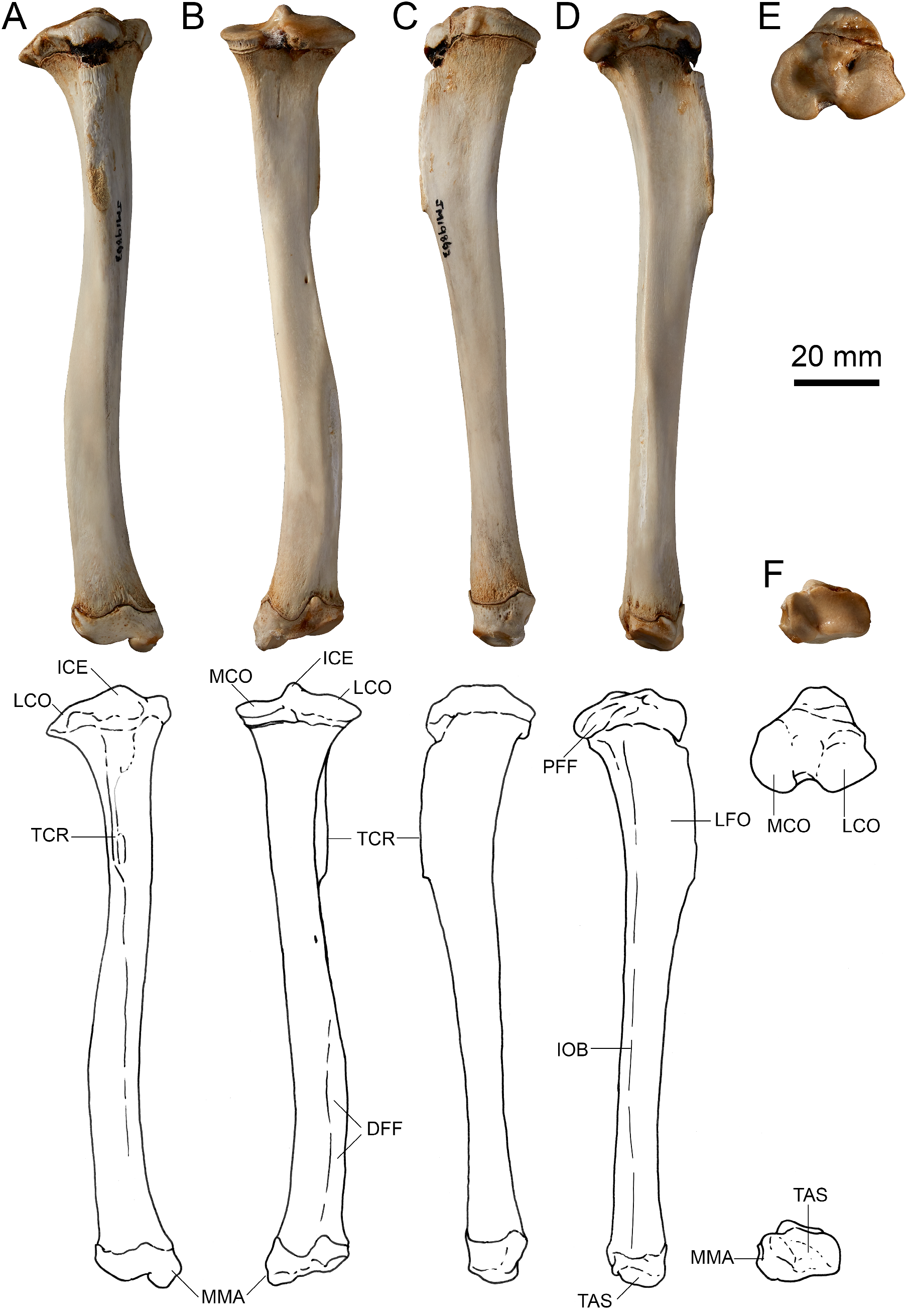
Tibia. The B. planei holotype preserves only the distal two-thirds of the tibia, the diaphysis of which is relatively straight, being very slightly sinuous from cranial view, and similarly in the cranial face of the proximal portion from the lateral view ( Figure 32B View FIGURE 32 ). Distally, the diaphysis is sub-rectangular in cross-section. The distal fibular facet is estimated to have extended for approximately the distal two-fifths of the tibial shaft. The interosseous border above the distal fibula facet is moderately rounded. The distal epiphysis is equal in width to the end of the diaphysis ( Figure 32C View FIGURE 32 ). The medial malleolus is robust, moderately long and aligned in the parasagittal plane ( Figure 32C View FIGURE 32 ). The talar articular facet is aligned square to the craniocaudal axis ( Figure 32E View FIGURE 32 ).
In overall shape, the tibial diaphysis and proportional length of the distal fibular facet of B. planei is very similar to that of B. illuminata . The rounded interosseous border is in contrast to the sharper interosseous crest observed in B. illuminata , B. nullarbora and the species of Dendrolagus ( Figure 8 View FIGURE 8 ) and Thylogale , but it is less rounded than in Setonix brachyurus . The distal epiphysis and talar articular facet are relatively narrower than in B. illuminata and species of Dendrolagus , and are not as laterally flared as in D. bennettianus . The medial malleolar process is more distally extended than in species of Dendrolagus , but narrower anteroposteriorly than in species of Thylogale and Setonix . In distal view, the process of the medial malleolus is more obliquely orientated than in other species of Bohra and Dendrolagus ( Figure 8F View FIGURE 8 ), being more similar to Thylogale in both this and in cranial view, in which it is relatively flat and horizontal.
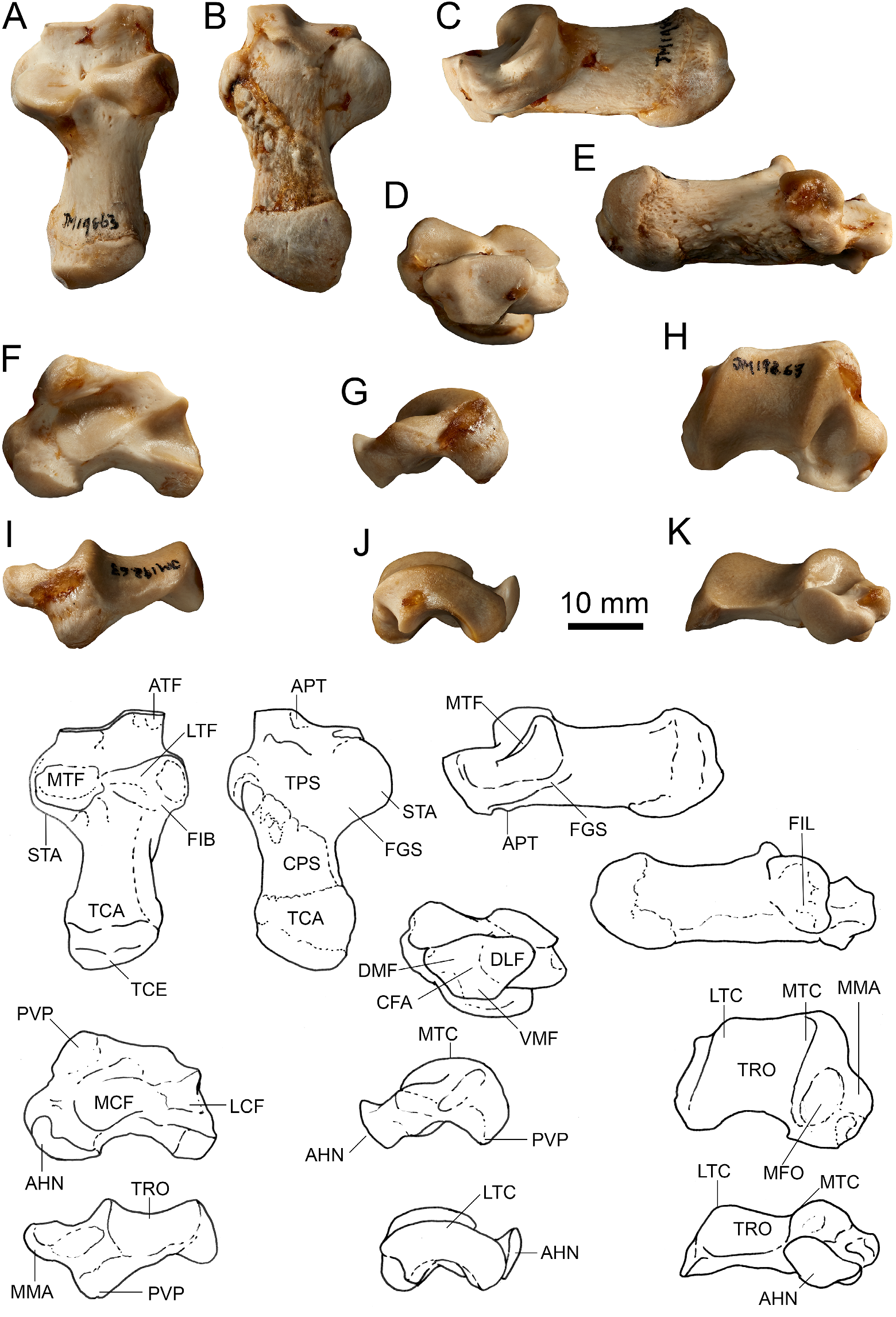
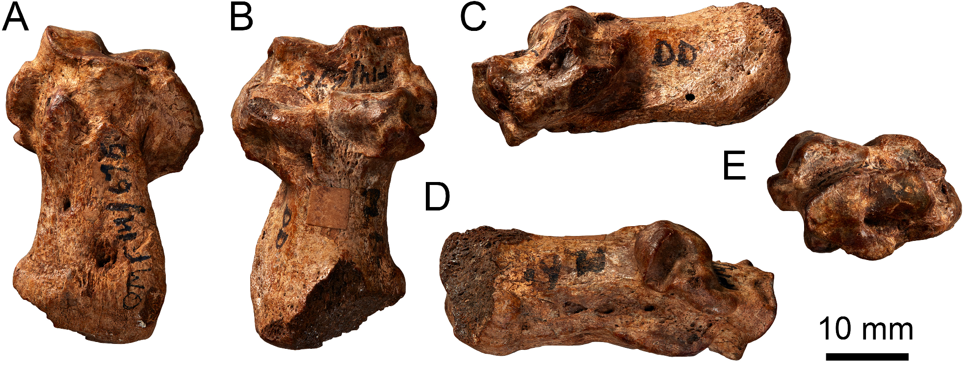
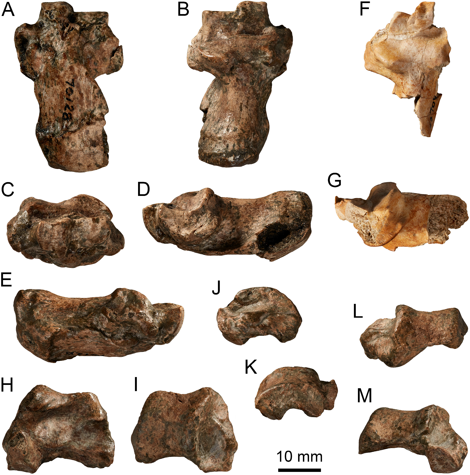
Calcaneus. The calcaneus is stout, robust, broad and dorsoventrally compressed ( Figure 33A–G View FIGURE 33 ). The tuber calcanei is expanded caudally and constricted cranially ( Figure 33B View FIGURE 33 ). The rugose plantar surface is very broad, and is expanded along the length of the medial margin by a distinct flange ( Figure 33A View FIGURE 33 ). The mid-lateral margin is particularly enlarged. The epiphyseal line on the rugose plantar surface is raised into a distinct, ventrally projected ridge. Paired tubercles (the lateral one much larger) on the craniolateral extremity of the plantar surface are positioned adjacent to the groove for the peroneal tendons, and are bounded on the opposite side by a projection cranial to the attachment site of the anterior calcaneofibular ligament. The transverse flexor sulcus is not well defined, being relatively shallow and with irregular margins. The sustentaculum tali is very broad, particularly caudally, extending well beyond the medial edge of the talar articular surface ( Figure 33D View FIGURE 33 ). The ventral surface is smoothly curved and the flexor groove relatively deep, accentuated by the expanded medial margin of the plantar surface. The lateral and medial facets for the talus are very broad; the lateral facet is smaller, mesially constricted and smoothly contiguous with the oval medial facet. The fibular facet is very small, though the tubercles for the fibular ligaments, particularly the calcaneocuboid ligaments, are strongly developed. The anteromedial facet for articulation with the talar head is small and distinct. The calcaneocuboid articulation is transversely broad and dorsoventrally compressed ( Figure 33C View FIGURE 33 ). The dorsomedial facet for the cuboid is rectangular, broader than deep, and convex in profile. The dorsolateral facet is narrower and subtriangular in shape, tapering from dorsal to ventral. The step between the two facets is distinct, only slightly smoothed and obliquely orientated in dorsal view ( Figure 33A–G View FIGURE 33 ). The ventromedial cuboid facet is abraded on the holotype, but was apparently relatively small and smoothly continuous with the dorsolateral facet, but separate from the dorsomedial facet.
The calcaneus of B. planei is similar overall in morphology to B. illuminata , B. nullarbora and the species of Dendrolagus ( Figure 9 View FIGURE 9 ). The distinct ridge formed at the plantar epiphyseal line distinguishes B. planei from all other species, but is most similar to D. bennettianus ( Figure 9B View FIGURE 9 ), and may reflect augmentation of the digital flexor muscles in the pes. The rugose tuberosities at the cranial margin of the plantar surface are similar to those observed in B. wilkinsonorum ( Figure 20A View FIGURE 20 ). The flaring of the medial plantar margin of the calcaneal tuber and flexor groove are developed to a greater extent than in other Bohra species. The morphology of the talar facets is most similar to that of B. paulae ( Figure 12A View FIGURE 12 ), but even more confluent and more dorsoventrally flattened. In all other species of Bohra , the margins of the medial and lateral facets are more clearly delineated, even where they abut each other. The calcaneocuboid step is deeper and more angular than in other species of Bohra and Dendrolagus , being most similar to B. wilkinsonorum , and also approaching the depth in species of Setonix and Thylogale . When viewed dorsally, the cranial extremity (cuboid articulation) is relatively much narrower (does not extend to the width of the fibular facet) in comparison to other species of Bohra . The cuboid facets are relatively deep and narrow compared to other species of Bohra . The separation of the ventromedial facet appears most like B. wilkinsonorum .
Talus. The talus is wider than long, dorsoventrally compressed (especially in cranial view) with low trochlear crests, which are subequal in height, and a shallow trochlea groove ( Figure 33H–M View FIGURE 33 ). The malleolar fossa is moderately wide and shallow. The talar neck is very short. The talar head is medially displaced relative to the medial trochlear crest and transversely quite broad in cranial view. The articular facet for the navicular is ovoid around an axis that is approximately 40° from a dorsoventral plane ( Figure 33M View FIGURE 33 ). The calcaneal facets are transversely broad, moderately deep and smoothly contiguous. The posterior plantar process is bulbous, transversely broad and dorsoventrally compressed.
The trochlear groove is shallower than in B. illuminata , B. nullarbora , D. bennettianus ( Figure 9H–I, K View FIGURE 9 ) and D. lumholtzi , but similar to that of B. sp. indet. 1. The medial trochlear crest is lower than in B. paulae , and also more similar to that of B. sp. indet. 1. The malleolar fossa is broad and shallower in B. planei than in other species of Bohra and species of Dendrolagus . The medial malleolus is broad and flattened, similar to the conditions seen in B. paulae and B. sp. indet. 1. It is relatively less projected than in B. nullarbora and species of Dendrolagus , Dorcopsulus , Setonix and Thylogale . The very short talar neck is similar to the morphology seen in species of Dendrolagus ( Figure 9H View FIGURE 9 ), and is shorter than in B. illuminata and B. nullarbora , but not as short as in B. paulae ( Figure 12K View FIGURE 12 ). The navicular facet of the talar head is set at a less oblique angle than in other species of Bohra , including B. paulae and especially B. illuminata and species of Dendrolagus , resulting in a narrower and more elongate outline. The lateral border of the navicular facet lies outside of, rather than in line with, the cranial extremity of the medial crest, as in B. paulae and D. bennettianus , D. lumholtzi . The talocalcaneal facets are smoothly conjoined and relatively shallow. The posterior plantar process is short and broad, as in B. nullarbora and B. illuminata , but proportionally smaller than in species of Dendrolagus .
Remarks. Bohra planei is the only species in this genus identified from New Guinea. It is one of only three species of fossil macropodoid, along with B. paulae and Rhizosthenurus flanneryi Kear, 2002 , initially described wholly or fundamentally on the basis of hindlimb elements. This reinforces past observations ( Flannery & Szalay 1982; Prideaux & Warburton 2008, 2009; Warburton & Prideaux 2010; Warburton et al. 2011), as well as those made in this paper, that demonstrate the high taxonomic utility of hindlimb material for recognising fossil tree-kangaroo remains. Along with Protemnodon otibandus Plane, 1967 , ‘Silvaroo’ buloloensis and Watutia novaeguineae , B. planei is the fourth extinct macropodine species identified from the Otibanda Formation ( Figure 1 View FIGURE 1 ) and described primarily on the basis of material from it.
The partial maxilla, AM F49468, has had a varied taxonomic history. It was first described and referred to ‘cf. Dorcopsis , large form’ by Plane (1967) in his monograph on the Otibanda Formation and Awe LF, but in another monograph published in the same series on the same day, Woodburne (1967) considered it more likely to belong to a taxon related to Dendrolagus . In part, this was because Woodburne inferred that the anteromesially orientated broken edge of what remains of the palate marks the maxilla–palatine suture, and that this orientation was more like that seen in Dendrolagus than in any dorcopsin. To our eyes, having examined multiple species of each, there is no consistent difference between species of Dendrolagus and dorcopsins in the shape of this suture: it is fundamentally anteroposteriorly orientated. However, an anteromesially orientated suture very obviously is a feature of Bohra nullarbora ( Figure 27E View FIGURE 27 ). For this reason, and given the marked similarities in molar morphology between AM F49468 and both B. nullarbora and B. bila , we confidently identify it as representing a species of Bohra . That Flannery et al. (1989) and Dawson (2004b) viewed the upper molars of AM F49468 as more like Dorcopsis than Dendrolagus is hardly surprising: it was not until 2008 that the first molars unequivocally referable to a species of Bohra were described and shown to resemble those of the species of Dorcopsis ( Prideaux & Warburton 2008) .
AM F49468 and the holotype partial hindlimb (UCMP 70128) come from different but nearby sites in the Otibanda Formation, yet we have chosen to allocate AM F49468 to the same species on size grounds: both AM F49468 and UCMP 70128 are a very close size match for the holotype of B. nullarbora ( Tables 6–8 View TABLE 6 View TABLE 7 View TABLE 8 ). Although a second Awe LF maxilla fragment preserving M3 (UCMP 70132) might possibly belong to another species of Bohra , the M3 is considerably smaller than that of AM F49468 and differs from it by lacking a stylar cusp C remnant and having a precingulum that terminates abruptly, well short of lingual side of the crown. These features are more suggestive of a large species of Dorcopsis than of Bohra , which is consistent with the original tentative identification ( Plane 1967).


| UCMP |
University of California Museum of Paleontology |
| V |
Royal British Columbia Museum - Herbarium |
| AM |
Australian Museum |
| T |
Tavera, Department of Geology and Geophysics |
No known copyright restrictions apply. See Agosti, D., Egloff, W., 2009. Taxonomic information exchange and copyright: the Plazi approach. BMC Research Notes 2009, 2:53 for further explanation.
|
Kingdom |
|
|
Phylum |
|
|
Class |
|
|
Order |
|
|
Family |
|
|
Genus |
Bohra planei
| Prideaux, Gavin J. & Warburton, Natalie M. 2023 |
Dorcopsis
| Black, K. H. & Archer, M. & Hand, S. J. 2012: 1050 |
Silvaroo sp.
| Dawson L. 2004: 284 |
Dorcopsis sp.
| Dawson L. 2004: 288 |
Dorcopsis
| Menzies, J. I. & Ballard, C. 1994: 131 |
Dorcopsis
| Flannery, T. F. & Mountain, M. - J. & Aplin, K. P. 1983: 77 |