
Anaphidna Gorochov & Cadena-Castañeda, 2012
|
publication ID |
https://doi.org/ 10.11646/zootaxa.5166.1.1 |
|
publication LSID |
lsid:zoobank.org:pub:17952A48-902C-47A0-A344-8B07490F3B28 |
|
DOI |
https://doi.org/10.5281/zenodo.6885804 |
|
persistent identifier |
https://treatment.plazi.org/id/03C0C519-CF0C-616B-D4A2-8E204E0FFF2F |
|
treatment provided by |
Plazi |
|
scientific name |
Anaphidna Gorochov & Cadena-Castañeda, 2012 |
| status |
|
Anaphidna Gorochov & Cadena-Castañeda, 2012 View in CoL
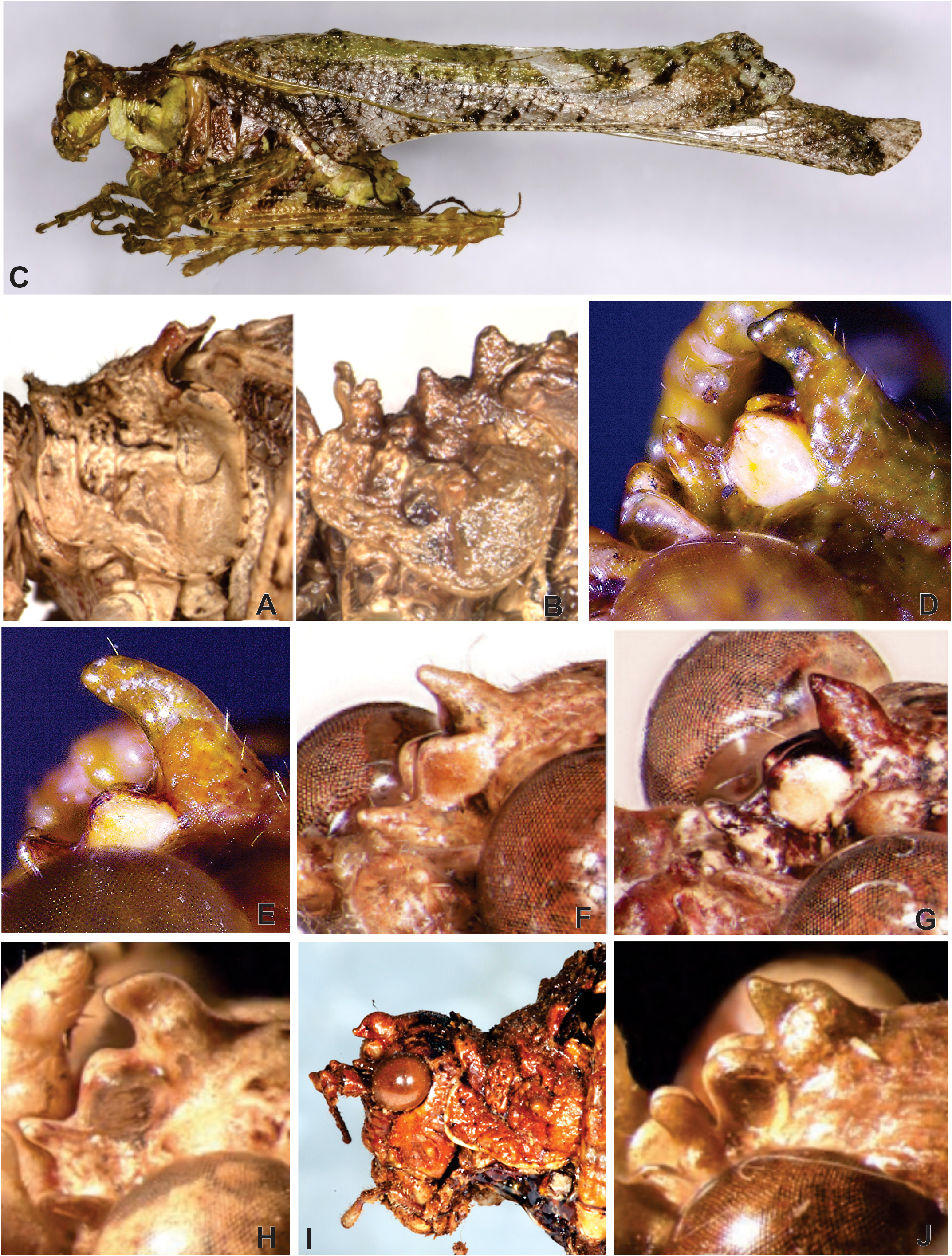
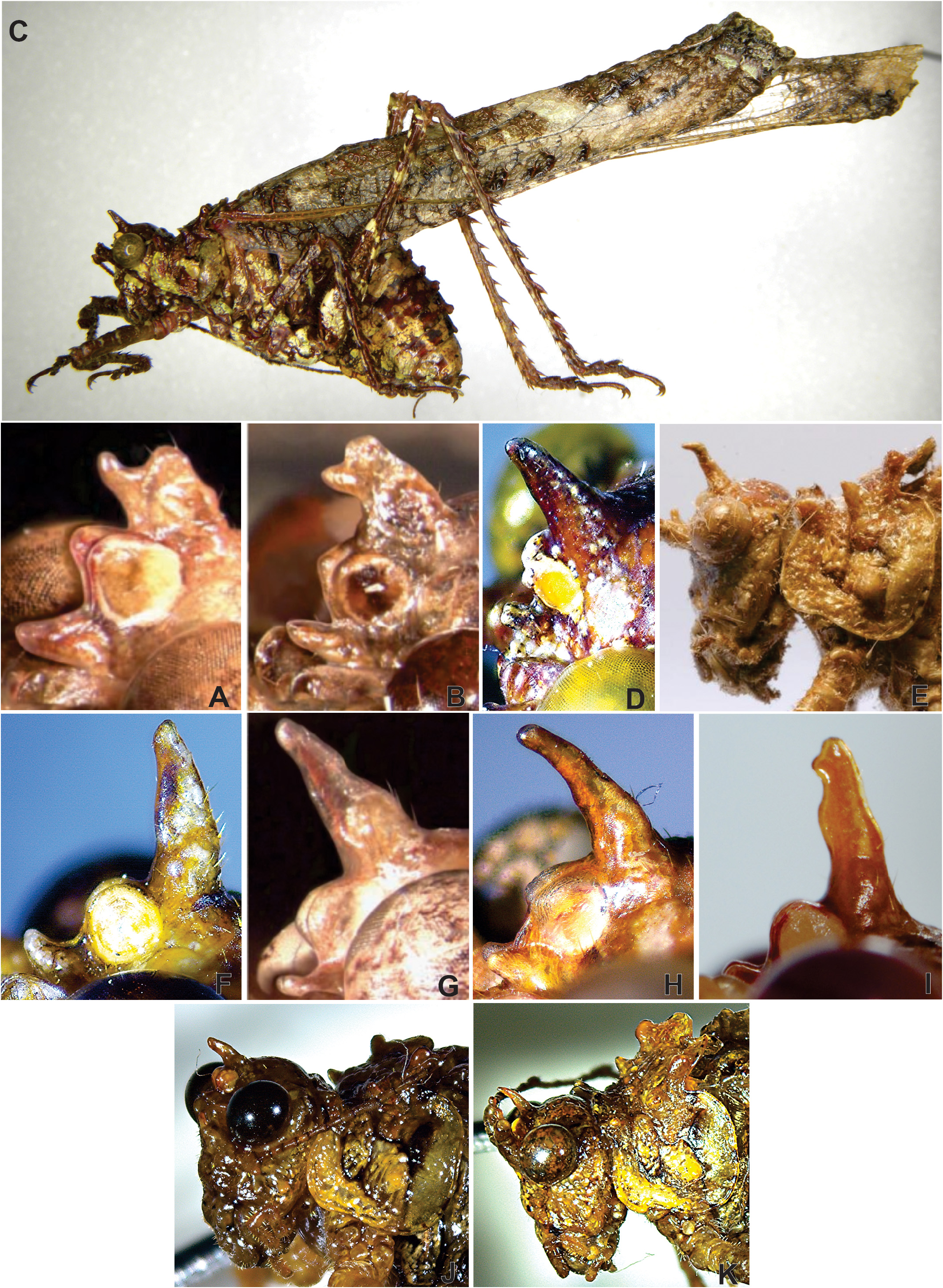
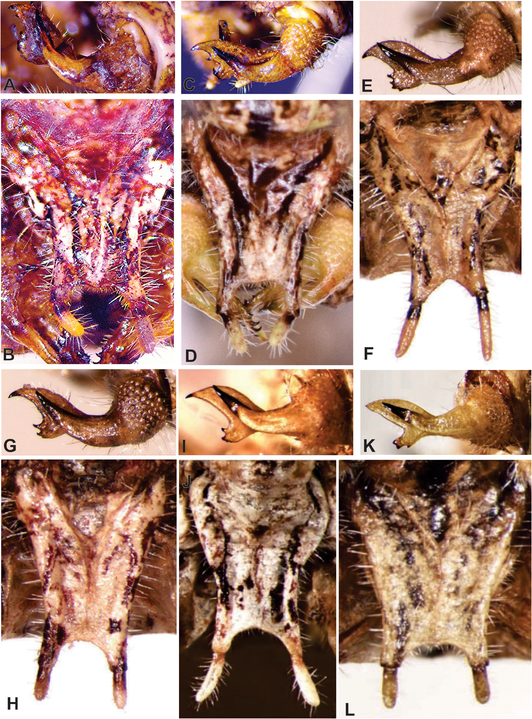
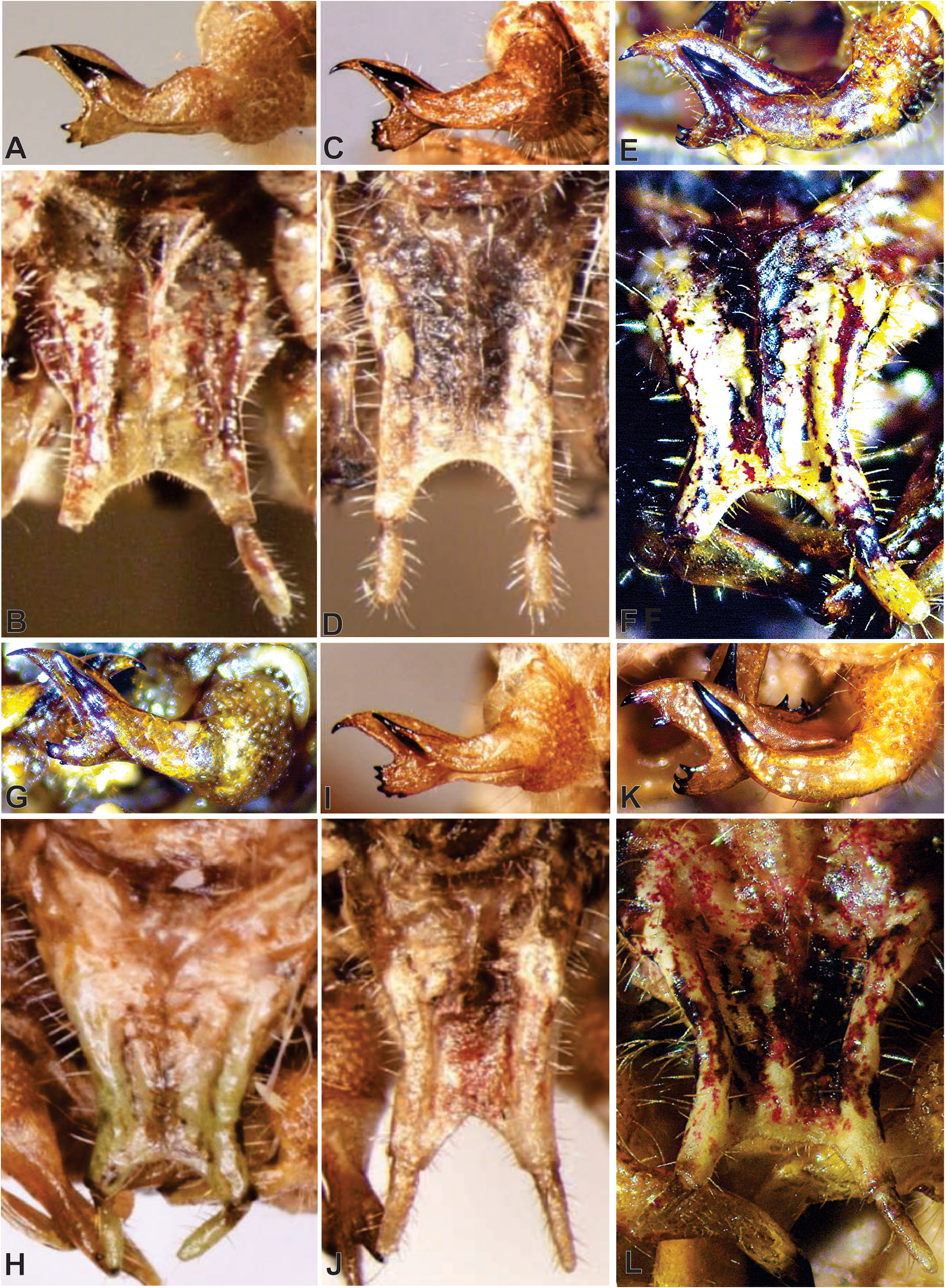
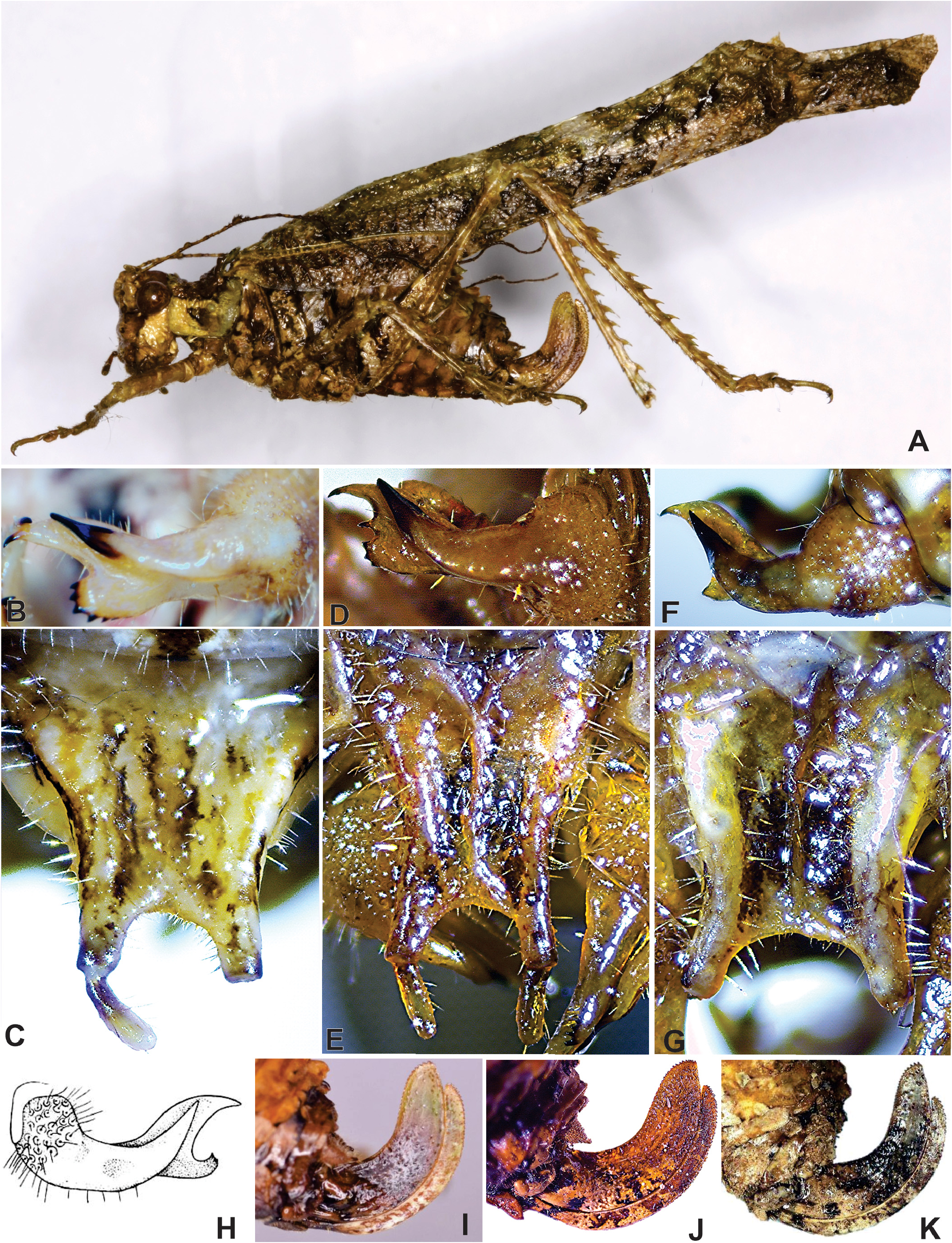
Diagnosis. Coloration predominantly greenish brown, some species reddish with white or whitish green stripes ( Figs. 28C View FIGURE 28 , 29C View FIGURE 29 , 32A View FIGURE 32 ). Structure on vertex variable, reduced and tubercle-shaped ( Figs. 28D, F, G, H, I, J View FIGURE 28 ) or elongated and cylindrical ( Figs. 29D, E, F, G, H, I View FIGURE 29 ). Median ocellus conspicuous, genae tuberculate ( Figs. 28I View FIGURE 28 , 29E, J, K View FIGURE 29 ). Pronotum usually tuberculate with four or six emarginations on lateral margins of pronotal disk ( Figs. 28 A, B View FIGURE 28 ). Male cerci with external branch rather long and spine-shaped, curved upward and with acute apex; inner branch longer, partially lamelliform and with two lobes, the upper hook-shaped, the lower one smaller and armed with tiny apical hooks ( Figs. 32H View FIGURE 32 ). Ovipositor as long as pronotum or slightly longer, curved from base upward, upper valve longer than ventral one, margins serrate ( Fig. 32I–K View FIGURE 32 ).
Type species. Paraphidnia (Anaphidna) mexicana Gorochov, 2012 View in CoL , by original designation.
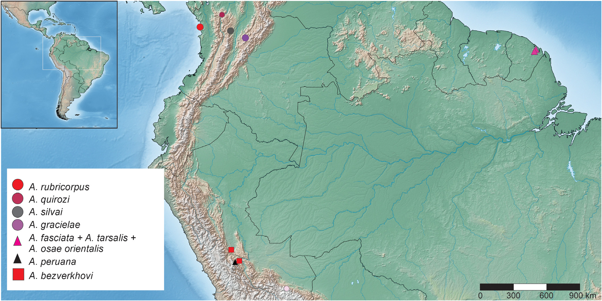
Distribution. From southern Mexico to northern Argentina ( Maps 14 View Map 14 and 15 View Map 15 ).
Comments. The species of this genus exhibit considerable phenotypic plasticity, which makes their identification difficult. Individuals of the same species, or even of the same population, may show variation in characters that are otherwise stable and useful for the delimitation of species in other genera of the Dysoniini , like shape of the face, pronotal tubercles, structure of the stridulatory vein, wing venation, spines on the legs, and male cerci. Specimens of the same species would mistakenly be considered as belonging to more than one species, unless this variability is taken into account. On the other hand, sympatry of related species among Neotropical Phaneropterinae is common: According to observations made in the course of this investigation in the canopy of Amazon rainforest (canopy walkway in Amacayacu, SE Colombia), it was found that two, three, and even more species of the same genus live together in the same area, and even on the same tree: Ceraia (with three species), Euceraia (four species), and Phylloptera (six species). Similar data were reported from lowland rainforest in northern Peru ( Nickle & Castner 1995) .
According to the above, an overestimation of the variability in a few sympatric species of the genus Anaphidna could lead to considering them erroneously a single very variable species. Conversely, an underestimation of the variability could lead to the false assumption of more different species than are actually present. Another problem with the taxonomy of the genus is the presence of numerous closely related species in the same distributional range, and lacking significant differences in copulatory structures and other characters. This may be the result of recent adaptive radiation. To delimit the species, acoustic and ethological studies would be useful. The following key to species is based on characters with low range of variability.
Key to species of Anaphidna View in CoL
1. Pronotal disc without tubercles, or when present, poorly developed ( Fig. 28A View FIGURE 28 ), usually with the posterior denticle of the crest reduced (the most developed denticle of the head) ( Figs. 28D–J View FIGURE 28 )................................................ 2
- Pronotal disc with conspicuous tubercles ( Fig. 28B View FIGURE 28 ), posterior denticle usually elongated (most developed denticle of the head) ( Figs. 29D–K View FIGURE 29 )........................................................................................ 8
2. Posterior denticle as long as scapus and pedicellus combined or longer, slender and three times as long as wide ( Figs. 28D– E View FIGURE 28 ).................................................................................................. ................................................................................................... 3
- Posterior denticle not exceeding scapus and pedicellus, robust and usually as long as wide ( Figs. 28F–J View FIGURE 28 )................ 4
3. Emargination of the male subgenital plate W-shaped ( Fig. 30B View FIGURE 30 )................ A. hernandezi (Cadena-Castañeda, 2012) View in CoL
- Emargination of male subgenital plate shallow and straight ( Fig. 30D View FIGURE 30 )................ A. osae (Cadena-Castañeda, 2012) View in CoL
4. Posterior denticle almost conical, more or less acute ( Figs. 28F–H View FIGURE 28 ); emargination of male subgenital plate U- or V-shaped ( Figs. 30F, H, J View FIGURE 30 )........................................................................................... 5
- Posterior denticle robust, strongly reduced and slightly bifurcate ( Figs. 28I–J View FIGURE 28 ); emargination of male subgenital plate almost straight ( Fig. 30L View FIGURE 30 )..................................................................................... 7
5. Emargination of male subgenital plate U-shaped ( Figs. 30H–J View FIGURE 30 ); tarsi unspecialized................................. 6
- Emargination of male subgenital plate V-shaped ( Fig. 30F View FIGURE 30 ); all tarsi with strongly inflated dorsal part of proximal segment and very large second segment........................................................ A. tarsalis ( Gorochov, 2014) View in CoL
6. Body coloration predominantly reddish, tubercles of scapus and pedicellus poorly developed...................................................................................................... A. fasciata ( Gorochov, 2014) View in CoL
- Body coloration predominantly greenish, tubercles of the scapus and pedicellus well developed................................................................................................. A. bezverkhovi (Gorochov, 2012) View in CoL
7. Anterior denticle slender ( Fig. 28I View FIGURE 28 ); emargination of male subgenital plate angled in shape of V with very little depth; inner branch of male cerci with lower margin of distal portion rounded and with more than three marginal denticles............................................................................ A. verrucosa ( Brunner von Wattenwyl, 1878)
- Anterior denticle dilated ( Fig. 28J View FIGURE 28 ); emargination of male subgenital plate straight with a very small medial prolongation ( Fig. 30L View FIGURE 30 ); inner branch of the cerci with lower margin of the distal portion reduced (rectangular) and with little denticulation ( Fig. 30K View FIGURE 30 )..................................................................... A. polestshuki (Gorochov, 2012) View in CoL
8. Posterior denticle not exceeding scapus and pedicellus, robust and usually as long as wide, apex bifurcate ( Figs. 29A–B View FIGURE 29 )... 9
- Posterior denticle as long as scapus and pedicellus or longer, slender and three or four times as long as wide ( Figs. 29D–K View FIGURE 29 ).. .................................................................................................. 10
9. Genicular lobe of hind femur armed with a small spinule, crest as in figure 30A............. A. peruana (Gorochov, 2012) View in CoL
- Genicular lobe of hind femur armed with a prominent spur, crest as in figure 30B.......... A. svetlanae (Gorochov, 2012) View in CoL
10. Predominant coloration of the body green and brown........................................................ 11
- Predominant coloration of the body red with white stripes ( Fig. 30C–D View FIGURE 30 , 31E–F View FIGURE 31 ).. A. rubricorpus (Cadena-Castañeda, 2012) View in CoL
11. Posterior denticle of the crest curved forward near apex...................................................... 12
- Posterior denticle of the crest straight or curved forward already from base....................................... 13
12. Posterior denticle of the crest bent forward near apex at almost right angle ( Fig. 29E View FIGURE 29 ), anterior denticle slender and slightly curved in lateral view............................................................. A. lankesteri ( Rehn, 1918) View in CoL
- Posterior denticle of the crest little curved near apex, anterior denticle robust and hook-shaped in lateral view ( Fig. 29F View FIGURE 29 )....................................................................... A. rhinoceros (Cadena-Castañeda, 2012) View in CoL
13. Emargination of male subgenital plate U- or V-shaped ( Figs. 31J View FIGURE 31 , 32C View FIGURE 32 ), posterior denticle of the crest moderately tapering, curving or not to the front ( Figs. 29G–I View FIGURE 29 ).................................................................. 14
- Emargination of male subgenital plate straight and shallow, posterior denticle of the crest curving forward ( Figs. 29J–K View FIGURE 29 ).. 16
14. Emargination of male subgenital plate U-shaped............................................................ 15
- Emargination of the male subgenital plate V-shaped.................................. A. mexicana (Gorochov, 2012) View in CoL
15. Posterior denticle of the crest tapering and progressively curving toward apex, without pre-apical undulations; styli of male subgenital plate cylindrical with thin and rounded apex........................... A. obrieni Cadena-Castañeda, 2016 View in CoL
- Posterior denticle of the crest with uniform diameter from base to apex, not curved and always erect, with pre-apical undulation at the front margin; styli of male subgenital plate thickened toward the apex........ A. gracielae (Cadena-Castañeda, 2012) View in CoL
16. Posterior denticle of the crest robust, not exceeding combined length of scapus and pedicellus ( Fig. 29J View FIGURE 29 ); inner lobe of male cerci with ventral projection rounded and with five or more marginal denticles ( Fig. 32D View FIGURE 32 ).................................................................................................... A. silvai (Cadena-Castañeda, 2012) View in CoL
- Posterior denticle of the crest slender, exceeding combined length of scapus and pedicellus ( Fig. 29K View FIGURE 29 ); male cerci with the lower projection of inner lobe distinctly smaller and with two small marginal denticles ( Fig. 32F View FIGURE 32 )............................................................................................. A. quirozi (Cadena-Castañeda, 2012) View in CoL
Genus group Dysoniae Rehn, 1950

No known copyright restrictions apply. See Agosti, D., Egloff, W., 2009. Taxonomic information exchange and copyright: the Plazi approach. BMC Research Notes 2009, 2:53 for further explanation.