
Achramorpha nivalis Jenkin, 1908
|
publication ID |
https://doi.org/ 10.11646/zootaxa.4615.2.1 |
|
publication LSID |
lsid:zoobank.org:pub:9B9884DA-18D5-4BC9-950F-0436E075AAF8 |
|
DOI |
https://doi.org/10.5281/zenodo.5584060 |
|
persistent identifier |
https://treatment.plazi.org/id/513F790D-FFD0-FFB0-E994-D2D3FE4D4933 |
|
treatment provided by |
Plazi |
|
scientific name |
Achramorpha nivalis Jenkin, 1908 |
| status |
|
Achramorpha nivalis Jenkin, 1908 View in CoL
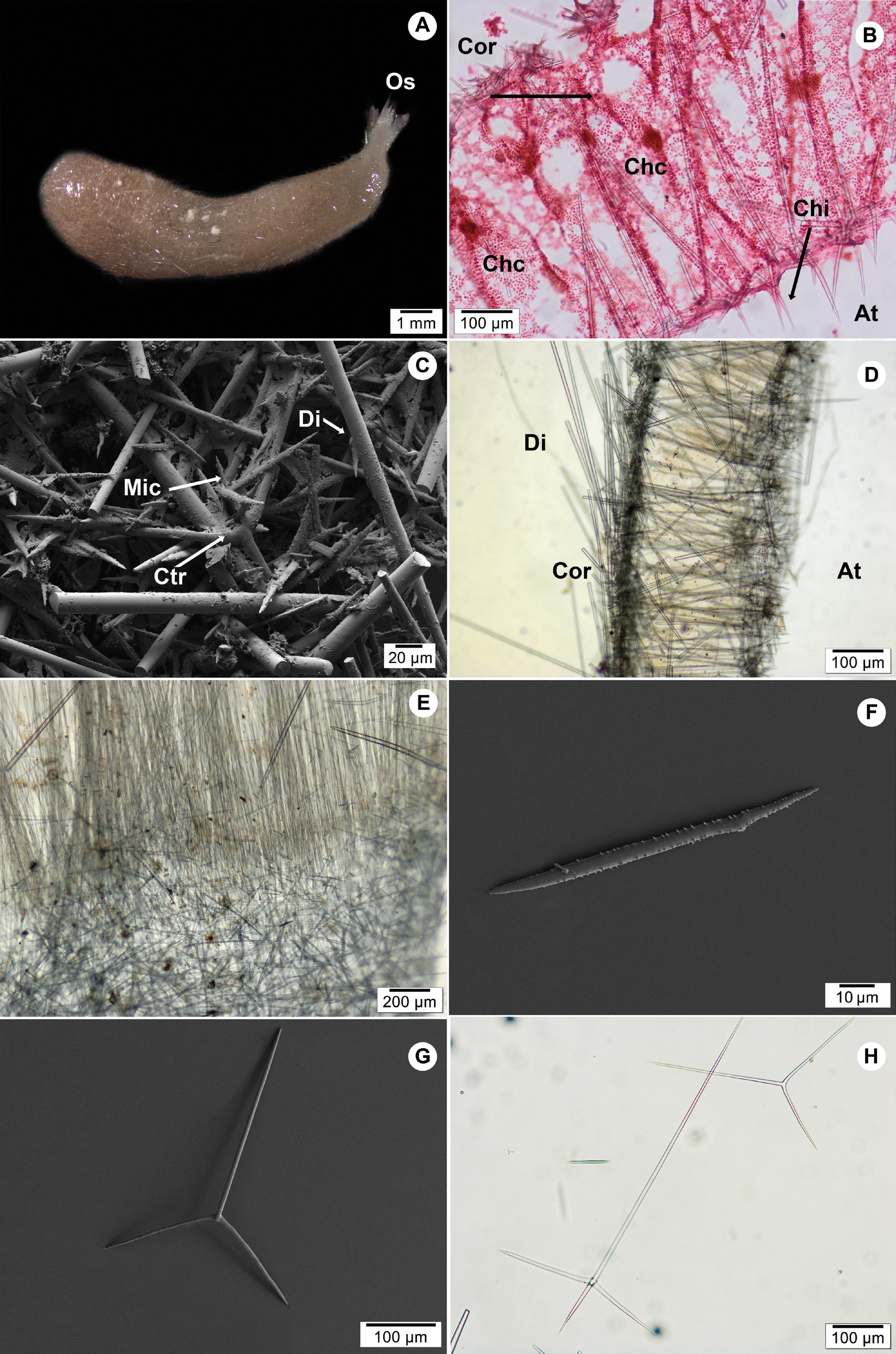
( Figs 1 View FIGURE 1 A–H; Table 2 View TABLE 2 )
Original description. Jenkin 1908, p. 33, pl. XXVII, figs 7–8, pl. XXXV and XXXVI, figs 105–112.
Type locality. Winter Quarters Bay , Antarctic .
Synonyms and citations. Achramorpha nivalis Dendy & Row 1913, p. 765 ; A. nivalis Hôzawa 1918, p. 542 ; A. nivalis, Brønsted 1931, p. 32 ; A. nivalis Burton 1963, p. 93 , 526 (fig 332).
Material examined. Holotype: BMNH-1907.8.6.122 (one section slide), Winter Quarters Bay , Antarctic, collection date 11.11.1902 . Paratype: BMNH-1907.8.6.111 (one complete specimen and four slides; see Table 1 View TABLE 1 ), Winter Quarters Bay , Antarctic, collection date 29.08.1903 . Additional material: BMNH-1907.8.6.119: one slide, National Antarctic Expedition ( HMS Discovery), collection date 08.09.1903. BMNH-1907.8.6.122-124: three slides, National Antarctic Expedition ( HMS Discovery). BMNH-1907.8.6.125: one slide, National Antarctic Expedition ( HMS Discovery), collection date 08.09.1903. BMNH-1907.8.6.128: one slide, National Antarctic Expedition ( HMS Discovery), collection date 24.10.1902. BMNH-1907.8.6.129: one slide, National Antarctic Expedition ( HMS Discovery), collection date 24.10.1902. BMNH-1926.10.26.49: one slide, British Antarctic Expedition 1910–1913 (Terra Nova).
Morphology. Sponge cylindrical, wider at the base and with well-developed oscular fringe at the narrow end ( Fig 1A View FIGURE 1 ). Surface hispid due to long diactines projecting from the choanosome. Colour light brown in ethanol. Aquiferous skeleton syconoid with elongated choanocyte chambers ( Fig 1B View FIGURE 1 ). The cotype is 12.95 mm high, 1.87– 2.94 mm wide and 0.61–0.86 mm thick ( Fig 1A View FIGURE 1 ).
Skeleton. The cortical skeleton is made up by triactines positioned tangentially, diactines and microdiactines ( Fig 1C View FIGURE 1 ). Diactines are very long and protruding, unevenly scattered and can cross through the body wall to the atrial cavity ( Fig 1D View FIGURE 1 ). Microdiactines are spined and organized around the ostia ( Figs 1C, 1F View FIGURE 1 ). Choanoskeleton inarticulated and mainly composed of the unpaired actines of the atrial chiactines and by the large diactines ( Figs 1B, 1D View FIGURE 1 ). Some cortical triactines can be observed in the middle of the choanosome ( Fig 1D View FIGURE 1 ). The chiactines are oriented perpendicularly to the atrium, with the long paired actines adjacent to the atrial wall and the apical actines projecting into the atrial cavity ( Fig 1B View FIGURE 1 ). The oscular region is composed of long trichoxeas which form the oscular fringe, and also by thin but large tetractines, which are positioned longitudinally with the unpaired actines pointing towards the base of the sponge ( Fig 1E View FIGURE 1 ).
Spicules. Diactines: long and straight with both ends sharp. Size: 688.4 ± 328.6 µm length, 14.2 ± 8.2 µm width ( Figs 1 View FIGURE 1 C–D; Table 2 View TABLE 2 ).
Microdiactines: small with hastate points and minute spines towards the hastate tip, which is slender than the other one. Some of these diactines are slightly curved. Size: 80.4 ± 11.5 µm length, 3.4 ± 0.9 µm width ( Figs 1C, 1F View FIGURE 1 ; Table 2 View TABLE 2 ).
Cortical triactines: sagittal with the unpaired actines straight and longer than the paired actines, which are slightly curved upwards forming a round bend. Paired actines of similar length. Size: unpaired actines 230.9 ± 73.1 µm length, 6.7 ± 2 µm width; paired actines 136.6 ± 43.8 µm length, 6.4 ± 2 µm width ( Fig 1G View FIGURE 1 ; Table 2 View TABLE 2 ). There are a few triactines with the paired angle almost straight, which probably are from the oscular region.
Chiactines: unpaired actines straight and longer than the paired actines. Apical actine straight and tapering to a sharp tip. Size: unpaired actines 462.1 ± 113.1 µm length, 9.4 ± 1.1 µm width; paired actines 175.2 ± 18.9 µm length, 8.8 ± 1.2 µm width; apical actine 102.5 ± 15.9 µm length, 7.7 ± 1 µm width ( Figs 1 View FIGURE 1 B–H; Table 2 View TABLE 2 ).
Oscular tetractines: the unpaired actines are longer and thinner than the paired actines. The apical actine is curved and pointing towards the osculum. Size: unpaired actines 259.7 ± 65.7 µm length, 9.4 ± 0.6 µm width; paired actines 167 ± 6.1 µm length, 12.7 ± 0.5 µm width; apical actines 49.9 ± 5 µm length, 7.7 ± 1.1 µm width ( Table 2 View TABLE 2 ).
Oscular trichoxeas: it was difficult to measure them in the spicule preparations, but according to Jenkin (1908) they are around 2.5 mm length and 6.0 µm width and minutely hastate at the distal end ( Fig 1E View FIGURE 1 ).
Molecular identification. Not available.
Distribution and depth. A. nivalis has been reported from two localities around the Antarctic: Winter Quarters Bay ( Jenkin 1908), and Winterstation in Wilkes Land in the East-Antarctic, at 350–385 m depth (Brønsted 1931).
Remarks. Jenkin (1908) mentioned that there were 14 specimens of A. nivalis in the collection at the BMNH. However, the material available in the museum collection was only one slide from the holotype, one specimen labelled as cotype and several spicules preparations and histological sections, probably from those specimens mentioned by Jenkin (1908).
Following the 4 th edition of the International Code of Zoological Nomenclature (ICZN) the term cotype is not recognize by the Code and should not be used in zoological nomenclature, especially e.g. in the sense of syntype or paratype (recommendation 73E). Therefore, the specimen BMNH-1907.8.6.111 which was labelled as cotype, is now erected as paratype.
All the examined slides from different specimens present the same long projecting diactines in the cortical skeleton, but these are shorter than what was reported in the original description (see Table 2 View TABLE 2 ), probably due to the fact that most of them were broken. Jenkin (1908) divided the tetractines in two categories according to the size (see Table 2 View TABLE 2 ). This may be associated to the position of the spicules in the oscular area, since it has been observed in other Achramorpha spp. that they are smaller the closer they are to the oscular fringe. However, as most tetractines were broken or not easily visible in the sections, we were only able to measure five complete ones.
A third type of diactines was mentioned by Jenkin (1908), who described them as “rather longer, small, straight hastate oxea” and of size 120–140 µm long, and 4 µm thick. However, we could not find this type of diactines in the material examined, but according to the figure presented by Jenkin (1908) they look similar to microdiactines, but slightly longer ( Table 2 View TABLE 2 ).
| HMS |
Embrapa Gado de Corte |
No known copyright restrictions apply. See Agosti, D., Egloff, W., 2009. Taxonomic information exchange and copyright: the Plazi approach. BMC Research Notes 2009, 2:53 for further explanation.