
Achramorpha grandinis Jenkin, 1908
|
publication ID |
https://doi.org/ 10.11646/zootaxa.4615.2.1 |
|
publication LSID |
lsid:zoobank.org:pub:9B9884DA-18D5-4BC9-950F-0436E075AAF8 |
|
DOI |
https://doi.org/10.5281/zenodo.5584066 |
|
persistent identifier |
https://treatment.plazi.org/id/513F790D-FFC4-FFA9-E994-D05BFC014B9C |
|
treatment provided by |
Plazi |
|
scientific name |
Achramorpha grandinis Jenkin, 1908 |
| status |
|
Achramorpha grandinis Jenkin, 1908 View in CoL
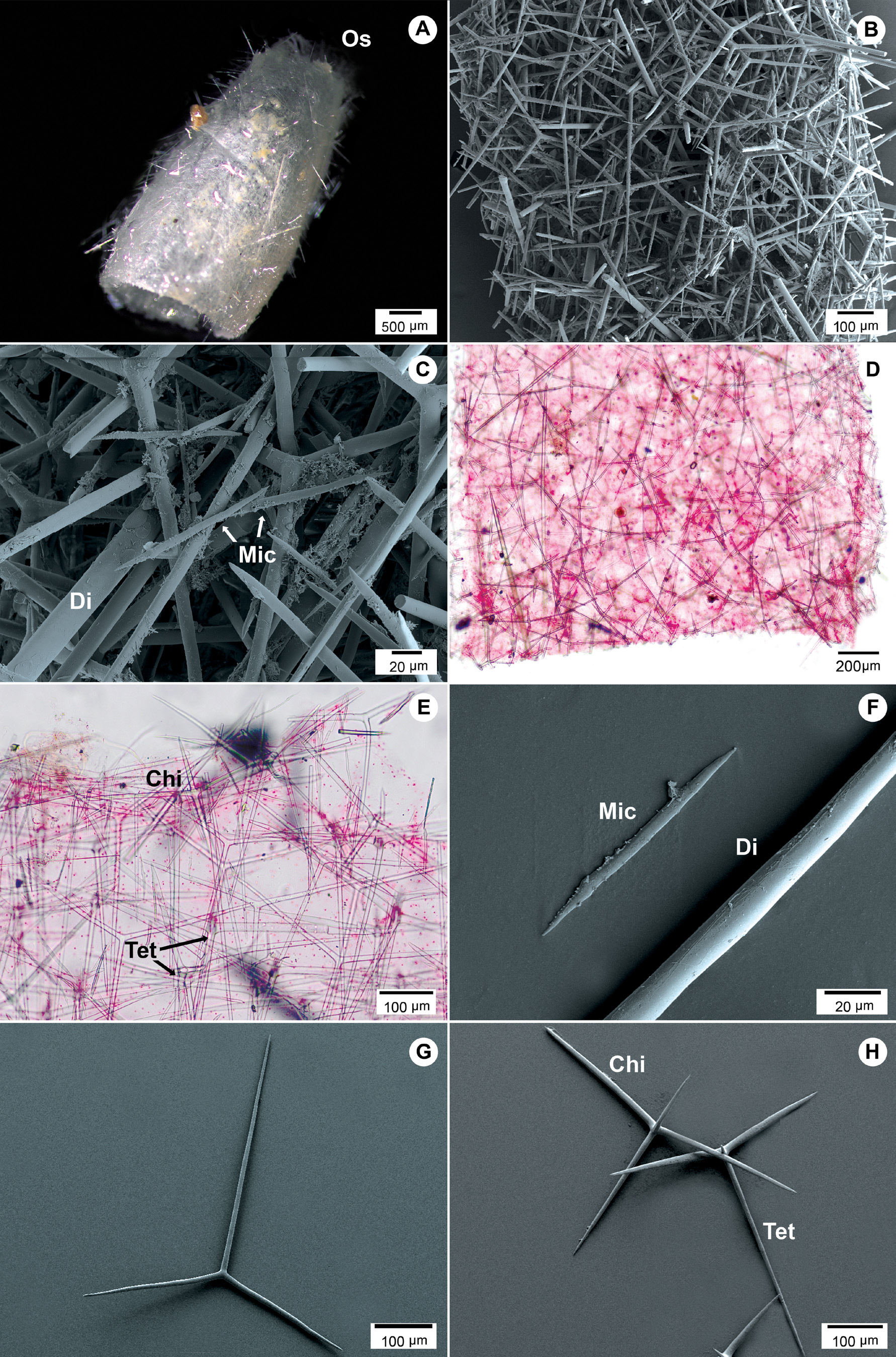
( Figs 6 View FIGURE 6 A–H; Table 6 View TABLE 6 )
Original description. Jenkin 1908, p. 32, pl. XXVII, figs 4, pl. XXXIV and XXXV, figs 103–104.
Type locality. Winter Quarters, Antarctic.
Synonyms and citations. Achramorpha grandinis, Dendy & Row 1913, p. 765 ; A. grandinis Hôzawa 1918, p. 542 ; A. grandinis, Brønsted 1931, p. 32 ; A. grandinis, Burton 1963, p. 93 , 525, fig 331.
Material examined. Holotype: BMNH-1907.8.6.108 (one fragment of the specimen and two slides).
Morphology. Based on the fragment examined, the specimen seems to be cylindrical and slender towards one end, which probably is where the osculum was ( Fig 6A View FIGURE 6 ). Surface hispid due to very long and scattered diactines that protrude the surface. The size of the holotype fragment is 6.0 mm in length and 2.5 mm wide. It was not possible to determine the type of aquiferous system of the species.
Skeleton. Cortical skeleton made up of tangential triactines, long protruding diactines, and microdiactines irregularly scattered ( Figs 6 View FIGURE 6 B–C). The long diactines can cross the atrial wall. Chiactines compose the atrial skeleton, and they are placed with the unpaired actines pointing and projecting the cortical skeleton. The apical actines project towards the atrial cavity ( Fig 6D View FIGURE 6 ). The oscular region is built up by tri- and tetractines placed with the unpaired actines pointing towards the base of the sponge. The short apical actines of the tetractines are bent upwards and crosses the atrial wall ( Figs 6E, 6H View FIGURE 6 ). There is no oscular fringe ( Fig 6E View FIGURE 6 ).
Spicules. Cortical diactines: long and straight diactines that taper in sharp points. Size: 879.1 ± 408.0 µm length, 15.4 ± 4.3 µm width ( Figs 6 View FIGURE 6 B–C; Table 6 View TABLE 6 ).
Microdiactines: small and nearly straight. One blunt tip and the other hastate. Surface with small scattered spines. Size: 93.6 ± 20.3 µm length, 4.0 ± 0.8 µm width ( Figs 6C, 6F View FIGURE 6 ; Table 6 View TABLE 6 ).
Cortical triactines: large, alate with straight unpaired actines, and shorter paired actines.All actines are tapering uniformly to sharp points. Size: unpaired actines 424.8 ± 91.9 µm length, 10.1 ± 1.0 µm width; paired actines 247.1 ± 26.6 µm length, 10.6 ± 1.5 µm width ( Fig 6G View FIGURE 6 ; Table 6 View TABLE 6 ).
Chiactines: large and straight unpaired actines, and slightly slender than the paired actines which are shorter and form a wide angle (around 160 º). Apical actine sharply pointed. Size: unpaired actines 410.1 ± 46 µm length, 11.1 ± 1.6 µm width; paired actines 231.4 ± 34.4 µm length, 11.9 ± 1.8 µm width; apical actine 120.3 ± 22.8 µm length, 10.1 ± 1.8 µm width ( Fig 6H View FIGURE 6 ; Table 6 View TABLE 6 ).
Oscular tetractines: alate and straight tetractines, with all actines tapering into sharp tips. Apical actine conical and shorter than the unpaired and paired actines. The length of the unpaired actines is most likely underestimated as they were often broken or not clearly visible in the slides ( Fig 6H View FIGURE 6 ; Table 6 View TABLE 6 ).
Distribution and depth. The species has been reported in two localities in Antarctic waters; Winter Quarters ( Jenkin 1908) and between “Winterstation” and the coast of Kaiser Wilhelm II Land, at 350–385 m depth (Brønsted 1931).
Molecular identification. Not available.
Remarks. Because the original description was based on one fragment of the specimen ( Jenkin 1908), some characters, such as aquiferous system and the arrangement of the choanoskeleton, are not mentioned. However, according to our observations the atrial skeleton seems to be made up only by chiactines with long unpaired actines that project the cortical surface. This skeletal organization suggests that A. grandinis has an inarticulated skeleton as in the rest of the Achramorpha spp. However, it will be necessary to include new material and additional histological sections to properly show the skeletal organization and aquiferous system of this species.
Jenkin (1908) mentioned that the osculum of A. grandinis differs considerably from the other species, because it does not have an oscular fringe nor special oscular ring/collar of tetractines at the edge of the sponge. According to Jenkin (1908) observations, it seems that A. grandinis presents a naked osculum, which could represent a distinctive characteristic of the species. Moreover, the absence of protruding trichoxeas in the cortical skeleton can also be considered as another difference of A. grandinis . However, we have to examine more specimens to conclude whether the absence of these morphological characters is consistent.
No known copyright restrictions apply. See Agosti, D., Egloff, W., 2009. Taxonomic information exchange and copyright: the Plazi approach. BMC Research Notes 2009, 2:53 for further explanation.