
Microporella curta, Almeida, Ana C. S., Souza, Facelucia B. C., Menegola, Carla & Vieira, Leandro M., 2017
|
publication ID |
https://doi.org/ 10.11646/zootaxa.4290.2.3 |
|
publication LSID |
lsid:zoobank.org:pub:0AE2706B-F77D-4903-B3A6-BB11891CD67B |
|
DOI |
https://doi.org/10.5281/zenodo.5701221 |
|
persistent identifier |
https://treatment.plazi.org/id/BF6087E4-8142-B51C-999D-FA03941A2AED |
|
treatment provided by |
Plazi |
|
scientific name |
Microporella curta |
| status |
sp. nov. |
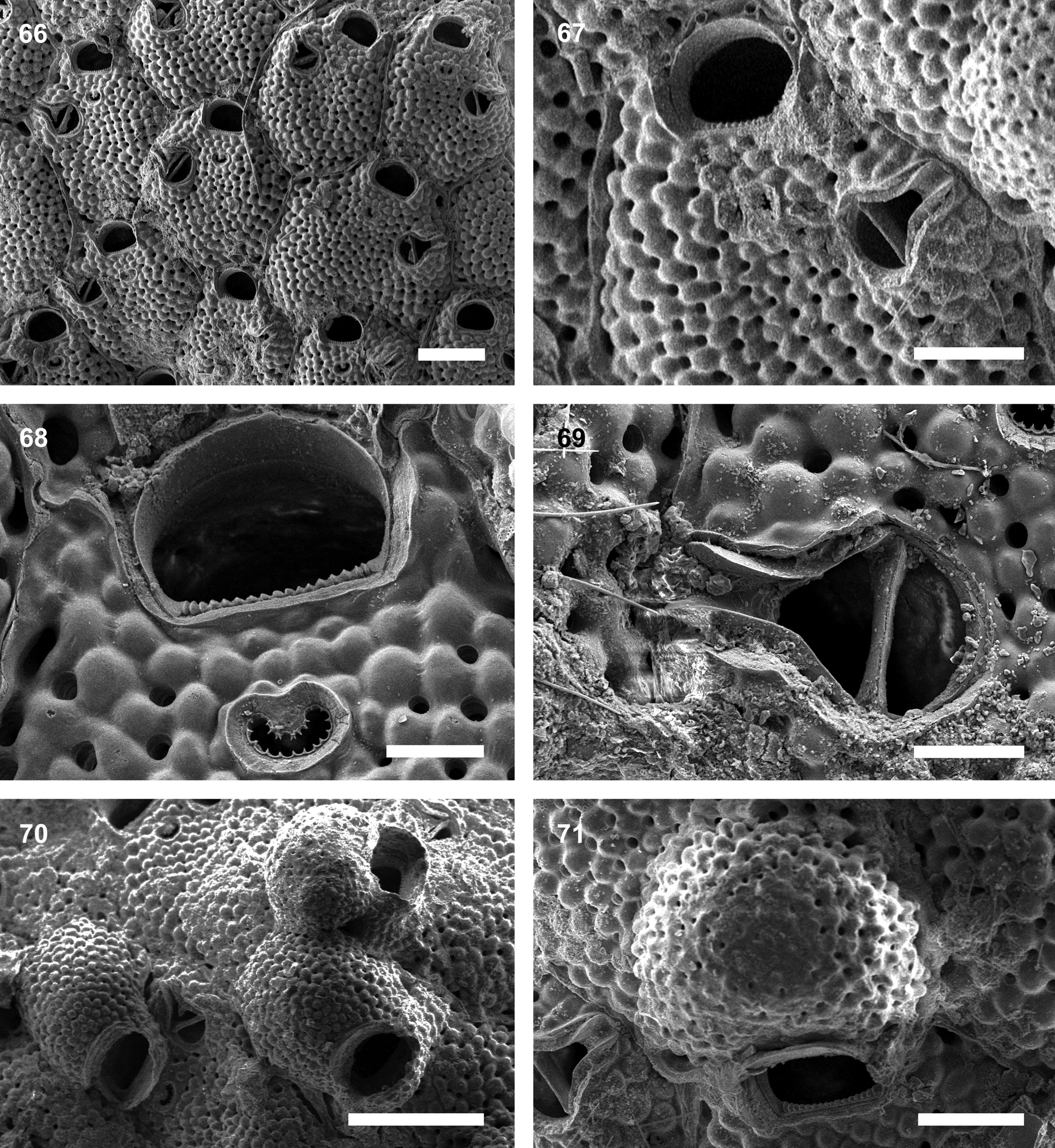
Microporella curta n. sp.
( Figs. 66–71 View FIGURES 66 – 71 ; Table 7)
Material examined. Holotype: UFBA 1580 , Todos os Santos Bay, 13°00’S, 38°32’W, 3–8 m, coll. 2013 (on sponge Callyspongia sp.) GoogleMaps . Paratype: UFBA 1582 , Todos os Santos Bay, 13°00’S, 38°32’W, 3–8 m, coll. 2013 (on sponge Callyspongia sp.). GoogleMaps
Type locality. Todos os Santos Bay, Bahia State, NE Brazil.
Etymology. From Latin curtus, short, alluding to the thin row of calcification around the primary orifice of non-ovicelled zooids.
Description. Colony encrusting, uni- to multilamellar. Autozooids large, irregularly polygonal to hexagonal, separated by raised walls. Frontal shield slightly convex, with small rounded nodes and uniformly punctured by numerous rounded pseudopores except for the area between ascopore and orifice. Marginal pores more elongate than pseudopores. Primary orifice almost D-shaped, distal edge smooth and proximal border serrated with 12–18 triangular denticles. Autozooids with a thin row of calcification around the primary orifice, not obscuring it. Nonovicelled zooids with three or four oral spines. Ascopore situated proximal to orifice at a distance about half of orifice length, reniform, surrounded by a thin rim, with a rounded median process and a crescentic (C-shaped) lumen, margins with regularly spaced sharp denticles. Avicularium single, placed proximolateral and below to the orifice and ascopore, directing distolaterally and slightly upwards, rostrum narrowing distinctly distal to complete crossbar, forming a chute-like strucure in distal half, uncalcified area proximal to crossbar semicircular. Sometimes the avicularium reaches the neighboring zooid. Ovicelled zooids with no visible oral spines; ooecia personate, i.e. with tall and thin granular collar, distally adjacent to ascopore but not obscuring it, raised over orifice and distally joined to thin, smooth, almost straight rim on proximal edge of ooecium forming a complete peristome. Remarkably large marginal pores easily seen around ooecia. Ovicells prominent; ooecium globose, closed by zooidal operculum; endooecial surface similar to autozooidal frontal shield. Aperture in ovicelled zooids transversally oval to quadrangular.
Remarks. Among the 92 valid Microporella species ( Bock 2016), Microporella curta n. sp. most resembles Microporella browni Harmelin, Ostrovsky, Cáceres-Chamizo & Sanner, 2011 , Microporella collaroides Harmelin, Ostrovsky, Cáceres-Chamizo & Sanner, 2011 , Microporella dentilingua Tilbrook, 2006 , Microporella harmeri Hayward, 1988 and Microporella orientalis ( Harmer, 1957) in having personate ovicells, a reniform denticulate ascopore, and a single avicularium placed the ascopore. Differences between Microporella curta n. sp. and M. browni are the number of oral spines (4 in Microporella curta n. sp. and 3–7 in M. browni ), and the primary orifice (with smooth distal edge and without condyles in Microporella curta n. sp., and with serrated distal edge and with low condyles in M. browni ). Microporella collaroides is distinct in having an orifice with a wavy distal edge and lateral condyles (primary orifice smooth distally and without condyles in M. curta n. sp.). Microporella curta n. sp. differs from M. dentilingua in having polygonal autozooids (roughly hexagonal in M. dentilingua ), 4 oral spines (3 in M. dentilingua ), and longer orifices (in M. dentilingua it is about 0.07 mm long). Microporella harmeri is distinct in having 2–5 oral spines that disappear in later astogeny (4 conspicuous oral spines in M. curta n. sp.), and a lanceolate avicularium mandible (setiform in M. curta n. sp.). Microporella orientalis can be distinguished from M. curta n. sp. in having 3 oral ephemeral spines (4 conspicuous in M. curta n. sp.).
At least 12 species of Microporella are recorded in the Western Atlantic Ocean, three of which are reported from Brazil: Microporella cucullata Canu & Bassler, 1928a , Microporella proxima Ramalho, Muricy & Taylor, 2011 and Microporella umbracula ( Audouin, 1826) . Microporella curta n. sp. is distinct from M. cucullata in the secondary calcification around the orifice in the autozooids (in M. cucullata the secondary calficiation forms a hood-like structure), avicularia placement and direction (below the ascopore and obliquely directed in M. curta n. sp., and above the ascopore and laterally directed in M. cucullata ), and smaller zooids (those from M. cucullata are 0.544 to 0.638 mm long and 0.438 to 0.700 mm wide). Microporella proxima differs in the frontal and ovicell calcification (smooth in M. curta n. sp. and pustulose in M. proxima ), avicularia (placed laterally and below the ascopore in M. curta n. sp., and near zooidal margins in M. proxima ), and in larger zooidal measurements (autozooids of Mi. proxima are 0.392 to 0.441 mm long and 0.294 to 0.343 mm wide, and orifices are 0.64 to 0.79 mm long and 0.93 to 0.107 mm wide). Microporella umbracula is readily distinguished from M. curta n. sp. by the paired, upward directed avicularia.
We found large colonies of M. curta n. sp. firmly attached to the basal surface of the sponge Callyspongia sp. Other Microporella species are commonly found encrusting shells, corals, algae and hydrozoans (e.g., Canu & Bassler 1928a; Marcus 1937; Winston 1984, 2005; Harmelin et al. 2011; Ramalho et al. 2011).
Distribution. Atlantic: Brazil (Bahia).
No known copyright restrictions apply. See Agosti, D., Egloff, W., 2009. Taxonomic information exchange and copyright: the Plazi approach. BMC Research Notes 2009, 2:53 for further explanation.
|
Kingdom |
|
|
Phylum |
|
|
Class |
|
|
Order |
|
|
Family |
|
|
Genus |