
Devendra Lehtinen, 1967
|
publication ID |
https://doi.org/ 10.11646/zootaxa.4362.1.3 |
|
publication LSID |
lsid:zoobank.org:pub:61A41EC1-2557-429D-B47C-595D12F2D8E3 |
|
DOI |
https://doi.org/10.5281/zenodo.5999802 |
|
persistent identifier |
https://treatment.plazi.org/id/03D38783-FF89-FF88-E6B3-6B10FD05601C |
|
treatment provided by |
Plazi |
|
scientific name |
Devendra Lehtinen, 1967 |
| status |
|
Devendra Lehtinen, 1967 View in CoL
Devendra LEhtInEn, 1967 View in CoL : 228 (TypE spEcIEs by OrIgInAl dEsIgnAtIOn: Campostichomma pardale SImOn, 1898). GrIswOld 1993: 7, FIgs 37–42; RAVEn & StumkAt 2005: 356. POlOtOw et al. 2015: 152; WSC 2017.
Note. The genus name Devendra View in CoL is masculine in the source language, Sanskrit, and a masculine a-stem noun in Latin (H. D. Cameron, in litt; WSC 2017), hence Devendra pardalis View in CoL , D. pumilus View in CoL and D. seriatus View in CoL .
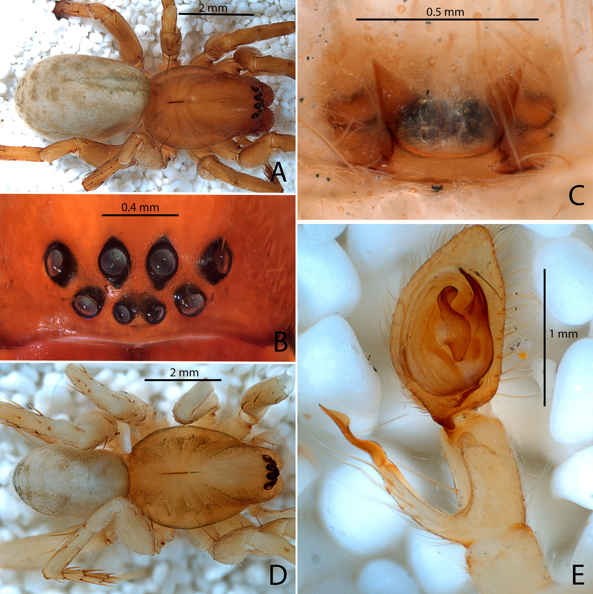
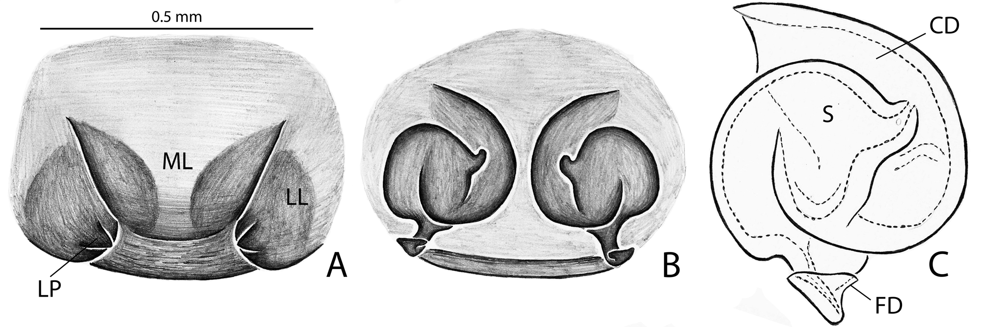
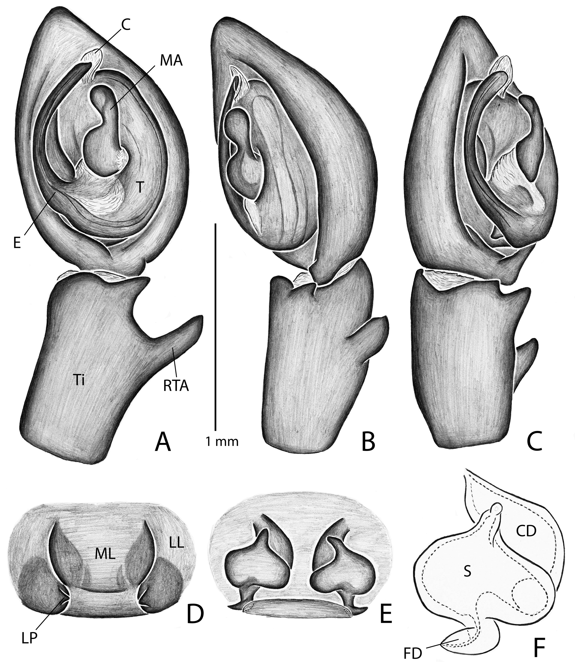
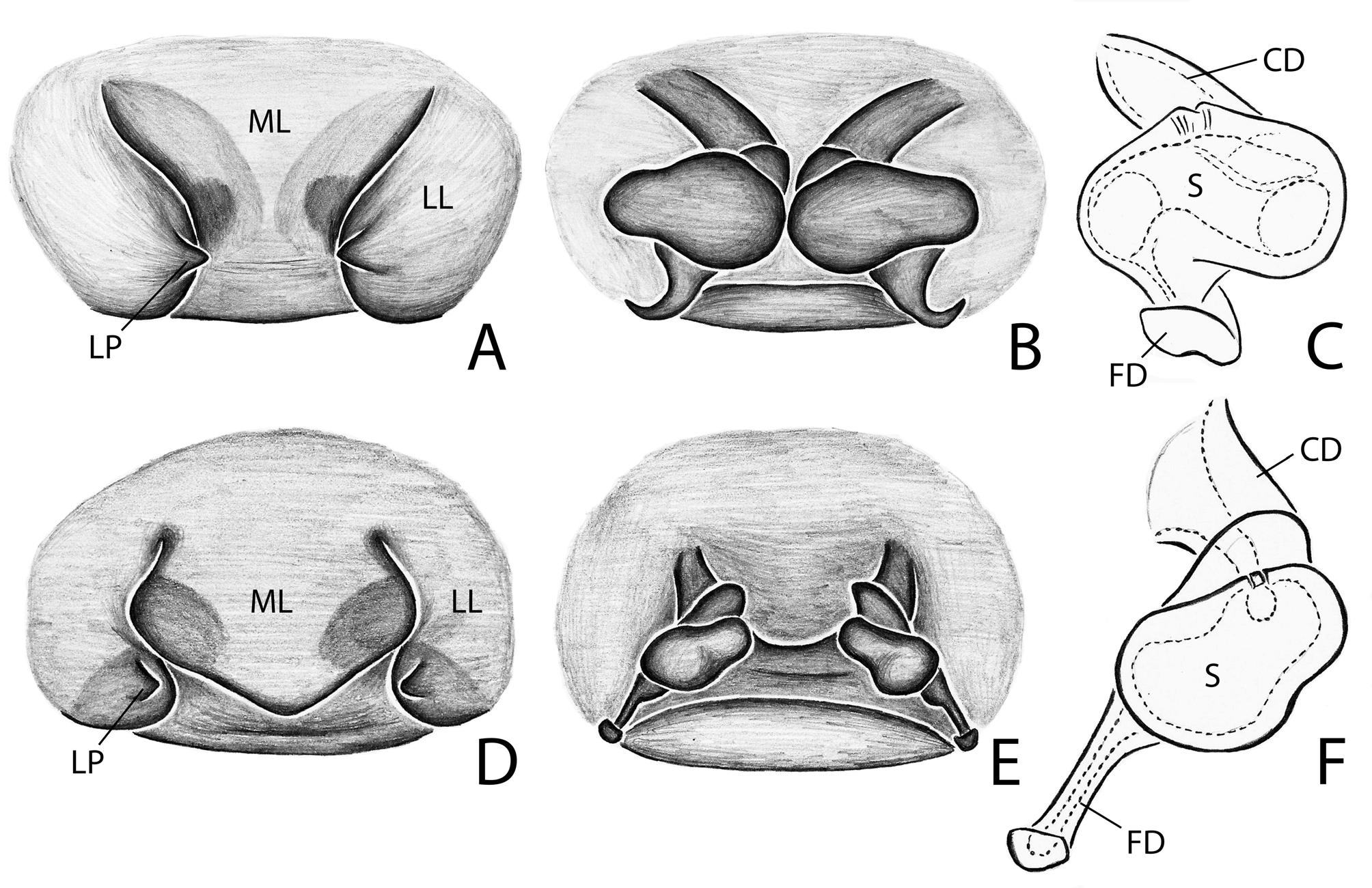
Diagnosis. Devendra are cribellate, RTA clade spiders with the PER usually straight, no claw tufts, leg tibiae of males with a visible, subcuticular transverse basal suture or crack (like in Fig. 3E View FIGURE 3 ). Males of Devendra can be distinguished from other zoropsid genera by characters of the male palp, as the presence of a S-shaped median apophysis ( Figs 11A View FIGURE 11 , 12A View FIGURE 12 ), by the presence of a ventral tibial apophysis and the RTA positioned distant from the tip of tibia ( Figs 9E View FIGURE 9 , 12A, B View FIGURE 12 ); in at least D. pumilus this process is surmounted by a spine ( Figs 9E View FIGURE 9 , 11A, B View FIGURE 11 ). The females have a simple epigynum with large teeth on the lateral lobes ( Figs 9C View FIGURE 9 , 10A View FIGURE 10 , 12D View FIGURE 12 , 13A, D View FIGURE 13 ) and can be distinguished by the shape of the epigynal structures, as the presence of a smooth and slightly sclerotized epigynal plate, with a small lateral projection. Internally, the vulva is simple with the head of the spermathecae very small, sessile or on a tubercle, the copulatory ducts are short, the spermathecae is large and the fertilization ducts are elongated ( Figs 10B, C View FIGURE 10 . 12E, F View FIGURE 12 , 13B, C, E, F View FIGURE 13 ).
DesCription. Medium sized-spiders, total length 3.90–7.10; sexual dimorphism slight ( Fig. 9A, D View FIGURE 9 ), males smaller with relatively longer legs. Carapace with darker band along each side of pars cephalica, each eye surrounded by black ( Fig. 9B View FIGURE 9 ), legs with faint annuli, especially on femora. Abdomen grey with anterodorsal cardiac mark, posteriorly with 2–3 paired chevrons ( Fig. 9A, D View FIGURE 9 ). Carapace pear-shaped in dorsal view, domed; fovea linear, deep ( Fig. 9A, D View FIGURE 9 ). Ocular area broad, ocular quadrangle trapezoidal, widest behind; eight eyes in 2 rows, anterior row slightly recurved, posterior row nearly straight ( Fig. 9B View FIGURE 9 ). Clypeus low ( Fig. 9B View FIGURE 9 ). Chelicerae stout with large boss, especially in female, pro- and retromargins of fang furrow with three and four stout teeth, respectively. Sternum broadly oval, bluntly curved or with small point posteriorly; labium with shallow basal notch; palpal coxae slightly converging anteriorly, serrula in a single row along outer margin. Legs moderately long; integument finely wrinkled ( Griswold 1993: fig. 77), with plumose setae (sensu Lehtinen 1975) and feathery scales ( Griswold 1993: figs. 75, 77); trochanters notched. Femora of legs and palpi with dorsal and lateral spines, patellae III and IV with lateral spines, tibiae I and II with 4 pairs of ventral spines and an additional proventral spine just behind apical pair, metatarsi I and II with 3 pairs of ventral spines, males usually with, females without, lateral spines on tibiae and metatarsi I and II; tibiae and metatarsi III and IV with dorsal, lateral, and ventral spines; male with sub-basal crack on leg tibiae; palpal femora with dorsal and lateral spines, tibiae and tarsi of females with spines, tibiae and tarsi of males with or without spines, cymbium usually lacking spines. Preening combs and scopulae absent. Superior tarsal claws pectinate, with inferior tarsal claws simple; claw tufts absent. Trichobothria on tibiae with a basal dorsolateral group and pro and retrolateral rows, retrolateral row extending to apex, metatarsi with dorsal, irregular row or dorsal row plus dorso-basal group, tarsi with dorso-basal row that divides into 2–3 rows apically; palpal tibiae with pro- and retrolateral rows, apparently absent from tarsi; trichobothrial base with transversely-ridged hood ( Griswold 1993: fig. 73). Tarsal organ median, capsulate, orifice keyhole-shaped ( Griswold 1993: fig. 72), with proximal seam. Male palpal tibia about as long as cymbium, tegulum simple; firmly attached embolus (E) arising on retrolateral side of tegulum and tapering to apex; median apophysis (MA) concave, attached in middle of tegulum ( Fig. 11A View FIGURE 11 ); conductor (C) hyaline, originating in the top of tegulum, fan-shaped and opposing but not embracing apex of embolus ( Figs 11A–C View FIGURE 11 , 12A–C View FIGURE 12 ); fundus in subtegulum, reservoir and ejaculatory duct simple, without loops or switchbacks, spiralling around outer margin of tegulum. Abdomen oval, without scuta ( Fig. 9A, D View FIGURE 9 ). Respiratory system comprising two anterior book lungs and a posterior tracheal spiracle. Epigynum divided by longitudinal epigynal folds into median lobe and lateral lobes; lateral lobes with small teeth ( Figs 10A View FIGURE 10 , 12D View FIGURE 12 , 13A, D View FIGURE 13 ). Vulva with elongated copulatory duct, oval spermathecae and small fertilization ducts ( Figs 10B, C View FIGURE 10 , 12E, F View FIGURE 12 , 13B, C, E, F View FIGURE 13 ). Colulus a fleshy triangular lobe set with several setae. Cribellum absent. Six spinnerets, anterior (ALS) and posterior laterals (PLS) two-segmented, posterior medians (PMS) one-segmented.
Composition. Five species: D. pardalis (Simon, 1898) , D. pumilus (Simon, 1898) , D. seriatus (Simon, 1898) , D. saama sp. nov. and D. amaiti sp. nov.
Natural history. All known specimens were collected in the late 19th century near Kandy and Nuwara Eliya, which are located in the highlands of southern Sri Lanka at above 800 m elevation ( Fig. 14 View FIGURE14 ). Today, this area in Central Province is largely cultivated for tea plantations but there are remnants of montane forest, especially near Nuwara Eliya and at Horton Plains National Park, at elevations from 1800 to more than 2200 m. Such a high species richness (five) in one small area suggests that Devendra species may be short-range endemics and that more species remain to be discovered in other montane regions.
Distribution. Endemic to Sri Lanka ( Fig. 14 View FIGURE14 ).


No known copyright restrictions apply. See Agosti, D., Egloff, W., 2009. Taxonomic information exchange and copyright: the Plazi approach. BMC Research Notes 2009, 2:53 for further explanation.
|
Kingdom |
|
|
Phylum |
|
|
Class |
|
|
Order |
|
|
Family |
Devendra Lehtinen, 1967
| Polotow, Daniele & Griswold, Charles 2017 |
Devendra LEhtInEn, 1967
| POlOtOw 2015: 152 |
| RAVEn 2005: 356 |
| GrIswOld 1993: 7 |
| LEhtInEn 1967: 228 |