
Dynastor napoleon, Doubleday, 1849
|
publication ID |
https://doi.org/10.5281/zenodo.5405005 |
|
persistent identifier |
https://treatment.plazi.org/id/03CD9A77-FF94-FFF9-7789-46C73E5A7840 |
|
treatment provided by |
Felipe (2021-08-06 16:50:46, last updated by Plazi 2023-11-03 19:03:32) |
|
scientific name |
Dynastor napoleon |
| status |
|
1 M*, Brazil, Santa Catarina, November 1954, 06-08 dissected by C.M.Penz ( MPM) ; 1 M*, Brazil, Santa Catarina, September 1964, 06-09 dissected by C.M.Penz ( MPM) ; 1 m, Brazil, Santa Catarina, 1956 ( MGCL) .
1 F*, Brazil, Rio de Janeiro, 1920, 07-09 dissected by I.Garzón (MPM); 1 F*, no data, 07-10 dissected by I.Garzón (MGCL).
Appendix 2. Character list.
1. Eye pubescence: absent (0), present (1). Fig. 1H View Figure 1 . Character 1 in Penz (2007).
2. Male FW anal margin: strongly bowed, distance between veins Cu2 and 1A+2A conspicuously decreasing toward distal margin (0), mildly bowed, distance between veins Cu2 and 1A+2A nearly homogeneous from medial area to distal margin (1). Fig. 1C, G View Figure 1 .
3 View Figure 3 . Dorsal FW blue iridescence below discal cell: absent (0), present (1). Fig. 1C, E View Figure 1 .
4. Edge of dorsal FW yellow scales located inside cell R5: bifid (0), trifid (1).
5. Male dorsal FW postmedial band reaching cell 3A: near tornus (0), near wing base (1). Note: Fig. 1 View Figure 1 shows that females have a different pattern than males. See Discussion for details. Fig. 1A, E View Figure 1 .
6. Dorsal FW yellow markings across costal margin: absent in males, present in females (0), present in both sexes (1). Fig. 1 View Figure 1 A-B, E-F.
7. Ventral FW eyespot in cell R5: absent (0), present (1). Fig. 1C, E View Figure 1 .
8. Dorsal HW medial band: well-developed (0), reduced (1). Fig. 1A, E View Figure 1 .
9. Male dorsal HW medial band: fully developed up to the costal margin (0), fading toward the costal margin (1). Fig. 1E View Figure 1 .
10. Dorsal HW medial band: same color as dorsal FW medial band (0), lighter color than dorsal FW medial band (1). Fig. 1C, E View Figure 1 .
11. Dorsal HW hairpencil at base of discal cell: absent (0), present (1). Fig. 1C View Figure 1 .
12. Color of the dorsal HW hairpencil at base of discal cell: pale-yellow (0), brown (1). Fig. 1C, G View Figure 1 .
13. Dorsal HW androconial patch at the Rs-M1 fork: absent (0), present (1). Fig. 1C View Figure 1 .
14. Color of the dorsal HW androconial patch at the Rs-M1 fork: pale-yellow (0), brown (1). Fig. 1C View Figure 1 .
15. Ventral HW eyespot in cell M1: absent (0), present (1). Fig. 1A, E View Figure 1 .
16. Ventral HW postmedial band: edges well defined (0); edges diffuse, interspersed by ripple-pattern (1). Fig. 1C, E View Figure 1 .
17. In ventral view, distal half of valva: gradually decreasing in width as compared to proximal half (0), strongly decreasing in width as compared to proximal half (1). Fig. 3E View Figure 3 .
18. In ventral view, distal portion of valva: strongly arched (0), moderately arched (1). Fig. 3G View Figure 3 .
19. Anterior ribs of the intersegmental sac between sternite 7 and sterigma: continuous (0), broken (1). Character 62 in Penz (2007). Fig. 3F View Figure 3 .
20. Posterior portion of sterigma: extended towards midline nearly enclosing the ostium bursae (0), further away from midline, not enclosing the ostium bursae (1). Fig. 3B View Figure 3 .
21. Posterior portion of sterigma, small midline extension: absent (0), present (1). Fig. 3F View Figure 3 .
Appendix 3. Character matrix.
1 22
0 01
Dynastor darius 01010? 0?0? 0?0?1?0001 0 Dynastor napoleon 01010? 0??? 0?0?1?0101 0 Dasyophthalma rusina 10100 0&1 0001 1111001011 1 Dasyophthalma geraensis 10100 0 0000 11111010 ??? Dasyophthalma creusa 10011 1 1110 10101?1010 0 Dasyophthalma vertebralis 10011 1 1010 1010111010 0
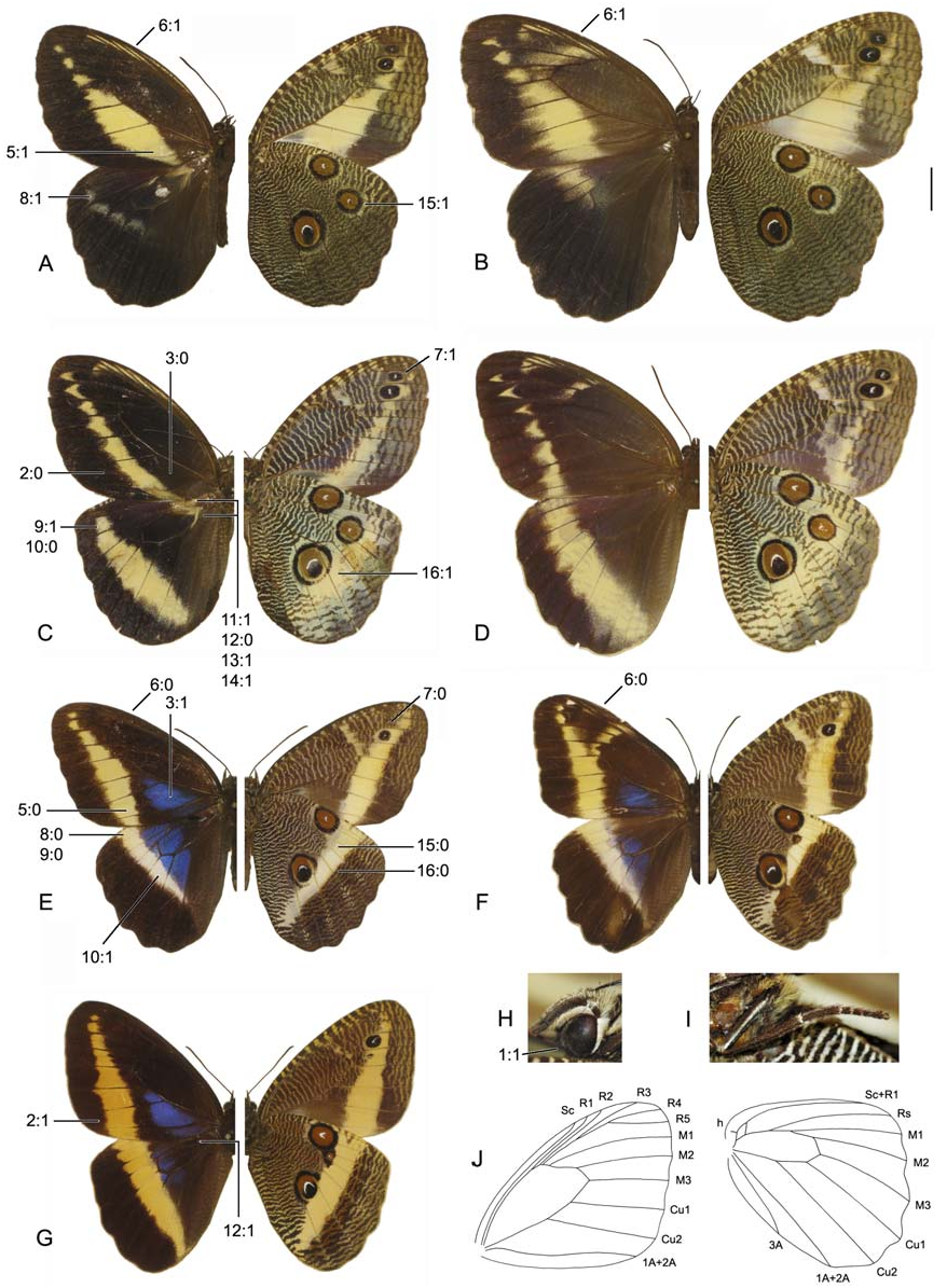
Figure 1. Dasyophthalma adults. Habitus with dorsal side on the left, ventral side on the right. Scale bar next to B = 1cm. A) D. creusa male, Brazil, Santa Catarina, São Bento do Sul. B) D. creusa female, Brazil, Santa Catarina, São Bento do Sul. C) D. verebralis male, Brazil, Espírito Santo. D) D. vertebralis female, [Brazil] East Amazonas. E) D. rusina male, South Brazil. F) D. rusina female, South Brazil. G) D. geraensis male, [Brazil] Minas Gerais. H) Detail of the head of D. rusina. I) Detail of the hindleg of D. rusina. J) Venation pattern of D. rusina, vein thickness is not shown.
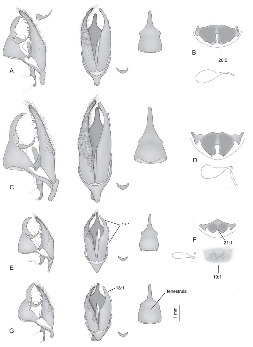
Figure 3. Dasyophthalma male genitalia in lateral and ventral views (setae omitted from right side of figure), with detail of the juxta and tegumen + uncus in dorsal view, plus female sterigma and corpus bursae. The line below sterigma represents the shape of the intersegmental sac. Scale bar next to G = 1mm. A) D. creusa male, South Brazil, 01-17 dissected by C.M.Penz. B) D. creusa female, South Brazil, 01-16 dissected by C.M.Penz. C) D. vertebralis male, Brazil, Espírito Santo, 07-170 dissected by C.M.Penz. D) D. vertebralis female, [Brazil] East Amazonas, no date, 07-171 dissected by C.M.Penz. E) D. rusina male, Brazil, Santa Catarina, 01-18 dissected by C.M.Penz. F) D. rusina female (with detail of the intersegmental sac below sterigma), Brazil, Santa Catarina, São Bento do Sul, 01-19 dissected by C.M.Penz. G) D. geraensis male, Brazil, Minas Gerais, 07-169 dissected by C.M.Penz.
| MPM |
Milwaukee Public Museum |
No known copyright restrictions apply. See Agosti, D., Egloff, W., 2009. Taxonomic information exchange and copyright: the Plazi approach. BMC Research Notes 2009, 2:53 for further explanation.
|
Kingdom |
|
|
Phylum |
|
|
Class |
|
|
Order |
|
|
Family |
|
|
Genus |