
Baryonychinae, Charig & Milner, 1986
|
publication ID |
https://doi.org/10.11646/zootaxa.5264.4.4 |
|
publication LSID |
lsid:zoobank.org:pub:0018046C-76BA-42AA-8F3D-171663333366 |
|
DOI |
https://doi.org/10.5281/zenodo.7836961 |
|
persistent identifier |
https://treatment.plazi.org/id/03BEE93D-FFB1-8F27-6FE6-EF99DCBAF34C |
|
treatment provided by |
Plazi |
|
scientific name |
Baryonychinae |
| status |
|
Baryonychinae gen. et sp. indet.
Fig. 2 View FIGURE 2
Morphological description: The three referred specimens LPUFS 5860, LPUFS 5870, and LPUFS 5871 are isolated crowns presenting the same general morphotype ( Fig. 2 View FIGURE 2 ). The teeth are well-preserved and have a similar taphonomical pattern of others at the same site ( Lacerda et al. 2023). The length of the crowns varies between 17.2 mm and 32.5 mm, and the specimens share: the conidont morphology, the main crown curvature being in the mesiodistal plane, the presence of labial and lingual enamel flutes, the denticulated mesial and distal carinae being centrally positioned, the absence of interdenticular sulci, veined enamel texture (wrinkled enamel), and the carinae reaching the cervix/root.
LPUFS 5860 ( Fig. 2 View FIGURE 2 A-H) is a completely preserved crown with a subcircular cross-sectional outline ( Fig. 2E View FIGURE 2 ). The tooth surface has eleven well-demarcated flutes in the lingual plane and nine less-demarcated flutes on the labial surface of the crown. The flutes, in general, are equally spaced, extending from the cervix to the apex ( Fig. 2F View FIGURE 2 ). The distal carina is better developed (more extensive) than the mesial one, being both serrated with diminutive denticles (being 7 denticles per millimetre) composed of both the dentin and enamel ( Fig. 2H View FIGURE 2 ) and lacking interdenticular sulci. The denticles have similar sizes along the carina and also when comparing both the mesial and distal carinae. The enamel ornamentation is arranged in a veined manner. Only a small wear surface is present at the apex of the crown ( Fig. 2E View FIGURE 2 ).
LPUFS 5870 ( Fig. 2 View FIGURE 2 I-P) is a more robust tooth crown, preserving the mid crown and a small portion of the root. The cross-section of the specimen is subcircular in outline ( Fig. 2M View FIGURE 2 ). The main crown curvature is noted in the mesiodistal plane; however, a subtle labiolingual curvature is also present. In the labial side the tooth has eleven flutes, and in the lingual plane are counted twelve flutes, some of which are also demarcated in the dentine below the enamel wear ( Fig. 2N View FIGURE 2 ), which are well-developed and equally spaced. Both carinae are well-developed, the mesial one extends slightly to the cervix, with its denticles (being 6 to 7 denticles per mm) composed of both dentin and enamel ( Fig. 2P View FIGURE 2 ). The individual minute denticles are equal in size, lacking interdenticular sulci. The main texturization of the enamel is veined arranged. In the apical-most portion, a round wear surface is noted in the tooth crown ( Fig. 2M View FIGURE 2 ).
LPUFS 5871 ( Fig. 2 View FIGURE 2 Q-X) is the smallest and more taphonomic damaged crown, lacking the root entirely. The cross-section is nearly circular in outline ( Fig. 2U View FIGURE 2 ) (however, see Multivariate analyses section and Table 2 View TABLE ). At least fourteen flutes are noted on the labial surface, some of which do not reach the apex (and therefore may overestimate the flute count); the lingual surface of the crown has at least eleven well-demarcated flutes composed by the dentin and enamel. The distal carina is poorly preserved in LPUFS 5871 specimen, however the mesial one is well-developed with equally sized diminutive denticles (being 7 denticles per mm) on the mid-to-apex portion of the carina distributed without the presence of interdenticular sulci ( Fig. 2X View FIGURE 2 ). The enamel ornamentation is arranged in a veined manner mainly noted in the base of the lingual surface. A small and round wear surface is present at the apex of the crown ( Fig. 2U View FIGURE 2 ).
Morphological comparisons: The three specimens LPUFS 5860, LPUFS 5870, and LPUFS 5871 present a conidont morphology (composed of a conical crown and flutes distributed on the labial and lingual enamel surfaces), different from the ziphodont morphology (comprising a narrow crown labiolingually distally curved), which is observed in some archosaurs and the vast majority of theropod species ( Andrade et al. 2010; Hendrickx et al. 2015a,b; 2019). In a complementary way, the materials described here present a feature considered typical in theropod dinosaurs, which is the mesiodistal plane as the main plane of curvature of the dental crown, a characteristic that allows the differentiation of these materials from conidont teeth of crocodyliforms (mainly Neosuchia), which generally have the main plain of curvature of the crown in the labiolingual direction ( Sánchez-Hernández et al. 2007; Buffetaut et al. 2008; 2019; Hone et al. 2010; Sales et al. 2017). Even ziphodont crocodyliforms (e.g., Riff & Kellner 2001) can be distinguished from the materials described here by the curvature of the crown and the size/ morphology of the denticles. Furthermore, the teeth described here can be distinguished from marine reptiles (e.g., plesiosaurs) on the basis of compression and enamel morphology, in addition to the presence of minute denticles on the carinae ( Hone et al. 2010; Buffetaut et al. 2019).
Unlike other theropods, the teeth of spinosaurids have a subcircular-elliptical cross-section, straighter crowns, and a deeply veined or anastomosed enamel ( Alonso & Canudo 2016; Hendrickx et al. 2015a; 2019). Based on the mentioned features, it is possible to differ the LPUFS specimens from non-archosaur reptiles; moreover, the conidont morphology, the mesiodistal plane of curvature as the main plane, the wrinkled enamel at the base of the tooth crown, the presence of mesiodistal carinae reaching the crown cervix, and the distribution of flutes on the labial and lingual sides (although variable in spinosaurids - Alonso & Canudo 2016), allow us to safely assign the specimens as belonging to spinosaurids.
As noted by Hendrickx et al. (2019), tooth enamel in spinosaurids may be veined in texture, as observed in Baryonyx , Suchomimus , Spinosaurus , and Iberospinus ( Mateus & Estraviz-López 2022) , or it may have an anastomosing texture, particularly noted in some Spinosaurus individuals. The materials described here present enamel with a veined texture, most similar to the morphology noted in baryonychines, as well as the presence of labial and lingual enamel flutes common in spinosaurids, albeit some unfluted teeth are also known ( Fowler 2007; Hone et al. 2010). Even with some degree of variation in Baryonyx , flutes are present on both sides of the crowns in Suchomimus and Suchosaurus ( Hendrickx et al. 2019) as well as in the materials described here. As described by Charig & Milner (1997), Baryonyx presents teeth with six to eight flutes; meanwhile Suchosaurus presents between ten and 12 flutes; and Suchomimus between two and ten ( Hendrickx et al. 2019). The largest number of flutes is noted in spinosaurines, approximately 24 in “ Sinopliosaurus ” ( Buffetaut et al. 2008), and 20 in materials referring to Spinosaurus ( Hendrickx et al. 2019) . The teeth described here show a number of flutes greater than that generally observed in baryonychines, but smaller than that observed in non-serrated crowns of spinosaurine (e.g., Buffetaut et al. 2019).
Compared with other baryonychine teeth (e.g., Fowler 2007; Canudo et al. 2008), the flutes approach the carinae more closely, and are “thinner” (less deep, and closer spaced) in the specimens described here. The morphofunction of the fluted crowns is probably broad, however, this structure may be related to some perforation/gripping function, in this way the sharp ridges allow the perforation of the skin and enlargement the flesh as the tooth penetrates, as well as keeping slippery prey trapped in the mouth ( Charig & Milner 1997; Sues et al. 2002; Hendrickx et al. 2019).
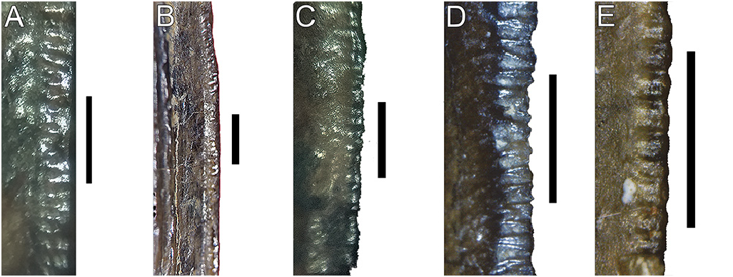
Although baryonychine teeth have a more pronounced distal curvature than in spinosaurine species ( Alonso & Canudo 2016), the LPUFS specimens have a less pronounced distal curvature of the crown ( Fig. 2 View FIGURE 2 ). Denticles are present in both carinae of LPUFS 5860, LPUFS 5870, and LPUFS 5871, and in general, the denticles are the same size. This feature differs from Iberospinus , which has smaller mesial denticles ( Mateus & Estraviz-López 2022). In Baryonyx , on the other hand, variation in the size of the denticles is more developed than in Suchomimus ( Hendrickx et al. 2019) . Furthermore, the presence of 6-7 minute denticles per millimetre and equal in size, distributed along both carinae lacking the interdenticular sulci ( Fig. 3 View FIGURE 3 ), represents the morphological condition observed only in baryonychine species ( Charig & Milner 1997; Sereno et al. 1998; Fowler 2007; Hone et al. 2010; Alonso & Canudo 2016; Hendrickx et al. 2019; Fig. 3 View FIGURE 3 ), which allows the taxonomic attribution of the specimens described here to baryonychines (see discussion).

No known copyright restrictions apply. See Agosti, D., Egloff, W., 2009. Taxonomic information exchange and copyright: the Plazi approach. BMC Research Notes 2009, 2:53 for further explanation.
|
Kingdom |
|
|
Phylum |
|
|
Class |
|
|
Order |
|
|
Family |