
Baryonyx walkeri Charig & Milner, 1986
|
publication ID |
https://doi.org/ 10.5281/zenodo.205679 |
|
DOI |
https://doi.org/10.5281/zenodo.6190565 |
|
persistent identifier |
https://treatment.plazi.org/id/03A287A3-FFB6-FFD2-FF2C-2C12FF52FA09 |
|
treatment provided by |
Plazi |
|
scientific name |
Baryonyx walkeri Charig & Milner, 1986 |
| status |
|
Baryonyx walkeri Charig & Milner, 1986
Holotype. NHM R9951, partial skull and associated postcranial skeleton.
Locality and horizon. The holotype is from the Upper Weald Clay (base of the Barremian, Lower Cretaceous) of Walliswook, England. The Portuguese specimen, ML1190, is from the Praia das Aguncheiras, Sesimbra Municipality (Papo Seco Formation; early Barremian; 38.44N 9.20W).
Synonyms. Possibly the nomina dubia Suchosaurus cultridens ( Owen, 1840–45) and Suchosaurus girardi Sauvage, 1897 –1898.
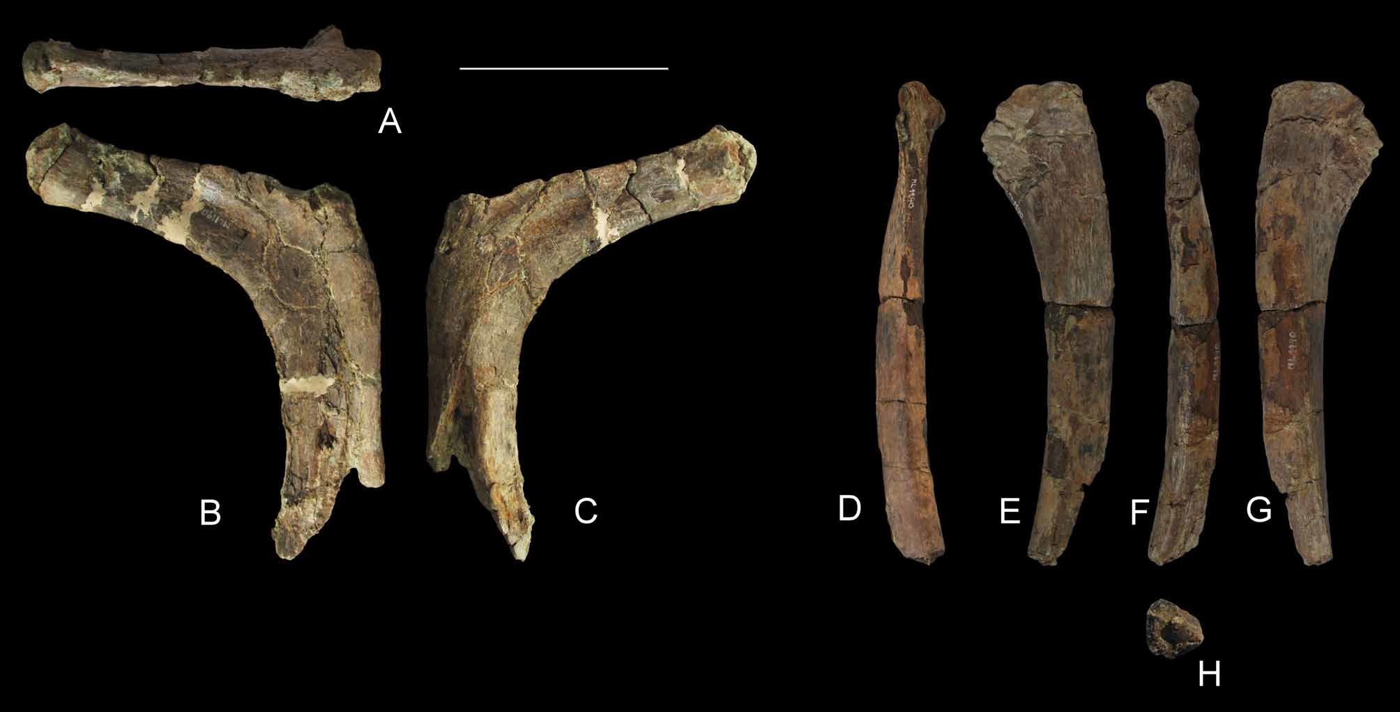
Referred specimen described here. One individual (ML1190) comprising a partial left dentary, two teeth, four dorsal neural arches, five caudal centra, fragments of chevrons, dorsal rib fragments, right scapula, right pubic shaft, possible pubic peduncle of left ilium, two calcanea, and one pedal ungual phalanx ( Figs 3–10 View FIGURE 3 View FIGURE 4 View FIGURE 5 View FIGURE 6 View FIGURE 7 View FIGURE 8 View FIGURE 9 View FIGURE 10 ).
Addition to the diagnosis: Besides the diagnostic features provided by Charig & Milner (1986, 1997), Sereno et al. (1998), and Martill & Hutt (1996), Baryonyx has an unique combination of characters of the teeth: carinae with high denticles density (6–7 denticles per millimetre), variable and non-gradual denticle size along the carinae, enamel surface with small and nearly vertical wrinkles (including at the base of the crown), and wrinkles forming a 45 degree angle near the carinae.
Description. Except for the mid-caudal vertebrae, all skeletal elements of Baryonyx walkeri ML1190 are also represented in NHM R9951, the holotype specimen of Baryonyx walkeri , thus enabling comparison. The bone dimensions are similar to those of NHM R9951; thus, the Portuguese specimen would have had a similar body size. Most of the bones of the Portuguese specimen ML1190 have damaged articular ends filled with sediment, and some have scratches on their surfaces, which may be marks of small scavengers. The disarticulation of ML1190 is indicative of transport, possibly from more terrestrial environments, due to the following factors: 1) the skeleton is incomplete; 2) the specimen is disarticulated but closely associated; 3) there was a significant loss of bone, indicative of disarticulation stage M (taphonomical terms from Heinrich 1999: 31).
The left dentary ( Fig. 4 View FIGURE 4 ), 162 mm long as preserved, comprises the symphysis with the 12 anterior-most alveoli. Most teeth are still present but with the crown broken off. The erupting replacement teeth are visible on the medial side of the dentary at the first, second, sixth and eighth alveoli. As in all spinosaurids, the anterior end of the dentary exhibits the tooth rosette, i.e., a dorsoventral expansion near the symphysis that results in a sigmoidal dorsal margin. As a result, the ninth and tenth teeth positions are in a more ventral position than the more anterior teeth. The dentary is laterally compressed and straight. The Meckelian groove is narrow (up to 3 mm deep dorsoventrally) and shallow. The preserved lateral view of the dentary bears 28 well defined and deep foramina for nutrient supply. The paradental groove is not visible, and it is unclear if it was present. The paradental plates are triangular and low, and nearly absent.
The specimen includes one complete isolated tooth with its root (but with damaged serrations; Fig. 3 View FIGURE 3 ) and several teeth within the left dentary. The cross section is eye-shaped or round (rather than D-shaped as in most theropods), resulting in a conical appearance, with only weak linguolabial compression. The tooth crowns in the dentary exhibit fluting on the lingual surface only. It has been shown that the presence of fluting in baryonychine teeth is highly variable ( Ruiz-Omeñaca et al. 1998: 206). Carinae are present on the mesial and distal margins of the teeth. The denticle density of the erupting teeth is about 6–7 denticles per millimetre, and the enamel is densely wrinkled (apicobasally extending micro-ridges). There is a small, posterior dentary fragment that bears four alveoli (7 mm in diameter anteroposteriorly and 6 mm lateromedially).
Three presacral neural arches, possibly of dorsal vertebrae, are preserved ( Fig. 5 View FIGURE 5 ). We describe here the most complete arch, which is identified as a posterior dorsal. It is fragmentary, missing the neural spine and diapophyses. Four laminae diverge from the diapophysis: the prezygadiapophyseal, the anterior and posterior centrodiapophyseal, and postzygadiapophyseal laminae. Vertical, small, auxiliary laminae support the posterior centrodiapophyseal lamina from beneath (synapomorphy of Spinosauridae : Sereno et al. 1998). The postzygapophyses do not bear epipophyses. The base of the neural spine is well compressed transversely and is supported posteriorly by spinopostzygapophyseal laminae.
Five caudal vertebrae with complete centra, and a sixth with a half centrum, are present ( Fig. 6 View FIGURE 6 ; Table 2). Taking into consideration the fact that few caudal vertebrae are preserved in the holotype of Baryonyx walkeri (NHM R9951), the exact position of these specimens within the tail is difficult to establish. However, we estimate their positions as one anterior, two mid-anterior, one mid-posterior, and one posterior caudal vertebra. All caudal vertebrae of Portuguese Baryonyx ML1190 are amphicoelous, although the posterior facet tends to be more shallowly concave. The anterior caudal centrum is hourglass-shaped in ventral view (but less so than in NHM R9951), while in posterior view, the centrum is sub-circular. The chevron facets are well visible, mainly on the ventroposterior margin of the centrum, giving a more squared shape to the outline in anterior and posterior views.
In all the caudal vertebrae, the ventral face of the centrum has two parallel ridges between which a deep and wide longitudinal groove extends along the midline. The groove is deepest posteriorly, where the ridges are confluent with the chevron facets.
Position in caudal series Length Anterior Height Anterior Width Anterior 95 107 101 Mid-anterior 105 81 71 Mid-anterior 104 79* 63 Mid-posterior 96 55 56 Posterior 82 48 49 Posterior - 52* 43*
* measured for the posterior facet due to the lack of preservation anteriorly.
The anterior caudal has an unfused centrum and neural arch. The neurocentral suture is unfused in the most anterior vertebrae but is fused and visible in middle caudal vertebrae and is fused and invisible in the most posterior vertebra. This suggests a posterior-to-anterior sequence of fusion. The unfused neurocentral suture is considered a young ontogenetic feature ( Brochu 1996) but is common in very large (and thus most likely adult) spinosaurids. The sutural area of the unfused centrum is much wider than the area for the neural canal itself, which is deep, narrow, and constricted in the middle. The anterior dorsal rim is prominent at the midline. The mid-posterior caudal vertebrae is the only tail vertebra to be preserved with a partial transverse process, which is placed on the posterior half of the vertebra, just above the neurocentral suture. The transverse process is a horizontal, platform-like projection supported by a centrodiapophyseal lamina.
The left side of the mid-posterior centrum has a 21 mm long elliptical pit (apparently produced post-mortem) that may correspond to an orthogonal tooth mark from a large predator or scavenger. More posterior centra have a more rectangular posterior outline, and are higher than wide. Part of the neural arch is preserved in the most posterior vertebrae. The prezygapophyses project anteroposteriorly at a 45 degree angle to the horizontal, with the small vertical prezygaphyseal facets positioned close to one another, while the postzygapophyses are partly confluent with the neural spine, which projects posterodorsally. In one posterior caudal vertebra only the posterior half of the centrum is preserved, showing the cross section of the vertebra with a hollow interior (now infilled with a calcite geode).
Several incomplete dorsal ribs are preserved ( Fig. 7 View FIGURE 7 ). The tuberculum is confluent with the shaft and the capitulum in ML 1190, whereas in NHM R9951 the tuberculum is more pronounced. The curve at the tuberculum area is pronounced, and the inner rim bears a sharp edge or keel, rather than the typical round margin present along the rest of the rib. Proximally, the shaft is broad, being convex anteriorly and concave posteriorly, but more distally, the shaft becomes rounder in cross section. The rib head is long and at its base there are two anterior shallow grooves running along its length that produce a distinctive crest near the tuberculum. Such features are not visible in Suchomimus tenerensis , Allosaurus fragilis, Lourinhanosaurus antunesi, and Ceratosaurus nasicornis , and less evident in NHM R9951 ( Gilmore 1920; Madsen 1976; Mateus 1998; Sereno et al. 1998).
The preserved portions of the right scapula ( Fig. 8 View FIGURE 8 ) are the proximal end and about one third of the blade, 327 mm long as preserved and 184 mm at the proximal expansion. Although only partially preserved, the scapula bears the typical curvature along its length, demonstrating that this bone would fit against the ribcage. The anterior and posterior margins of the blade are subparallel. The anterior margin is slightly thicker than the posterior margin. Proximally, the scapula is expanded relative to the blade, bearing the acromion process posteriorly and the glenoid fossa anteriorly. There is a prominent posteroventral lip that is widely distributed among theropods including Majungasaurus crenatissimus and Allosaurus fragilis as well as NMH R9951 ( Madsen 1976; Charig & Milner 1997; Carrano 2007). The mediolateral thickness of the blade tends to decrease distally. The acromion process is not complete, and thus it cannot be determined whether it is of the typical subretangular shape present in Suchomimus tenerensis ( Sereno et al. 1998) . ML1190 shares with Baryonyx walkeri the well-formed peg-and-notch scapular attachment with the coracoid on its proximal surface ( Charig & Milner 1997: fig. 31; autapomorphy of Baryonyx walkeri according to Sereno et al. 1998: 1302).
Only the proximal middle part of the right pubis ( Fig. 8 View FIGURE 8 ) shaft is preserved; the acetabular and distal portions are missing entirely. The preserved element measures 295 mm in length. The pubic apron is not preserved, but its medial sinuous outline, to which the main shaft of the pubis was connected, is preserved. As in NHM R9951, the middle part of the pubis is straight and compressed lateromedially at its proximal end ( Charig & Milner 1997: 49) and at its distal fracture has a teardrop shaped cross section. The lateral surface of the bone is slightly concave and anteriorly there is a small mound-like process with longitudinal striations. The dorsal surface bears longitudinal striations on the distal part and forms a well-defined, rounded edge towards the proximal end. The rounded edge of the ventral surface tapers proximally.
Two calcanea are present in ML1190, each measuring 110 mm in maximun expansion ( Fig. 9 View FIGURE 9 ). The right calcaneum of NHM R9951 was figured by Charig & Milner (1997) and was used for comparison. The calcanea of ML1190 are unfused to the astragali or tibiae. Both articulation surfaces for the tibia and fibula are preserved, concave, and equivalent in area to one another. The distal and anterior surfaces are rugose with longitudinal striations. The tibial articular facet is damaged.
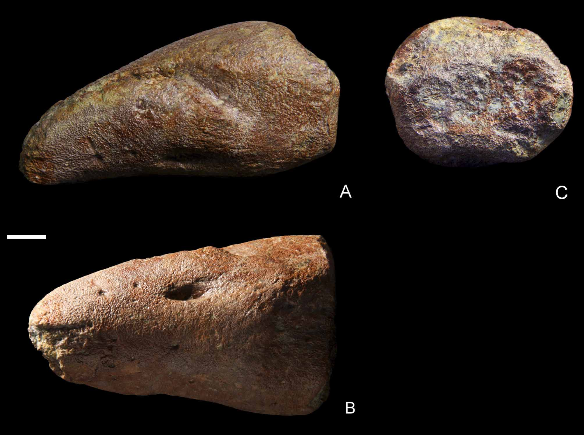
A single pedal ungual phalanx is preserved ( Fig. 10 View FIGURE 10 ), measuring 44 mm transversely and 78 mm in length. The shape of its proximal articular contour is roughly ellipsoidal (but slightly depressed forming a sigmoid). Thus, its overall shape might be triangular if complete. The proximoventral flexor tubercle is very reduced: it is only a smooth eminence visible in lateral view. The collateral grooves extend from the very tip of the phalanx until the distal third of the bone. Due to the relative orientation of the collateral grooves, it is presumed that this element comes from the left side of the specimen, because the lateral groove is placed more dorsally than the medial groove.


No known copyright restrictions apply. See Agosti, D., Egloff, W., 2009. Taxonomic information exchange and copyright: the Plazi approach. BMC Research Notes 2009, 2:53 for further explanation.