Cylapus, Say, 1832
publication ID |
https://doi.org/10.11646/zootaxa.5074.1.1 |
publication LSID |
lsid:zoobank.org:pub:7B3C6765-F0D2-4846-BB95-200258ECC0E1 |
DOI |
https://doi.org/10.5281/zenodo.5760400 |
persistent identifier |
https://treatment.plazi.org/id/039587FB-AE7F-FF90-FF51-11574347FA5D |
treatment provided by |
Plazi (2021-12-06 08:45:40, last updated 2024-11-28 20:55:07) |
scientific name |
Cylapus |
status |
|
Cylapus View in CoL View at ENA complex
Cylapus View in CoL complex: Carvalho & Fontes, 1968: 273; Gorczyca 2006a: 12.
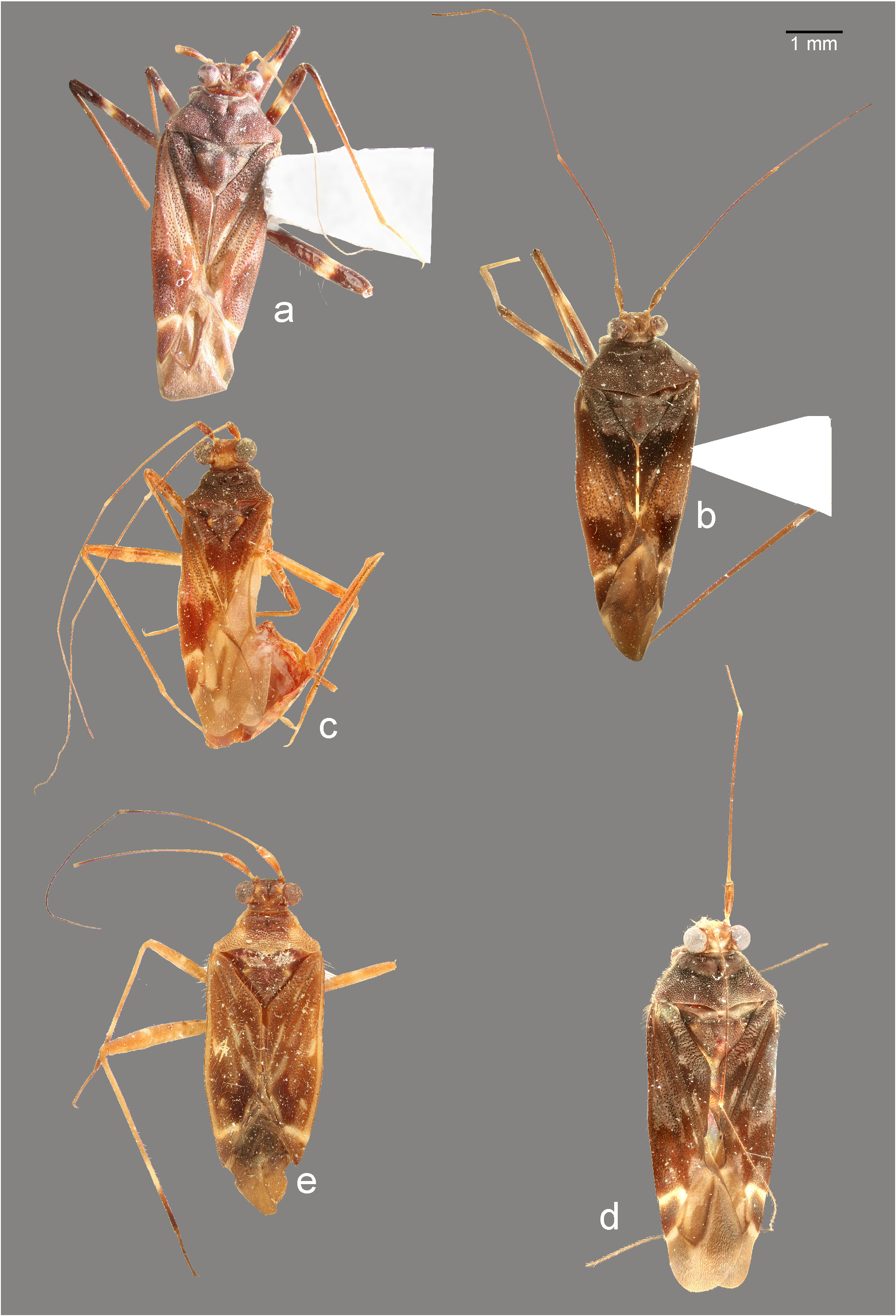
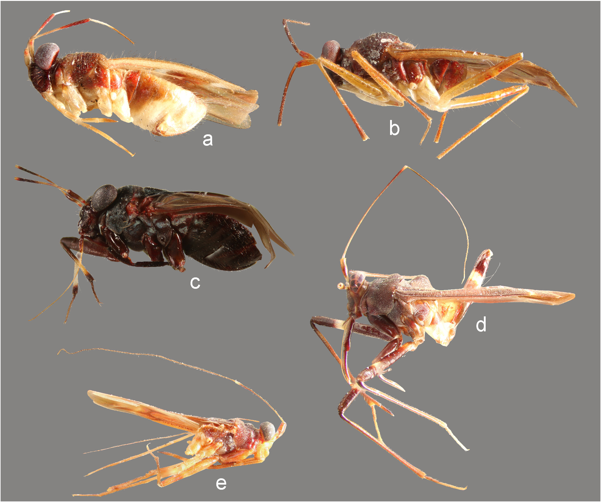
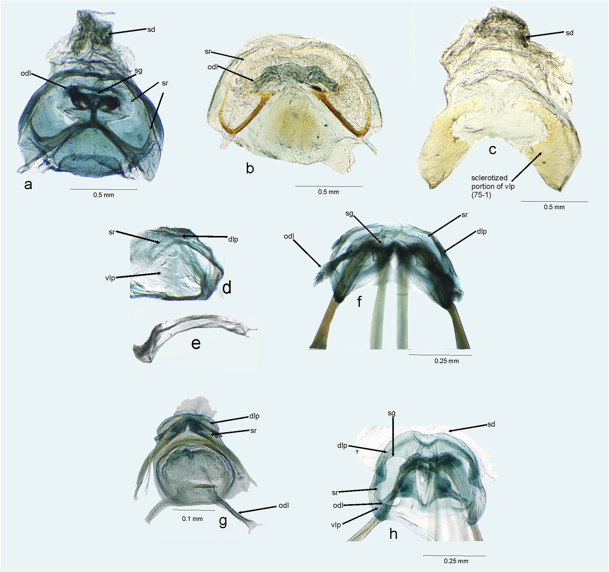
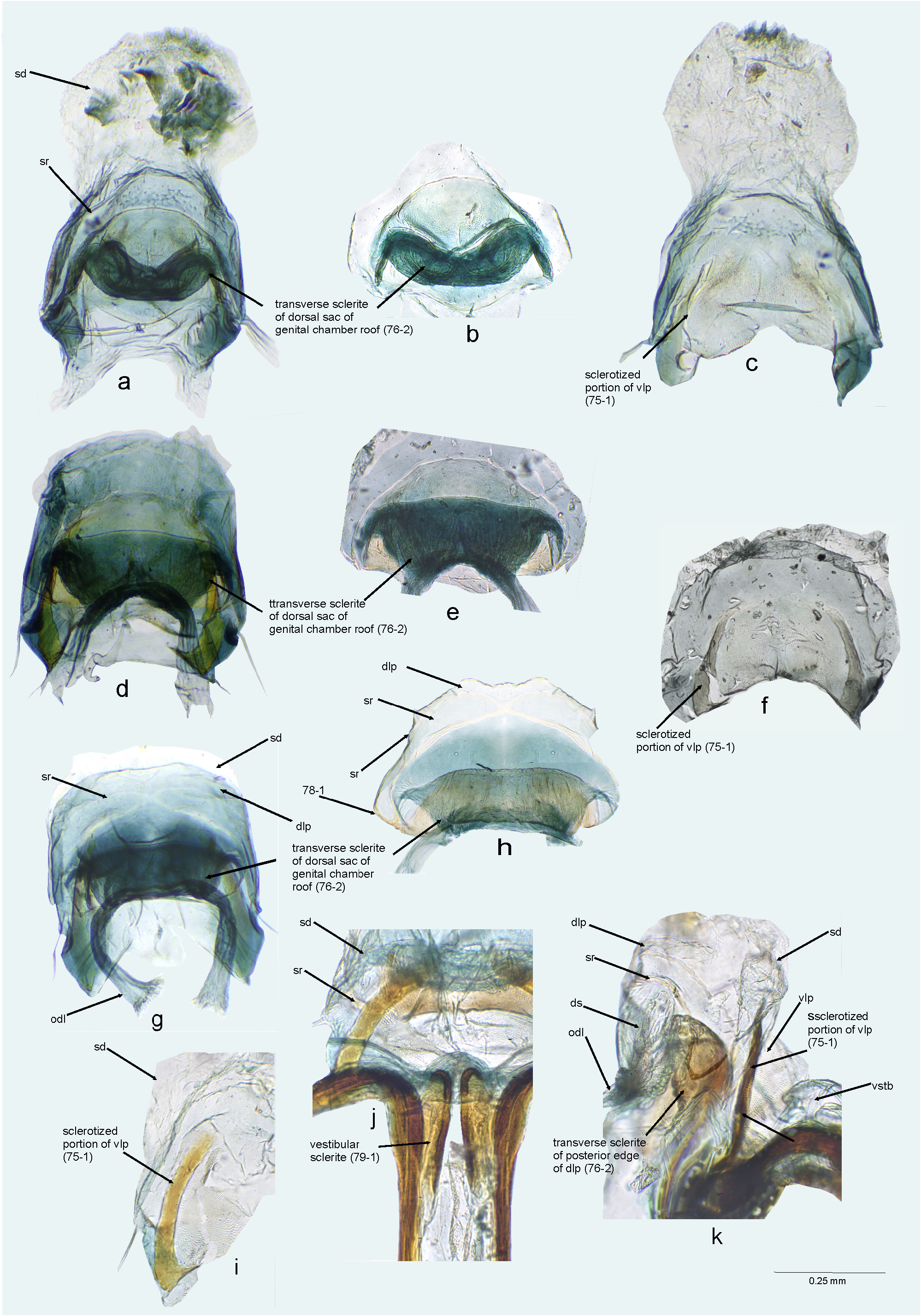
Diagnosis. The Cylapus complex is easily recognized by the following characters: dorsum densely punctuate ( Figs 4–6 View FIGURE 4 View FIGURE 5 View FIGURE 6 ; 9a View FIGURE 9 , 10a View FIGURE 10 , 11g, m View FIGURE 11 ); frons flattened, vertical, perpendicular to vertex ( Figs 7d, e View FIGURE 7 , 8c–h View FIGURE 8 , 9a View FIGURE 9 , 11n View FIGURE 11 ); vertex strongly depressed along midline, V–shaped in anterior view ( Figs 8c–h View FIGURE 8 , 9g View FIGURE 9 ); antennal fossae situated well above the suture between maxillary and mandibular plates ( Figs 8c–h View FIGURE 8 , 9g View FIGURE 9 ); clypeal base strongly removed from ventral margin of eyes ( Figs 8c–h View FIGURE 8 , 9g View FIGURE 9 ); suture separating maxillary and mandibular plates curved, strongly depressed, long, reaching antennal fossae ( Figs 9a, g View FIGURE 9 ; Wolski 2017: figs 9, 123; Wolski et al. 2020: figs 28, 31); eyes strongly pedunculate (8c–h, 9g); aedeagus with ductus seminis relatively broad and short, secondary gonopore with microsculpture ( Figs 12j, p View FIGURE 12 , 13a, g, m View FIGURE 13 , 14a, f, l, r View FIGURE 14 ; Wolski 2017: figs 18, 66, 71; Wolski et al. 2020: figs 40, 48); bursa copulatrix voluminous, anterior margin distinctly removed from first gonapophyses ( Figs 15a, b View FIGURE 15 , 16a, d, g, j View FIGURE 16 , 17c, g, j View FIGURE 17 , 18b View FIGURE 18 , 19a, d View FIGURE 19 ; Wolski et al. 2020: figs 61, 66); ventral labiate plate broad, with more or less developed sclerotized portion medially ( Figs 15c View FIGURE 15 , 16c, f, i View FIGURE 16 , 17e, i, m View FIGURE 17 , 18d View FIGURE 18 , 19c View FIGURE 19 ); vestibulum with well-developed sclerite, adjacent to base of first gonapophysis ( Figs 20a, d–h View FIGURE 20 ).
Remarks. Carvalho & Fontes (1968) proposed establishing the Cylapus complex for the taxa currently included in the New World genera Amapacylapus , Cylapus , Peltidocylapus , and Valdasus . The results of the current phylogenetic analysis, revealing a strongly supported grouping of those genera, fully justifies the recognition of Cylapus complex. The New World genera Microcylapus Carvalho , Valdasoides Carvalho , and Duckecylapus Carvalho were not included in the present analysis due to lack of specimens, and it’s therefore difficult to indicate whether they belong to the Cylapus complex in this phylogenetic analysis. The placement of Microcylapus within the group is doubtful, as it possesses an impunctate body ( Carvalho 1989). Based on the shared characters such as the punctate dorsum, pedunculated eye, distinct longitudinal depression on vertex or long antennae occurring in the genera Valdasoides Carvalho and Duckecylapus Carvalho (Carvalho 1982, 1989), it can be hypothesized that they may belong to the Cylapus complex. However, this placement remains uncertain until the structure of the thoracic pleura and male and female genitalia are thoroughly examined. Therefore, the Cylapus complex as defined herein is composed of the genera Amapacylapus , Cylapus , Peltidocylapus , and Valdasus ( Tab. 2 View TABLE 2 ). Due to the limited access to the material, some species currently belonging to the genera Amapacylapus and Cylapus could not be carefully studied, and their generic placement remains uncertain ( Wolski 2017). This is also the case for the genus Peltidocylapus (see below).
Carvalho, J. C. M. & Fontes, A. V. (1968) Mirideos neotropicais, CI: Revisao do complexo Cylapus Say, com descricoes de gene- ros e especies novos (Hemiptera). Revista Brasileira de Biologia, 28, 273 - 282.
Carvalho, J. C. M. (1989) Mirideos neotropicais, CCCVI: novos generos e especies da tribo Cylapini Kirkaldy (Hemiptera). Boletim do Museu Paraense Emilio Goeldi, Zoologia, 5, 79 - 94.
Gorczyca, J. (2006 a) The catalogue of the subfamily Cylapinae Kirkaldy, 1903 of the World (Hemiptera, Heteroptera, Miridae). Monographs of the Upper Silesian Museum. Vol. 5. Department of Natural History, Upper Silesian Museum, Bytom, 100 pp.
Wolski, A. (2017) Taxonomic review of the plant bug genera Amapacylapus and Cylapus with descriptions of two new species and a key to the genera of Cylapini (Hemiptera: Heteroptera: Miridae). Acta Entomologica Musei Nationalis Pragae, 57 (2), 399 - 455. https: // doi. org / 10.1515 / aemnp- 2017 - 0084
Wolski, A., Cherot, F. & Carpintero, L. C. (2020) Review of the genus Valdasus Stal, 1860 (Heteroptera, Miridae, Cylapinae), with descriptions of four new species from Brazil, Ecuador and French Guiana. Zootaxa, 4849 (2), 187 - 206. https: // doi. org / 10.11646 / zootaxa. 4869.2.2
FIGURE 4. Dorsal habitus photographs. a. Peltidocylapus calyciformis (paratype, ♂); b. Peltidocylapus caudatus (holotype); c. Peltidocylapus cerbereus (lectotype); d. Peltidocylapus ecuadorensis (holotype); e. Peltidocylapus festinabundus (♀, Peru).
FIGURE 5. Dorsal habitus photographs. a. Peltidocylapus nubilus (lectotype); b. Peltidocylapus pallidus (holotype); c. Peltidocylapus parallelus (holotype); d. Peltidocylapus picatus (holotype); e. Peltidocylapus politus (holotype).
FIGURE 6. Dorsal habitus photographs. a. Peltidocylapus rugosus (lectotype); b. Peltidocylapus scutellaris (♂, Costa Rica); c. Peltidocylapus simplex (holotype); d. Peltidocylapus spinosus (holotype).
FIGURE 7. Lateral view. a. Cylapinus minusculus (♀); b. Cylapinus yasunagai (paratype); c. Cylapoides unicolor (♀); d. Peltidocylapus calyciformis (paratype); e. Peltidocylapus cerbereus (lectotype).
FIGURE 8. Head and pronotum, anterior (a–j, l, m) and lateral (k) views. a. Cylapinus yasunagai (paratype); b. Cylapoides unicolor (♀); c. Cylapus ruficeps Bergroth (♂); d. Cylapus tenuicornis (♂); e. Peltidocylapus calyciformis (paratype); f. Peltidocylapus caudatus (paratype); g. Peltidocylapus rugosus (holotype); h. Peltidocylapus simplex (paratype); i. Cylapomorpha sp. (♂); j, k. Vanniusoides clypeatus (paratype); l. Fulvius pallens (♂); m. Rhinocylapus vittatus (♀).
FIGURE 9. Scanning electron micrographs. a–f. Head and pronotum (left lateral view); g–i. Head (anterior view); j, k. Labium; l. Body (left lateral view). a. Valdasus flavinotum; b, h. Corcovadocola sp.; c, i. Vannius podager; d, k. Bothriomiris dissimulans; e. Fulvius pallens; f. Rhinomiris sp.; g. Valdasus schoechnerri; j. Cylapus tenuicornis Say; l. Cylapocoris sp..
FIGURE 10. Scanning electron micrographs. a. Pronotum (left lateral view); b–h. Thoracic pleura; i–m. Metatarsus. n, o. Pretarsal claw. a. Valdasus schoechnerri; b. Amapacylapus amapariensis; c, i. Cylapus tenuicornis; d. Peltidocylapus scutellaris; e, j. Valdasus henryi; f. Bothriomiris dissimulans; g. Cylapocoris sp.; h. Psallops sp.; k, o. Vannius sp.; l. Fulvius sp.; m. Cylapocoris sp.; n. Cylapinus minusculus.
FIGURE 11. Scanning electron micrographs. a, e, g, m. Dorsal habitus; b, n. Lateral habitus; h, l. Scutellum; c, f, i. Thoracic pleura; d, j, o. Tarsus. k. Pretarsal claw. a–d. Cylapinus minusculus; e, f. Cylapoides unicolor; g–j. Peltidocylapus caudatus; k. Peltidocylapus calyciformis; l. Peltidocylapus ecuadorensis; m–o. Peltidocylapus scutellaris.
FIGURE 12. Male genitalia. a, d, g, j, p. Endosoma (dorsal view); b, e, h, m, s. Left paramere (right lateral view). c, f, i, o, u. Right paramere (left lateral view); n, t. Left paramere (dorsal view). k, q. Transparent portion of distal sclerotized portion of ductus seminis inside endosoma; l, r. Endosoma (ventral view). a–c. Cylapinus minusculus; d–f. Cylapinus yasunagai; g–i. Cylapoides unicolor; j–o. Peltidocylapus calyciformis; p–u. Peltidocylapus caudatus. ap = apical process; bp = basal process; dss = distal part of ductus seminis inside endosoma; pb = paramere body; sl = sensory lobe. Scale bars 0.2 mm.
FIGURE 13. Male genitalia. a, g, m. Endosoma (dorsal view); d, j, p. Left paramere (right lateral view). e, k, q. Left paramere (dorsal view). f, l, r. Right paramere (left lateral view). b, h, o. Endosoma (ventral view); c, i, n. Endosoma (lateral view). a–f. Peltidocylapus ecuadorensis; g–l. Peltidocylapus pallidus; m–r. Peltidocylapus parallelus. dss = distal part of ductus seminis inside endosoma. Scale bars 0.2 mm.
FIGURE 14. Male genitalia. a, f, l, r. Endosoma (dorsal view); b, h, n. Endosoma (ventral view); g, m. Endosoma (right lateral view); c, i, o, s. Left paramere (right lateral view). d, j, p, t. Left paramere (dorsal view). e, k, q, u. Right paramere (left lateral view). a–e. Peltidocylapus rugosus; f–k. Peltidocylapus scutellaris; l–k. Peltidocylapus simplex; r–u. Peltidocylapus spinosus. dss = distal part of ductus seminis inside endosoma. Scale bars 0.2 mm.
FIGURE 15. Female genitalia. a, b, f, g, h. Bursa copulatrix (dorsal view); c, d. Ventral labiate plate; e. Sclerotized rings of dorsal labiate plate. a. Amapacylapus amapariensis; b, c. Amapacylapus unicolor; d–f. Cylapinus minusculus; g. Cylapoides unicolor; h. Cylapomorpha sp. dlp = dorsal labiate plate; odl = lateral oviduct; sd = seminal depository; sg = spermathecal gland; sr = sclerotized ring; vlp = ventral labiate plate.
FIGURE 16. Female genitalia. a, d, g. Bursa copulatrix (dorsal view); b, e, h. Bursa copulatrix (dorsal view, ventral labiate plate dissected). c, f, i. Ventral labiate plate and seminal depository (ventral view, with remainder of bursa copulatrix dissected). j. First gonapophyses and bursa copulatrix (ventral view); k. Bursa copulatrix (left lateral view). a–c. Cylapus amazonicus; d–f. Cylapus antennatus; g–k. Cylapus citus. dlp = dorsal labiate plate; odl = lateral oviduct; sd = seminal depository; sg = spermathecal gland; sr = sclerotized ring; vlp = ventral labiate plate; vstb = vestibulum.
FIGURE 17. Female genitalia. a, c, g, j, Bursa copulatrix (dorsal view); b. Bursa copulatrix (ventral view); d, k. Bursa copulatrix (dorsal view, with ventral labiate plate and seminal depository dissected); e, i, m. Ventral labiate plate and seminal depository (ventral view, with remainder of bursa copulatrix dissected); f, h, l. Bursa copulatrix (ventral view, with ventral labiate plate and seminal depository dissected). a, b. Cylapus marginicollis; c–f. Cylapus ruficeps; g–i. Cylapus tenuicornis; j–m. Cylapus tucuruiensis. dlp = dorsal labiate plate; odl = lateral oviduct; sd = seminal depository; sg = spermathecal gland; sr = sclerotized ring; vlp = ventral labiate plate.
FIGURE 18. Female genitalia. a–c. Bursa copulatrix (dorsal view); d. Ventral labiate plate (ventral view, with remainder of bursa copulatrix dissected). a. Peltidocylapus calyciformis; b–d. Peltidocylapus ecuadorensis. dlp = dorsal labiate plate; odl = lateral oviduct; sd = seminal depository; sg = spermathecal gland; sr = sclerotized ring; vlp = ventral labiate plate.
FIGURE 19. Female genitalia. a, b, d, e. Bursa copulatrix (dorsal view); c. Ventral labiate plate (ventral view, with remainder of bursa copulatrix dissected); f, g. Bursa copulatrix, posterior wall. a–c. Peltidocylapus parallelus; d, e, f. Peltidocylapus simplex; g. Cylapus tenuicornis. dlp = dorsal labiate plate; odl = lateral oviduct; sd = seminal depository; sg = spermathecal gland; sr = sclerotized ring; vlp = ventral labiate plate.
No known copyright restrictions apply. See Agosti, D., Egloff, W., 2009. Taxonomic information exchange and copyright: the Plazi approach. BMC Research Notes 2009, 2:53 for further explanation.
Kingdom |
|
Phylum |
|
Class |
|
Order |
|
Family |
|
Tribe |
Cylapini |
Cylapus
Wolski, Andrzej 2021 |
Cylapus
Gorczyca, J. 2006: 12 |
Carvalho, J. C. M. & Fontes, A. V. 1968: 273 |
1 (by plazi, 2021-12-06 08:45:40)
2 (by ExternalLinkService, 2021-12-06 08:57:06)
3 (by carolina, 2021-12-15 18:31:45)
4 (by ExternalLinkService, 2021-12-15 18:50:25)
5 (by ExternalLinkService, 2021-12-15 19:12:56)
6 (by ExternalLinkService, 2021-12-15 19:46:04)
7 (by plazi, 2023-11-08 10:55:03)