
Fridericia samurai Degtyarev, 2022
|
publication ID |
https://doi.org/10.11646/zootaxa.5094.2.7 |
|
publication LSID |
lsid:zoobank.org:pub:6358595D-87A2-4263-8D8F-D727ABF8DEAF |
|
DOI |
https://doi.org/10.5281/zenodo.6301639 |
|
persistent identifier |
https://treatment.plazi.org/id/0390C679-2353-1602-0A97-1FF0A9BE710D |
|
treatment provided by |
Plazi |
|
scientific name |
Fridericia samurai Degtyarev |
| status |
sp. nov. |
Fridericia samurai Degtyarev , sp. nov.
( Figure 2 View FIGURE 2 )
Holotype. ZMMU 1262 View Materials , adult specimen, preserved in 96% ethanol. Samur forest near Samur river , 41.8465° N, 48.5696° E, soil at 5 cm depth, 20.10.2020, Magaramkentsky District, Republic of Dagestan, Russia. M. Degtyarev leg. GoogleMaps
Paratypes. Two specimens (private collection of M. Degtyarev) from type locality, same date and collector .
Further material. Six specimens from loc. F4 (two of them chosen for DNA sequencing), two specimens from loc. F3 . Vouchers not preserved .
Etymology. The species name is derived from the shape and position of the spermathecal diverticula which resemble a “maedate”, the wooden crest decoration of a samurai helmet; also after the locality where it was found (the Samur forest near Samur river); noun in apposition.
Diagnosis. The new species can be recognized by the following combination of characters: (1) max. 4 chaetae per bundle; (2) clitellum saddle-shaped; (3) five preclitellar pairs of nephridia; (4) coelomo-mucocytes “ type a”; (5) oesophageal appendages simple, short; (6) spermathecae without ectal glands, ampullae open separately into oesophagus, two long variously bent diverticula, lumen filled with sperm.
Description. Medium-sized Fridericia species, body length 9–10 mm, width 270–310 µm at VIII and 320–340 µm at clitellum. Segment number 38–45. Chaetal formula 3,4 – 4,3,(2): 3,4 – 4,3,(2). Chaetae within all bundles arranged in pairs: larger outer chaetae (length c. 50 µm in lateral bundles and c. 45 µm in ventral bundles; diameter c. 3 µm) and smaller inner chaetae (length 20–25 µm, diameter c. 2 µm. In few caudal segments only 2 chaetae per bundle. Head pore at 0/1. Dorsal pores from VII. Epidermal gland cells pale, arranged in 3–4 (usually 3) rows per segment. Subneural glands absent.
Body wall c. 25 µm thick, cuticle thin, less than 1 µm in thickness. Septa 6/7–9/10 thickened, 3–3.5 µm wide. Brain truncate posteriorly, 125–130 µm long, c. 90 µm wide. Oesophageal appendages simple, short, confined to IV and V. Pharyngeal glands in IV–VI, all three pairs without dorsal connection, in all pairs both dorsal and ventral lobes present. Chylus cells in XIII–XIV, occupying two segments. Chloragocytes from V, brown in transparent light. Midgut pars tumida not seen. Nephridia 5 pairs in preclitellar segments, from 6/7 to 10/11, length ratio anteseptale: postseptale 1: 1.5–2. Dorsal blood vessel rising in XVIII, often inconspicuous. Blood colorless. Two types of coelomocytes: coelomo-mucocytes ellipsoid, hyaline, without refractile vesicles, “ type a” ( Möller 1971), 40–50 µm long and c. 25 µm wide; coelomo-lenticytes minute, c. 5–6 µm long and c. 3 µm wide.
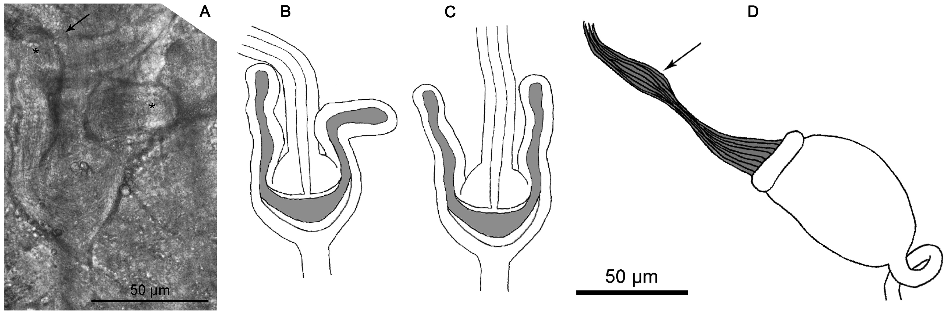
Clitellum in XII–1/3XIII, saddle-shaped; cells in c. 25 regular rows, elevated. Testes and sperm funnels in XI. Mature spermatozoa about 100 µm long, aligned on top of sperm funnel. Sperm funnels barrel-shaped, small, about 75–90 µm long and 40–50 µm wide, collar as wide as funnel body (or slightly narrower) ( Fig. 2D View FIGURE 2 ). Vasa deferentia confined to XII in a dense coil, 8–9 µm wide. Seminal vesicle in XI, occupying only one segment. Male copulatory organs c. 80 µm long and 50–55 µm wide, almost the same size as sperm funnels. Bursal slit mostly longitudinal. Spermathecae: no ectal glands; ectal ducts 240–265 µm long, 12–13 µm wide, ectal duct proximally projecting into ampulla, canal 4 µm wide. Ampulla almost spherical, 45–50 µm long and c. 40 µm wide, with 2 long variously bent diverticula (oriented ectad) ( Figs. 2A–B View FIGURE 2 , 3 View FIGURE 3 ). Lumen of diverticula and distal part of ampulla forming common Ushaped sperm-containing chamber. Length of diverticula c. 80 µm, width 13–18 µm. Ampullae open separately into oesophagus in V. One mature egg at a time.
Distribution and habitat. Russian Federation, Republic of Dagestan, Magaramkentsky District, Samur forest. Relic of a temperate-subtropical forest with the predominance of Quercus robur and Populus sp. ; soil type cambisol.
Remarks. There are a many Fridericia species with two spermathecal diverticula and a maximum of four chaetae in ventral preclitellar bundles, i.e., those belonging to the group “E” in Schmelz (2003). Within this group, only F. gamotheca Issel, 1905 and F. christeri Rota & Healy, 1999 combine simple unbranched oesophageal appendages with 5 pairs of preclitellar nephridia, coelomo-mucocytes “ type a” and the absence of spermathecal ectal gland. However, spermathecal ampullae of both sides in F. gamotheca are fused into one common chamber, and the clitellum is girdle-shaped, while in F. samurai sp. nov. ampullae open separately into the oesophagus, and the clitellum is absent ventrally. In F. christeri , sperm funnels and seminal vesicles are larger than that of F. samurai sp. nov., and subneural glands are present. Also, most specimens of F. christeri do not have spermathecae.
F. roembkei Schmelz & Collado, 2013 and F. ciliotheca Schmelz & Collado, 2013 resemble F. samurai sp. nov. in the chaetal pattern, small sperm funnels, absence of spermathecal ectal glands and, to some degree, shape of spermathecal ampullae and diverticula. However, in F. roembkei and F. ciliotheca lumen of the diverticula is subdivided into a peripheral sperm-containing chamber and ciliated sub-chamber, which is a conspicuous morphological feature in Fridericia ( Schmelz & Collado 2010) . Also, both species have some branches on the oesophageal appendages, and a seminal vesicle is absent.
F. samurai sp. nov. is morphologically most similar to F. brachiata Rota, 1994 , which has a very similar shape of spermathecal diverticula. The location where F. brachiata was found and described is also important, since Western Anatolia (Köppen’s Csa climate type) is climatically close enough to Eastern Dagestan (Cfa type) ( Beck et al. 2018). However, F. brachiata differs by the presence of spermathecal ectal glands and the possession of up to 6 chaetae in ventral preclitellar bundles. Also, the ectal duct of F. brachiata is much shorter, and the seminal vesicle is much larger than that of F. samurai sp. nov.

| M |
Botanische Staatssammlung München |
No known copyright restrictions apply. See Agosti, D., Egloff, W., 2009. Taxonomic information exchange and copyright: the Plazi approach. BMC Research Notes 2009, 2:53 for further explanation.
|
Kingdom |
|
|
Phylum |
|
|
Class |
|
|
Order |
|
|
Family |
|
|
Genus |