
Goniusa Casey, 1906
|
publication ID |
https://doi.org/10.5281/zenodo.156407 |
|
DOI |
https://doi.org/10.5281/zenodo.6273695 |
|
persistent identifier |
https://treatment.plazi.org/id/E310B73C-FF98-FFFC-1E2A-F9FC1392D39E |
|
treatment provided by |
Plazi (2016-04-02 11:13:45, last updated 2023-10-25 03:07:55) |
|
scientific name |
Goniusa Casey, 1906 |
| status |
|
Goniusa Casey, 1906 View in CoL ( Figs. 156 View FIGURES 1 5 View FIGURES 6 11 View FIGURES 12 15 View FIGURES 16 19 View FIGURES 20 24 View FIGURES 25 29 View FIGURES 30 36 View FIGURES 37 41 View FIGURES 42 47 View FIGURES 48 51 View FIGURES 52 56 )
Goniusa Casey, 1906: 348 View in CoL (in tribe Bolitocharini Thomson, 1859 View in CoL ).
Goniusa: Casey 1911: 208 View in CoL (in tribe Bolitocharini View in CoL ).
Goniusa: Fenyes, 1918: 19 View in CoL (in subtribe Athetina Casey, 1910 View in CoL of tribe Myrmedoniini Thomson, 1867 ).
Goniusa: Fenyes, 1920: 235 View in CoL .
Goniusa: Bernhauer & Scheerpeltz, 1926: 597 View in CoL (in subtribe Athetina View in CoL ).
Goniusa: Blackwelder, 1952: 174 View in CoL .
Goniusa: Kistner, 1976: 84 View in CoL (in tribe Zyrini Bradley, 1930).
Goniusa: Seevers, 1978: 133 View in CoL (in Goniusa View in CoL group of tribe Athetini). Goniusa: Ashe View in CoL in Newton, Thayer, Ashe & Chandler, 2000: 371 (in tribe Athetini, not assigned to subtribe).
Diagnosis. Goniusa can be distinguished from the other athetine genera by the combination of the following characters: body broad; antennal articles 510 slightly elongate ( Fig. 14 View FIGURES 12 15 ) or slightly transverse; in dry specimens gaps between antennal articles inconspicuous; ligula split apically ( Fig. 6 View FIGURES 6 11 ); labial palpus with setae,, and present; pronotum strongly transverse, 1.51.6 times as wide as long, with microsetae directed posteriorly along the midline; in lateral portions of the disc microsetae directed posteriorly and obliquely laterally (Type V, Benick & Lohse 1974) ( Fig. 12 View FIGURES 12 15 ); pronotal macrosetae short; pronotal hypomera fully visible in lateral view; medial macroseta of mesotibia inconspicuous, shorter than tibial width; tarsal formula 455; metatarsal segment 1 slightly shorter than segment 2; one empodial seta; abdominal sterna with numerous semierect macrosetae, sternum 8 with 3060 macrosetae ( Figs. 21, 24 View FIGURES 20 24 ); male pronotum with broad medial impression that is half as wide as pronotum, deeper in the posterior half, posterolateral portions of the impression with less dense microsculpture, without punctation and pubescence; male sternum 8 with broad apical emargination, apical portion of the sternum membranous ( Fig. 21 View FIGURES 20 24 ); aedeagus with narrow but blunt apex ( Figs. 25 View FIGURES 25 29 , 48 View FIGURES 48 51 ); medial lamellae of internal sac absent ( Figs. 30 View FIGURES 30 36 , 56 View FIGURES 52 56 ); copulatory piece troughshaped, with pointed apex ( Figs. 3034 View FIGURES 30 36 ); proximal portion of spermatheca with 23 coils ( Figs. 3536 View FIGURES 30 36 ).
Goniusa View in CoL differs from Notothecta Thomson, 1858 View in CoL in having abdominal sternum 8 with numerous (3060) macrosetae ( Figs. 21, 24 View FIGURES 20 24 ), male pronotum with broad medial impression, male sternum 8 with broad apical emargination, and different shape of the aedeagus and spermatheca.
Goniusa View in CoL can be distinguished from similarlooking genera of the tribe Lomechusini View in CoL by having longer and narrower mesothoracic process ( Figs. 13 View FIGURES 12 15 ; 1718); by shorter galea with its apical lobe only slightly projecting beyond the apex of lacinia ( Figs. 8 View FIGURES 6 11 ; 16, 19) and stronger setae on its internal margin ( Figs. 11 View FIGURES 6 11 ; 16).
Description. Length 3.24.2 mm, pronotal width 0.841.07 mm. Body broad, reddish brown to dark brown with darker head and brownish red appendages.
Head transverse; eyes large, temple length to eye length ratio 0.61.0; infraorbital carina very weak, complete, incomplete or absent altogether. Antennal article 2 as long as article 3, article 4 elongate, 510 slightly elongate or slightly transverse, apical article without coeloconic sensilla, longer than articles 9 and 10 combined ( Fig. 14 View FIGURES 12 15 ). In dry specimens gaps between antennal articles inconspicuous. Labrum ( Fig. 1 View FIGURES 1 5 ) transverse, with straight anterior margin. Adoral surface of labrum (epipharynx) as in Fig. 2 View FIGURES 1 5 . Mandibles ( Figs. 35 View FIGURES 1 5 ) broad, right mandible with small medial tooth; dorsal molar area with velvety patch consisting of very small denticles (poorly visible at 400x). Maxilla ( Figs. 811 View FIGURES 6 11 ) with galea projecting slightly beyond apex of lacinia; apical lobe of galea covered with numerous fine and short setae; internal margin of galea with long subapical setae ( Fig. 11 View FIGURES 6 11 ); apical 1/7 of lacinia with row of closely spaced spines, middle portion produced medially and covered with numerous setae ( Figs. 910 View FIGURES 6 11 ). Labium as in Figs. 67 View FIGURES 6 11 , 15 View FIGURES 12 15 ; ligula split apically; medial area of prementum without pores but with 5 pseudopores, lateral areas with 2 pores, single setose pore and 1216 pseudopores ( Fig. 6 View FIGURES 6 11 ). Hypopharyngeal lobes as in Fig. 7 View FIGURES 6 11 . Labial palpus with setae,, and present. Mentum ( Fig. 15 View FIGURES 12 15 ) with concave anterior margin.
Pronotum ( Figs. 12 View FIGURES 12 15 , 3741 View FIGURES 37 41 ) strongly transverse, with microsetae directed posteriorly in midline; in lateral portions of disc microsetae directed posteriorly and obliquely laterally (Type V, Benick & Lohse 1974); macrosetae short; hypomera fully visible in lateral view. Meso and metasternum as in Fig. 13 View FIGURES 12 15 , mesosternal process narrow, extending about ½ length of mesocoxal cavities, metasternal process short, mesosternum and mesosternal process not carinate medially; relative lengths of mesosternal process: isthmus: metasternal process in ratio of about 2:1:1; mesocoxal cavities margined posteriorly; mesocoxae narrowly separated. Medial macroseta of mesotibia inconspicuous, shorter than tibial width. Tarsal segmentation 455, metatarsal segment 1 slightly shorter than segment 2. One empodial seta, half as long as claws. Posterior margin of elytra slightly concave near posterolateral angle. Wings fully developed.
Abdominal terga 35 with moderate basal impressions. Tergum 7 1.11.2 times as long as tergum 6. Punctation on terga 67 sparser than on terga 35. Tergum 7 with wide white palisade fringe. Abdominal sterna with numerous semierect macrosetae, sternum 8 with 3060 macrosetae.
Male pronotum with broad medial impression that is half as wide as pronotum, deeper in posterior half, posterolateral portions of the impression with less dense microsculpture, without punctation and pubescence. Compared to female, male pronotum matte, with stronger microsculpture and weaker punctation. Male sternum 8 with broad apical emargination, apical portion of the sternum membranous ( Fig. 21 View FIGURES 20 24 ); aedeagus with narrow but blunt apex ( Figs. 25 View FIGURES 25 29 , 48 View FIGURES 48 51 ); medial lamellae of internal sac absent ( Figs. 30 View FIGURES 30 36 , 56 View FIGURES 52 56 ); copulatory piece troughshaped, with pointed apex ( Figs. 3034 View FIGURES 30 36 ); proximal portion of spermatheca with 23 coils ( Figs. 3536 View FIGURES 30 36 ).
Type species. Goniusa caseyi Gusarov , sp. n., by subsequent designation (see Discussion below), fixed under provisions of Article 70.3.
Discussion. When proposing the new generic name Goniusa, Casey (1906) included in a single species, Euryusa obtusa LeConte, 1866 , and this species would be the type of the genus by monotypy. However, my examination of the holotype of Euryusa obtusa (MCZ) and the specimens labeled as “ Goniusa obtusa ” in the Casey collection, as well as my analysis of characters mentioned by Casey (1906) demonstrated that Casey’s concept of the species does not agree with that of LeConte (1866). In fact the holotype of Euryusa obtusa belongs to the genus known as Lypoglossa Fenyes, 1918 while Casey's specimens agree with current usage of the name " Goniusa obtusa ". The status of E. obtusa LeConte (nec Casey) will be discussed in a separate paper on the genus Lypoglossa . Since the type species of the genus Goniusa Casey, 1906 was misidentified by Casey (1906) the provisions of the Article 70.3 apply to it and the type species needs to be fixed ( ICZN, 1999). To best serve stability and universality of nomenclature I select to fix as the type species of Goniusa the taxonomic species actually involved in the misidentification (Article 70.3.2). The type species of the genus Goniusa Casey, 1906 is now fixed as Goniusa caseyi Gusarov , sp. n., misidentified as Euryusa obtusa LeConte, 1866 in the original description by Casey (1906).
Fenyes (1918) placed Goniusa in the subtribe Athetina and noted the similarity between Goniusa and the myrmecophilous genus Notothecta Thomson, 1858 ( Fenyes 1920) . Kistner (1976) disagreed with Fenyes and argued that based on "the structure of the maxillae with their setigerous lacinia and galea" Goniusa should be placed in Zyrini Bradley, 1930 (spelled as Zyrasini ), a junior synonym of Lomechusini Fleming, 1821 ( Newton & Thayer 1992). However, in Goniusa the galea is relatively short and it is only slightly projecting beyond the apex of lacinia ( Fig. 8 View FIGURES 6 11 ) in comparison to the members of Lomechusini (e. g., Drusilla Leach in Samouelle, 1819 and Zyras Stephens, 1835 ; Figs. 16, 19 View FIGURES 16 19 ). In Goniusa subapical setae of the internal margin of the galea are strong, and both galea and lacinia are very much like in other members of Athetini. Additionally, in Goniusa the mesosternal process is narrow and long ( Fig. 13 View FIGURES 12 15 ) compared to the members of Lomechusini ( Figs. 1718 View FIGURES 16 19 ). Presented arguments confirm the view of Fenyes (1918): Goniusa is not related to Lomechusini and is a member of Athetini.
Both known species of Goniusa are associated with the ants of the genus Formica and have been collected inside the ant nests.
Benick, G. & Lohse, G. A. (1974) 14. Tribus: Callicerini (Athetae). In: Freude, H., Harde, K. W. & Lohse, G. A. (Eds.), Die Kafer Mitteleuropas. Band 5, Staphylinidae II (Hypocyphtinae und Aleocharinae). Pselaphidae. Goecke & Evers Verlag, Krefeld, pp. 72 - 220.
Bernhauer, M. & Scheerpeltz, O. (1926) Staphylinidae VI. In: Junk, W. & Schenkling, S. (Eds.), Coleopterorum Catalogus, Pars 82. W. Junk, Berlin, pp. 499 - 988.
Blackwelder, R. E. (1952) The generic names of the beetle family Staphylinidae, with an essay on genotypy. U. S. National Museum Bulletin, 200, 1 - 483.
Bradley, J. C. (1930) A manual of the genera of beetles of America north of Mexico. Daw, Illston & Co., Ithaca, 360 pp.
Casey, T. L. (1906) Observations of the staphylinid groups Aleocharinae and Xantholinini, chiefly of America. Transactions of the Academy of Sciences of St. Louis, 16 (6), 125 - 434.
Casey, T. L. (1910) New Species of the Staphylinid Tribe Myrmedoniini. Memoirs on the Coleoptera I. The New Era Printing Company, Lancaster, pp. 1 - 183.
Casey, T. L. (1911) New American species of Aleocharinae and Myllaeninae. Memoirs on the Coleoptera II. The New Era Printing Company, Lancaster, pp. 1 - 245.
Fenyes, A. (1918) Coleoptera: Fam Staphylinidae, subfam. Aleocharinae. In: Wytsman, P. (Ed.), Genera Insectorum, Fasc. 173 A. L. Desmet-Verteneuil, Bruxelles, pp. 1 - 110.
Fenyes, A. (1920) Coleoptera. Fam. Staphylinidae, subfam. Aleocharinae. In: Wytsman, P. (Ed.), Genera Insectorum, Fasc. 173 B. L. Desmet-Verteneuil, Bruxelles. Pp. 111 - 414.
Fleming, J. (1821) Insecta. In: Supplement to the fourth, fifth and sixth editions of the Encyclopaedia Britannica, with preliminary dissertations on the history of the sciences. Vol. 5. Archibald Constable and Company, Edinburgh, pp. 41 - 56.
ICZN (1999) International Code of Zoological Nomenclature. Fourth Edition. The International Trust for Zoological Nomenclature, London, xxix + 306 pp.
Kistner, D. H. (1976) Revision and Reclassification of the Genus Goniusa Casey with a Larval Description and Ant Host Records (Coleoptera: Staphylinidae). Sociobiology, 2 (1), 83 - 95.
LeConte, J. L. (1866) Additions to the coleopterous fauna of the United States. No. 1. Proceedings of the Academy of Sciences of Philadelphia, 18, 361 - 394.
Newton, A. F. & Thayer, M. K. (1992) Current Classification and Family-Group Names in Staphyliniformia (Coleoptera). Fieldiana: Zoology, 67, 1 - 92.
Newton, A. F., Thayer, M. K., Ashe, J. S. & Chandler, D. S. (2000) Staphylinidae Latreille, 1802. In: Arnett, R. H., Thomas, M. C. (Eds.), American Beetles. Vol. 1. Archostemata, Myxophaga, Adephaga, Polyphaga: Staphyliniformia. CRC Press, Boca Raton, pp. 272 - 418.
Samouelle, G. (1819) The Entomologist's useful compendium; or an introduction to the knowledge of British insects, comprising the best means of obtaining and preserving them, and a description of the apparatus generally used; together with the genera of Linne, and the modern method of arranging the classes Crustacea, Myriapoda, spiders, mites, and insects from their affinities and structure, according to the views of Dr. Leach. Also an explanation of the terms used in entomology; a calendar of the times of appearance, and usual situations of near 3000 species of British insects; with instructions for collecting and fitting up objects for the microscope. Thomas Boys, London, 496 pp.
Seevers, C. H. (1978) A generic and tribal revision of the North American Aleocharinae (Coleoptera: Staphylinidae). Fieldiana: Zoology, 71, vi + 275 pp.
Stephens, J. F. (1835) Illustrations of British entomology; or, a synopsis of indigenous insects; containing their generic and specific distinctions; with an account of their metamorphoses, times of appearance, localities, food, and economy, as far as practicable. Mandibulata. 5. Baldwin and Cradock, London, pp. 369 - 448.
Thomson, C. G. (1858) Forsok till uppstallning af Sveriges Staphyliner. Ofversigt af Kongl. Vetenskaps-Akademiens Forkhandlingar, 15, 27 - 40.
Thomson, C. G. (1859) Skandinaviens Coleoptera, synoptiskt bearbetade. Tom 1. Berlingska Boktryckeriet, Lund, v + 290 pp.
Thomson, C. G. (1867) Skandinaviens Coleoptera, synoptiskt bearbetade, Tom 9. Lundbergska Boktryckeriet, Lund, 408 pp.
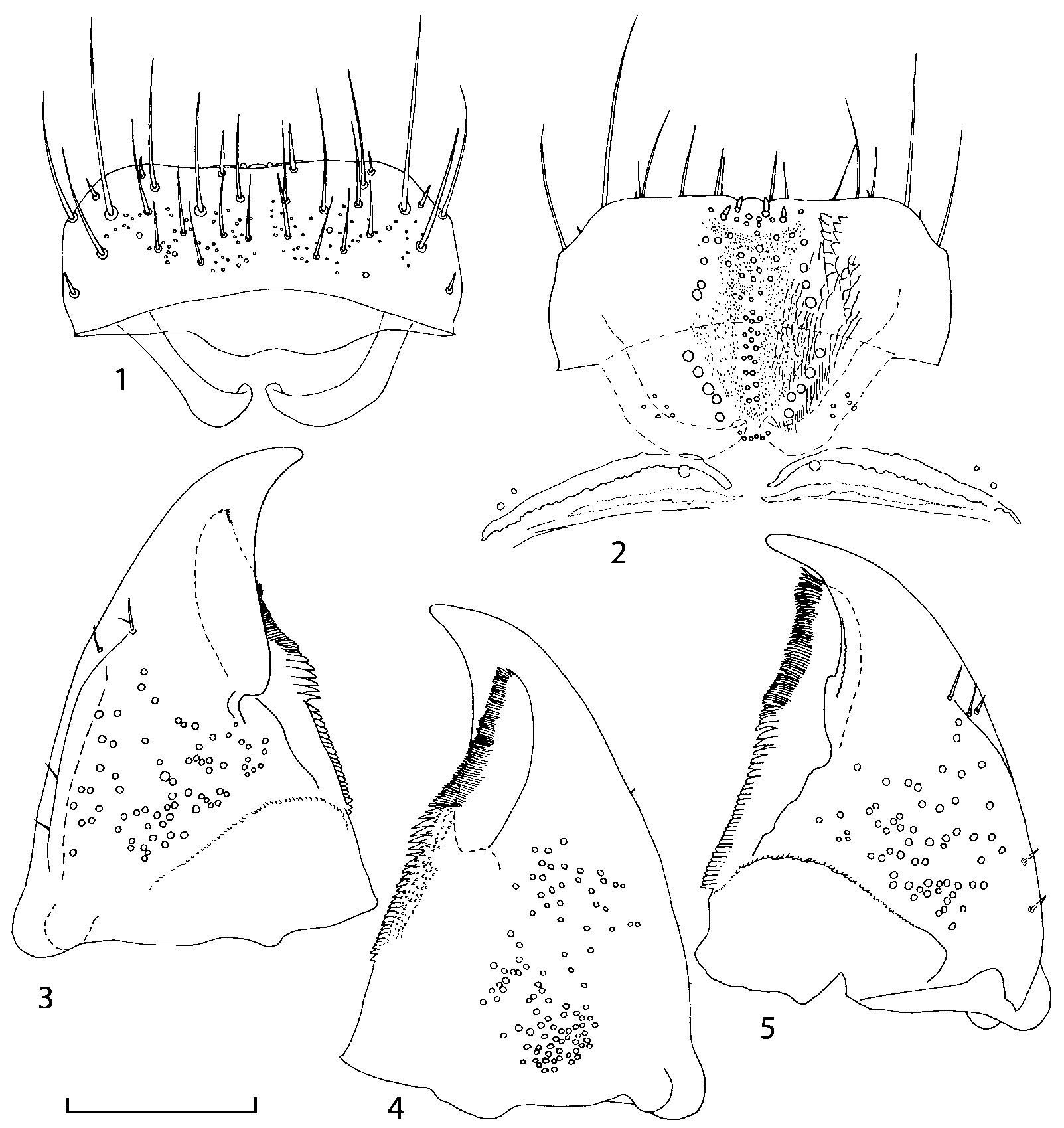
FIGURES 1 5. Mouthparts of Goniusa caseyi Gusarov, sp. n. (paratype from Aweme, Manitoba). 1 – labrum; 2 – epipharynx; 3 – left mandible, dorsal view; 4 – left mandible, ventral view; 5 – right mandible, dorsal view. Scale bar 0.1 mm.
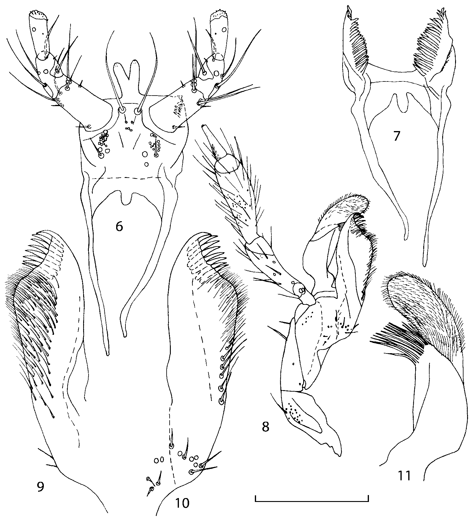
FIGURES 6 11. Mouthparts of Goniusa caseyi Gusarov, sp. n. (paratype from Aweme, Manitoba). 6 – prementum; 7 – hypopharynx; 8 – right maxilla, ventral view; 9 – right lacinia, dorsal view; 10 – right lacinia, ventral view; 11 – right galea, dorsal view. Scale bar 0.1 mm (6 7, 9 11), 0.2 mm (8).
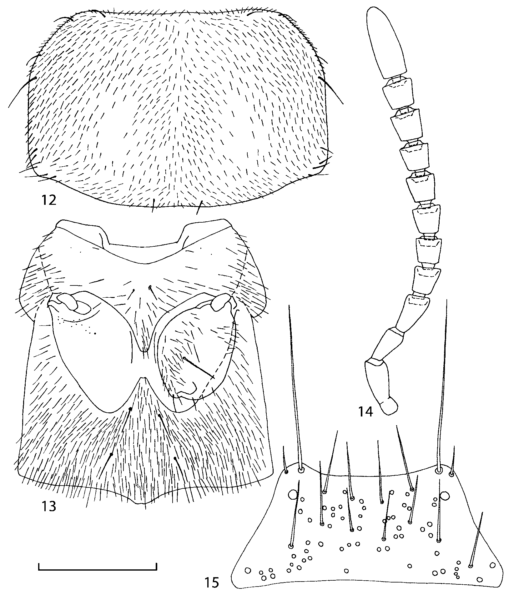
FIGURES 12 15. Details of Goniusa caseyi Gusarov, sp. n. (paratype from Aweme, Manitoba). 12 – pronotum; 13 – meso and metathorax, ventral view; 14 – right antenna; 15 – mentum. Scale bar 0.1 mm (15), 0.2 mm (12), 0.4 mm (13 14).
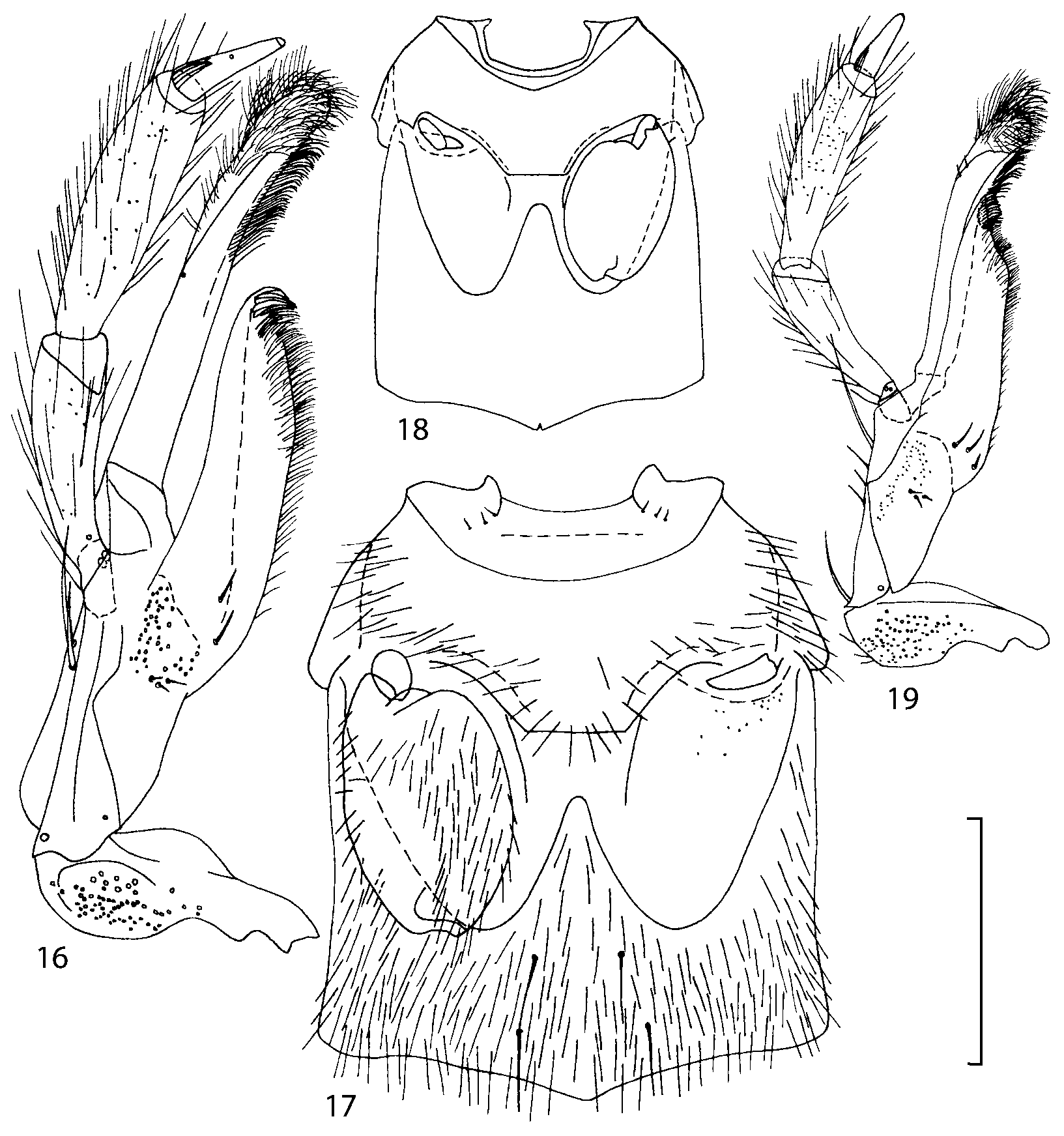
FIGURES 16 19. Details of Drusilla canaliculata (Fabricius) (female from Kiev, Ukraine (16); male from 15 km N of Magadan, Russia (17 )) and Zyras humeralis (Gravenhorst) (female from Briançon, France). 16, 19 – right maxilla, ventral view; 17 – meso and metathorax, ventral view; 18 – meso and metathorax, ventral view (setation not shown). Scale bar 0.2 mm (16), 0.4 mm (17, 19), 1.0 mm (18).
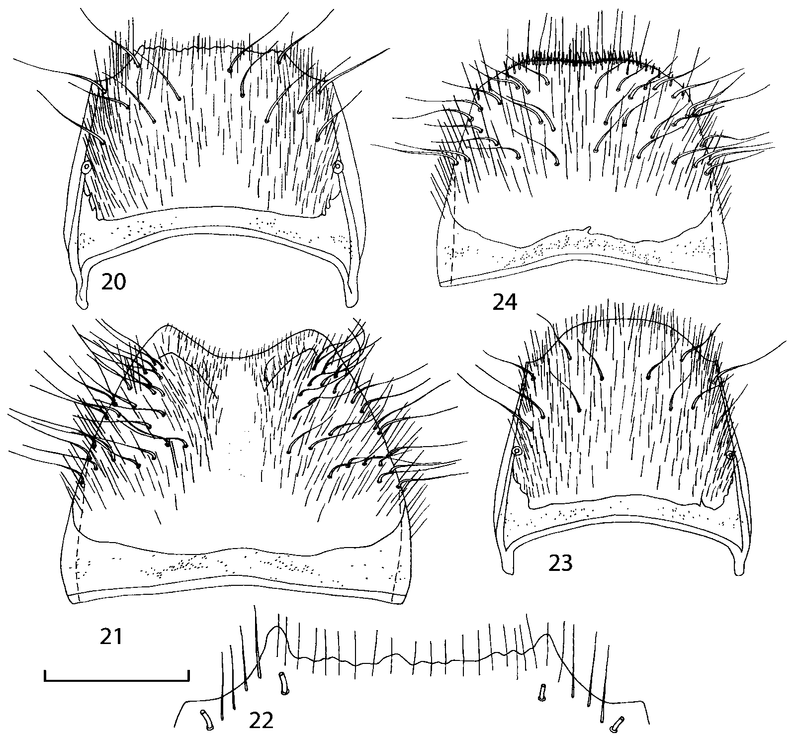
FIGURES 20 24. Abdominal segment 8 of Goniusa caseyi Gusarov, sp. n. (paratypes from Washington, D. C. (20 21); Aweme, Manitoba (22); and Stonewall, Manitoba (23 24 )). 20 – male tergum 8; 21 – male sternum 8; 22 – apex of male tergum 8; 23 – female tergum 8; 24 – female sternum 8. Scale bar 0.4 mm (20 21, 23 24), 0.2 mm (22).
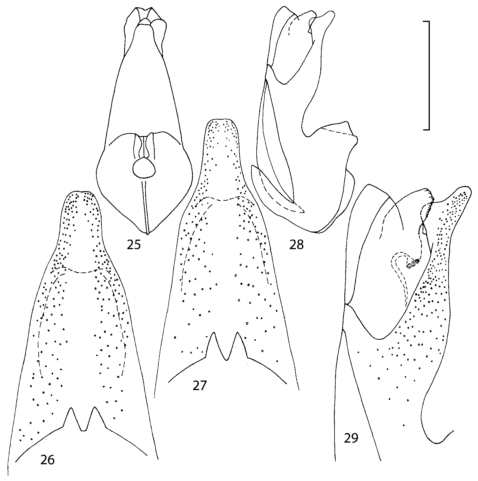
FIGURES 25 29. Median lobe of aedeagus of Goniusa caseyi Gusarov, sp. n. (paratypes from Washington, D. C. (25 26, 28 29); and Sherborn, Massachusetts (27 )). 25 – median lobe, parameral view; 26 27 – apex of median lobe, parameral view; 28 – median lobe, lateral view; 29 – apex of median lobe, lateral view. Scale bar 0.4 mm (25, 28), 0.2 mm (26 27, 29).
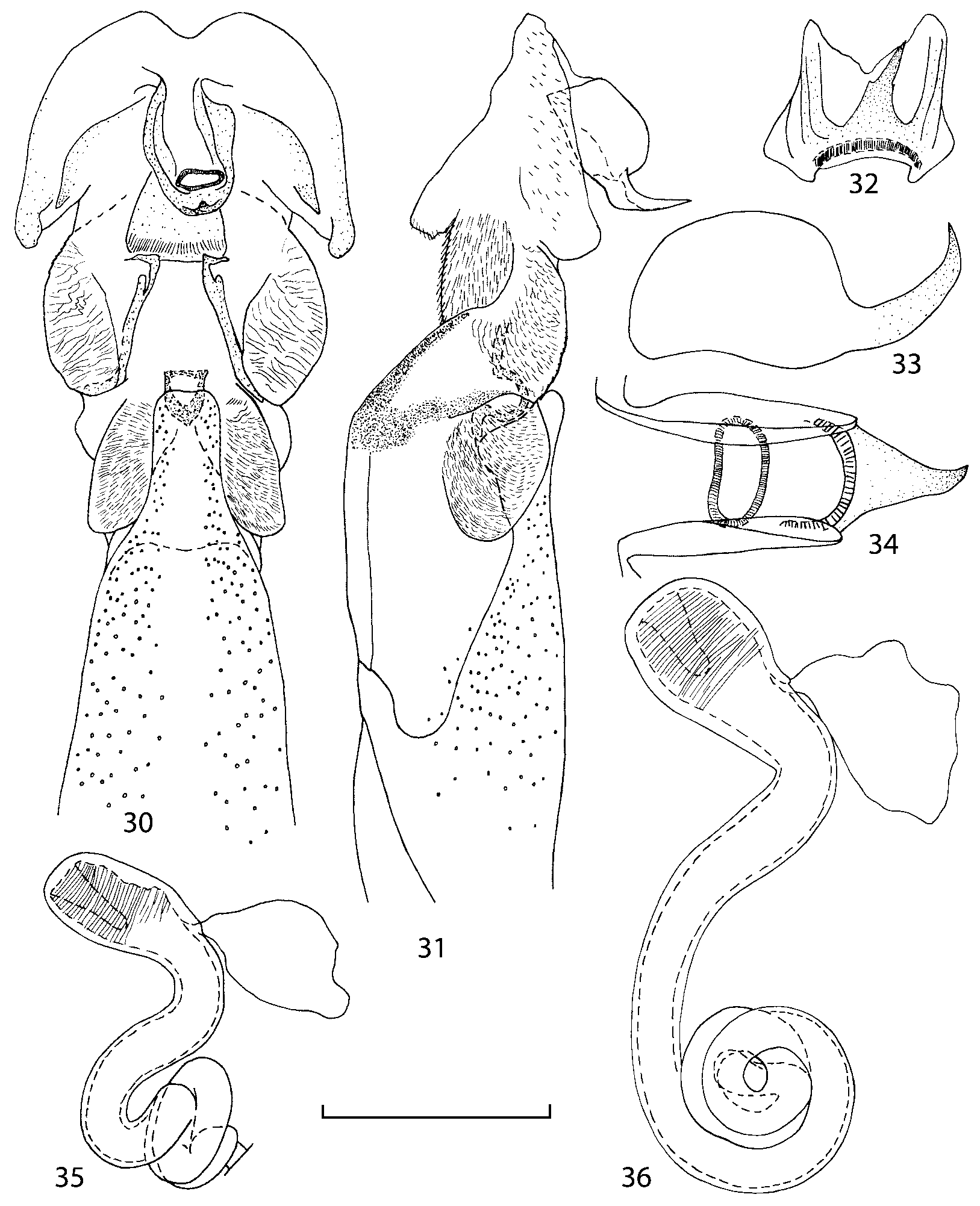
FIGURES 30 36. Genitalia of Goniusa caseyi Gusarov, sp. n. (paratypes from Aweme, Manitoba (30 31); Texas (32 34); Sherborn, Massachusetts (35); and Stonewall, Manitoba (36 )). 30 – everted internal sac of aedeagus, parameral view; 31 – everted internal sac of aedeagus, lateral view; 32 – copulatory piece, apical view; 33 – copulatory piece, lateral view; 34 – copulatory piece, abparameral view; 35 36 – spermatheca. Scale bar 0.1 mm (32 34), 0.2 mm (30 31, 35 36).
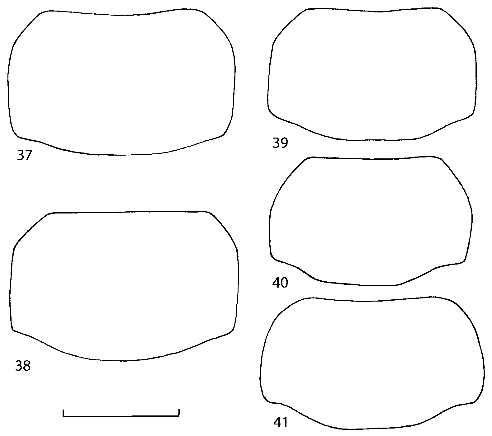
FIGURES 37 41. Pronotum of Goniusa caseyi Gusarov, sp. n. (paratypes from Aweme, Manitoba (37); and Stonewall, Manitoba (38 )) and G. alperti Kistner (holotype (39); female from San Bernardino Mts., California (40), paratype from 10 mi. E of Coville, Washington (41 )). 37, 39 – male pronotum; 38, 40 41 – female pronotum. Scale bar 0.5 mm.
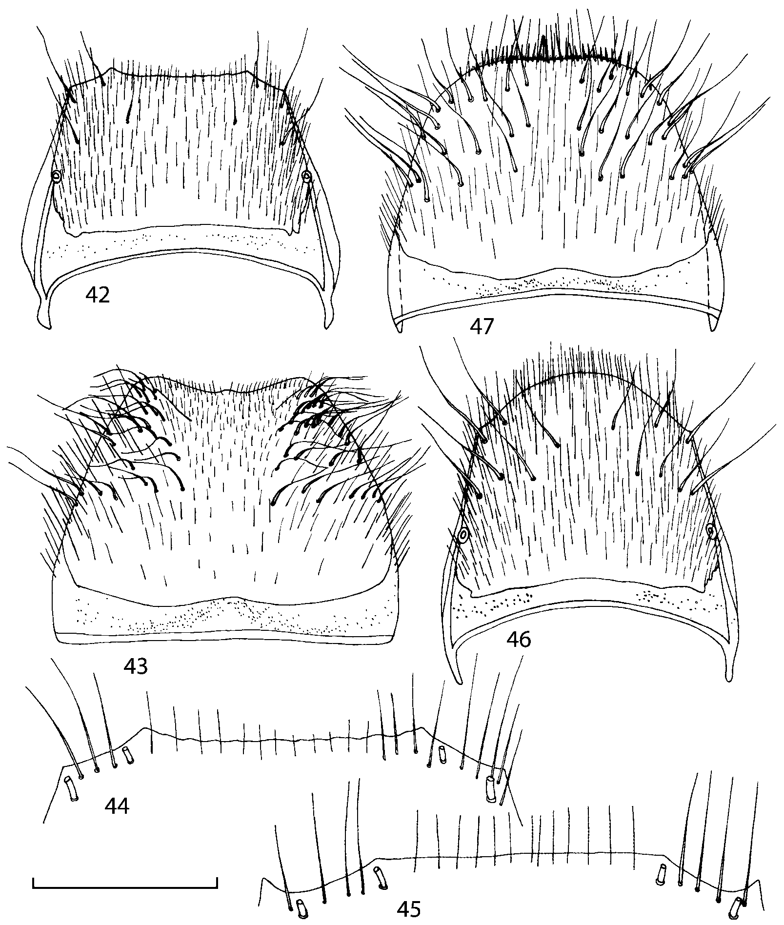
FIGURES 42 47. Abdominal segment 8 of Goniusa alperti Kistner (holotype (42 44); male from San Bernardino Mts., California (45); female from 16 km NW of Granby, Colorado (46 47 )). 42 – male tergum 8; 43 – male sternum 8; 44 45 – apex of male tergum 8; 46 – female tergum 8; 47 – female sternum 8. Scale bar 0.4 mm (42 43, 46 47), 0.2 mm (44 45).
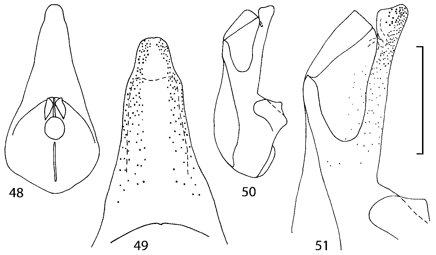
FIGURES 48 51. Median lobe of aedeagus of Goniusa alperti Kistner (male from West Point, Nebraska). 48 – median lobe, parameral view; 49 – apex of median lobe, parameral view; 50 – median lobe, lateral view; 51 – apex of median lobe, lateral view. Scale bar 0.4 mm (48, 50), 0.2 mm (49, 51).
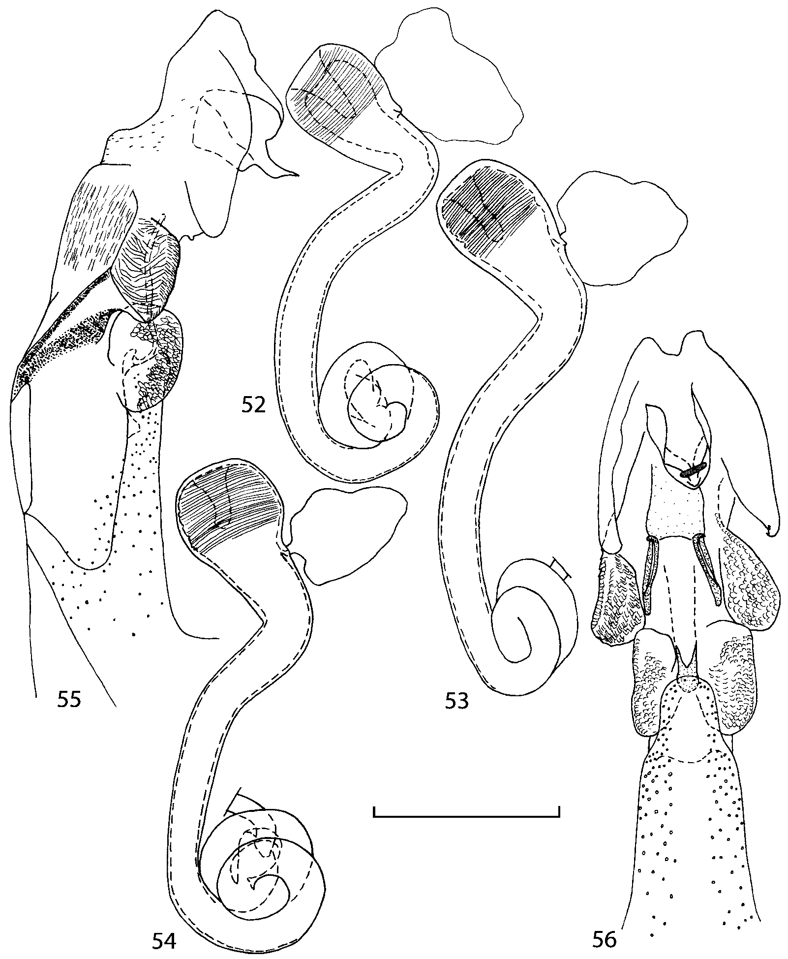
FIGURES 52 56. Genitalia of Goniusa alperti Kistner (paratype from 10 mi. E of Coville, Washington (52); female from Tahoe City, California (53); female from 16 km NW of Granby, Colorado (54); holotype (55 56 )). 52 54 – spermatheca; 55 – everted internal sac of aedeagus, lateral view; 56 – everted internal sac of aedeagus, parameral view. Scale bar 0.2 mm.
No known copyright restrictions apply. See Agosti, D., Egloff, W., 2009. Taxonomic information exchange and copyright: the Plazi approach. BMC Research Notes 2009, 2:53 for further explanation.
|
Kingdom |
|
|
Phylum |
|
|
Class |
|
|
Order |
|
|
Family |
Goniusa Casey, 1906
| Gusarov, Vladimir I. 2003 |
Goniusa:
| Newton 2000: 371 |
| Seevers 1978: 133 |
Goniusa:
| Kistner 1976: 84 |
Goniusa:
| Blackwelder 1952: 174 |
Goniusa:
| Bernhauer 1926: 597 |
Goniusa:
| Fenyes 1920: 235 |
Goniusa:
| Fenyes 1918: 19 |
Goniusa:
| Casey 1911: 208 |
Goniusa
| Casey 1906: 348 |
1 (by plazi, 2016-04-02 11:13:45)
2 (by ImsDioSync, 2016-09-27 22:34:58)
3 (by ImsDioSync, 2016-11-26 03:54:59)
4 (by ImsDioSync, 2016-11-26 03:59:41)
5 (by ImsDioSync, 2017-06-24 13:24:17)
6 (by ExternalLinkService, 2019-09-26 22:45:33)
7 (by ExternalLinkService, 2022-01-31 01:03:15)
8 (by ExternalLinkService, 2022-02-25 06:59:45)
9 (by plazi, 2023-10-25 02:56:33)