
Pseudexechia
|
publication ID |
https://doi.org/10.5281/zenodo.186691 |
|
DOI |
https://doi.org/10.5281/zenodo.6214051 |
|
persistent identifier |
https://treatment.plazi.org/id/937787FF-FFB6-FFCE-FF5C-FDBF519EFAB4 |
|
treatment provided by |
Plazi (2016-04-19 08:51:10, last updated 2023-10-29 23:14:23) |
|
scientific name |
Pseudexechia |
| status |
|
Key to European males of Pseudexechia View in CoL View at ENA
1 Terminalia large, stout ( Fig. 6 View FIGURE 6 A); dorsal branch of the gonostylus with a characteristically smooth whitish apex; ventral branch forming a thin plate shaped more or less like a spatula ( Fig. 6 View FIGURE 6 B); hypandrial lobe heavily sclerotized, bud-like by elaborate folding, triangular to subrectangular ( Fig 6 View FIGURE 6 A).......................................................................... 2
- Terminalia more slender ( Fig. 14 View FIGURE 14 A), with elongated dorsal and ventral branches of the gonostylus ( Fig. 14 View FIGURE 14 B); hypandrial lobe narrow rectangular, forming parallel branches medially ( Fig. 14 View FIGURE 14 A) ................................. trivittata group 8
2 Median ocellus small, but distinctly present ( Fig. 4 View FIGURE 4 A); vein R4+5 distinctly downcurved (C> D in Fig. 3A); hypandrial lobe stout, subrectangular ( Fig. 6 View FIGURE 6 A) ........................................................................................ trisignata View in CoL group 3
- Median ocellus absent or at most tiny and vestigial; vein R4+5 almost straight (C ≤ D in Fig. 3B&D); hypandrial lobe
conic, triangular ( Fig. 9 View FIGURE 9 A) or enlarged and diverging apically ( Fig. 11 View FIGURE 11 D) ...................................... canalicula View in CoL group 5 3 A dull brown species with a distinct greyish dusting ( Fig. 2 View FIGURE 2 A); hypandrial lobe with rounded corners ( Fig. 6 View FIGURE 6 A); ventral branch of gonostylus with angular basodorsal corner making the spatula broadest basally ( Fig. 6 View FIGURE 6 B); smooth whitish tip of dorsal branch of gonostylus evenly thick ( Fig. 6 View FIGURE 6 B)............... Pseudexechia trisignata ( Edwards, 1913) View in CoL
- Moderately to distinctly bi-coloured species without greyish dusting; hypandrial lobe sharply pointed apically ( Fig. 7 View FIGURE 7 A); ventral branch of gonostylus without angular basodorsal corner; smooth whitish tip of dorsal branch of gonostylus slightly dilated or constricted ............................................................................................................................... 4
4 Broad pale band along whole margin mesonotum; ventral branch of gonostylus forming small, symmetric and round spatula ( Fig. 7 View FIGURE 7 B); smooth whitish tip of dorsal branch of gonostylus slightly constricted ( Fig. 7 View FIGURE 7 B) ............................ .......................................................................................................... Pseudexechia pectinacea ( Ostroverkhova, 1979) View in CoL
- Without or at most narrow pale band along margin of mesonotum; ventral branch of gonostylus forming large, asymmetric spatula that is broadest apically ( Fig. 8 View FIGURE 8 B); smooth whitish tip of dorsal branch of gonostylus slightly dilated ( Fig. 8 View FIGURE 8 B) ......................................................................................................... Pseudexechia tuomikoskii View in CoL sp. n.
5 Larger species, distinctly bi-coloured in dark greyish brown and yellow, with sharply defined mesonotal stripes ( Fig. 2 View FIGURE 2 B) ............................................................................................................................................................................... 6
- Smaller species, weakly bi-coloured to dull brown, with indistinct or fused mesonotal stripes ................................. 7
6 Gonostylus large, exposed ( Fig. 9 View FIGURE 9 A); dorsal branch distinctly dilated apically ( Fig. 9 View FIGURE 9 B); ventral branch forming large, slightly asymmetrical spatula ( Fig. 9 View FIGURE 9 B) ......................................... Pseudexechia canalicula ( Johannsen, 1912)
- Gonostylus small, retracted ( Fig. 10 View FIGURE 10 A); dorsal branch not dilated apically ( Fig. 10 View FIGURE 10 B); ventral branch forming narrow, angular boot-shaped spatula ( Fig. 10 View FIGURE 10 B) ..................................................... Pseudexechia aurivernica Chandler, 1978 View in CoL
7 Hypandrial lobe enlarged, diverging apically ( Fig. 11 View FIGURE 11 A&D); dorsal branch of gonostylus apically strongly dilated, campanulate ( Fig. 11 View FIGURE 11 B); dorsointernal branch small ( Fig. 11 View FIGURE 11 B); ventral branch narrow, angular boot-shaped ( Fig. 11 View FIGURE 11 B) ........................................................................................... Pseudexechia monica Kjaeandsen & Chandler, 2006 View in CoL
- Hypandrial lobe not enlarged, conic triangular ( Fig. 12 View FIGURE 12 A); dorsal branch of gonostylus apically dilated, but not campanulate ( Fig. 12 View FIGURE 12 B); dorsointernal branch large ( Fig. 12 View FIGURE 12 B); ventral branch forming medium sized spatula with acute angled apicodorsal corner ( Fig. 12 View FIGURE 12 B)............................................................ Pseudexechia parallela ( Edwards, 1925) View in CoL
8 Dorsal branch of gonostylus subrectangular, with sharply truncated, whitish tip ( Fig 13 View FIGURE 13 A&B); dorsointernal branch forming narrow, elongated lamellate fan ( Fig. 13 View FIGURE 13 B); hypoproct reduced, hyaline ( Fig. 13 View FIGURE 13 C) ....................................... ......................................................................................... Pseudexechia latevittata Chandler & Blasco-Zumeta, 2001 View in CoL
- Dorsal branch of gonostylus oblong to lanceolate, without sharply truncated, whitish tip ( Fig. 14 View FIGURE 14 A&B); dorsointernal branch reduced to hyaline knob basally on dorsal branch ( Fig. 14 View FIGURE 14 B); hypoproct sclerotized, pointed ( Fig. 14 View FIGURE 14 C). ...................................................................................................................................................................................... 9
9 A distinctly bi-coloured species in dark greyish brown and yellow, with sharply defined thoracic stripes and triangular pale bands on abdominal tergites; dorsal branch of gonostylus shorter and smaller than ventral branch which has medial surface covered with setae and no brush apically ( Fig. 14 View FIGURE 14 B) ........ Pseudexechia tristriata Stackelberg, 1969 View in CoL
- A dull brown species with fused, hardly discernible thoracic stripes and indistinct, narrow (if any) pale bands on abdominal tergites; dorsal branch of gonostylus longer than ventral branch which has medial surface devoid of setae but a narrow brush of strong setae apically ( Fig. 15 View FIGURE 15 B) ................................... Pseudexechia trivittata ( Staeger, 1840) View in CoL
Chandler, P. J. (1978) Notes on the Holarctic Species of Pseudexechia Tuomikoski (Diptera: Mycetophilidae), with the Description of a New British Species. Entomologist's Record and Journal of Variation, 90, 44 - 51.
Chandler, P. J. & Blasco-Zumeta, J. (2001) The fungus gnats (Diptera, Bolitophilidae, Keroplatidae and Mycetophilidae) of the Monegros region (Zaragoza, Spain) and five other new European species of Pyratula Edwards and Sciophila Meigen. Zapateri, Revista Aragonesa de Entomologia, 9, 1 - 24.
Edwards, F. W. (1913) Notes on British Mycetophilidae. Transactions of the Royal Entomological Society of London, 1913, 334 - 382.
Edwards, F. W. (1925) British Fungus-Gnats (Diptera, Mycetophilidae). With a revised Generic Classification of the Family. Transactions of the Entomological Society of London, 73, 505 - 670.
Johannsen, O. A. (1912) The Mycetophilidae of North America. Part IV (Conclusion). Maine Agricultural Experimental Station Orono, Bulletin No. 200, 57 - 146.
Kjaerandsen, J. & Chandler, P. J. (2006) On the identity of Pseudexechia parallela (Edwards, 1925) and description of a new related species from Great Britain (Diptera, Mycetophilidae). British Journal of Entomology and Natural History, 19, 41 - 49.
Ostroverkhova, G. P. (1979) Fungus-gnats (Diptera, Mycetophiloidea) of Siberia. Tomsk.
Stackelberg, A. A. (1969) Family Bolitophilidae. In: G. Y. Be i- B ie n ko (E d), Key to the Insects of the European Part of the USSR. Volume V Diptera and Siphonaptera Part I., Leningrad, pp. 247 - 257.
Staeger, R. C. (1840) Systematisk fortegnelse over de i Danmark hidtil fundne Diptera. Naturhistorisk Tidskrift, 3, 228 - 288.
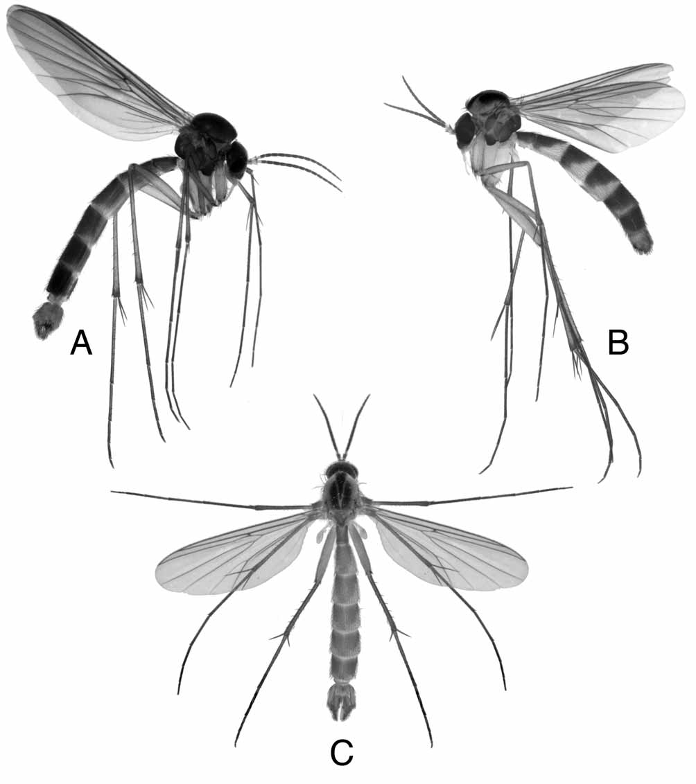
FIGURE 2. Habitus photos of Pseudexechia. — A. P. trisignata (Edwards, 1913) in lateral view, a dull brown species with a distinct greyish dusting, with fused thoracic stripes and narrow apical pale bands on abdomen. — B. P. aurivernica Chandler, 1978 in lateral view, a distinctly bi-coloured species in dark greyish brown and yellow, often with a reddish tinge, with distinct thoracic stripes and with large, triangular apical pale bands on abdomen — C. P. canalicula (Johannsen, 1912) in dorsal view, yet another bi-coloured species in greyish brown and yellow with distinct thoracic stripes.
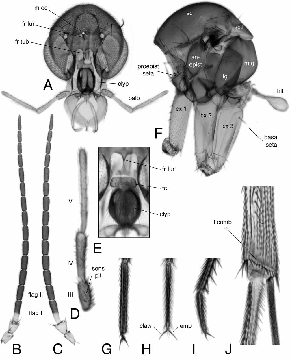
FIGURE 4. Morphology of Pseudexechia [A – D + F – J = P. trisignata (Edwards, 1913), E = P. pectinacea (Ostroverkhova, 1979)]. — A. Head in dorsal view. — B. Antenna, dorsal view. — C. Antenna, lateral view — D. Segment III – V of maxillary palp. — E. Face enlarged. — F. Thorax, lateral view. — G. Fore tarsus V. — H. Mid tarsus V. — I. Hind tarsus V. — J. Apex of hind tibia, medial view. Abbreviations: anepist = anepisternum; clyp = clypeus; cx 1 = forecoxa; cx 2 = midcoxa; cx 3 = hindcoxa; emp = empodium; fc = face; flag I = first flagellar segment; flag II = second flagellar segment; fr fur = frontal furrow; fr tub = frontal tubercle; htl = halter; ltg = laterotergite; m oc = medial ocellus; mtg = mediotergite; proepist seta = proepisternal seta; sc = scutum; sctl = scutellum; sens pit = sensory pit; t comb = tibial comb.

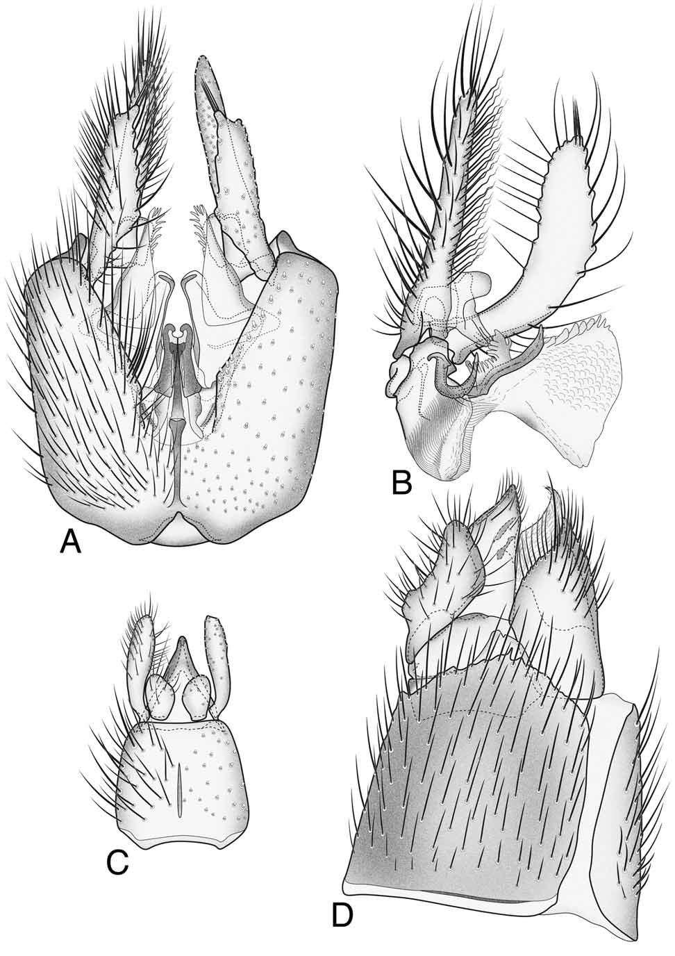
FIGURE 6. Pseudexechia trisignata (Edwards, 1913). — A. Male terminalia, ventral view. — B. Male gonostylus, internal view. — C. Male tergite IX and cerci, dorsal view. — D. Female terminalia, lateral view.

FIGURE 7. Pseudexechia pectinacea (Ostroverkhova, 1979). — A. Male terminalia, ventral view. — B. Male gonostylus, internal view, enlarged. — C. Male tergite IX and cerci, dorsal view. — D. Female terminalia, lateral view.
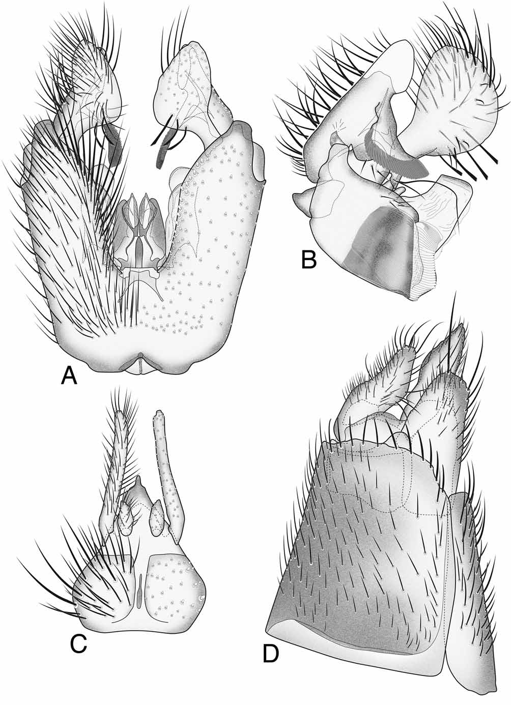
FIGURE 8. Pseudexechia tuomikoskii sp. n. — A. Male terminalia, ventral view. — B. Male gonostylus, internal view. — C. Male tergite IX and cerci, dorsal view. — D. Female terminalia, lateral view.
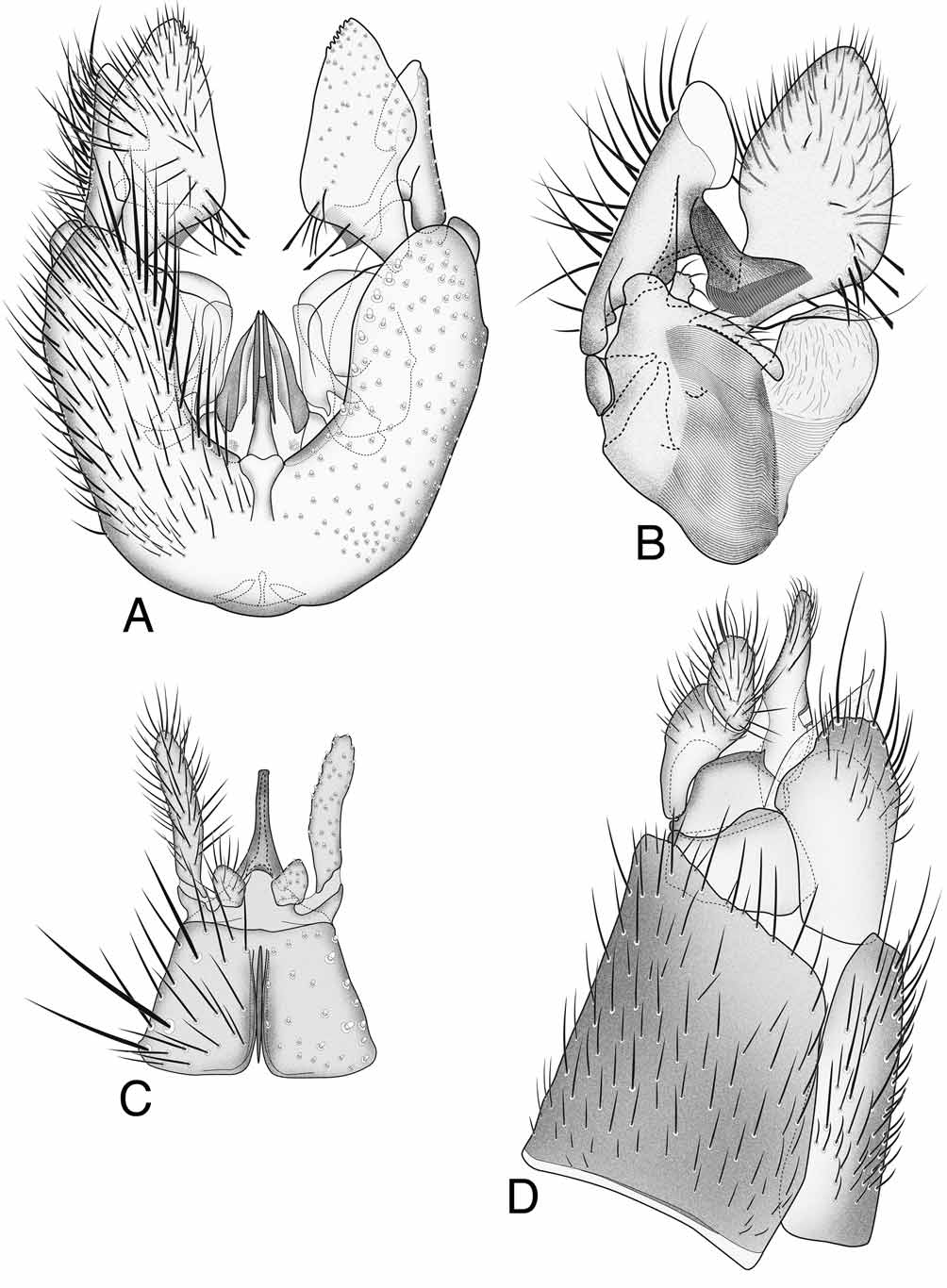
FIGURE 9. Pseudexechia canalicula (Johannsen, 1912). — A. Male terminalia, ventral view. — B. Male gonostylus, internal view, enlarged. — C. Male tergite IX and cerci, dorsal view. — D. Female terminalia, lateral view.
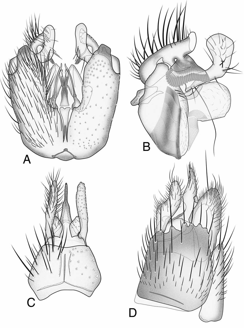
FIGURE 10. Pseudexechia aurivernica Chandler, 1978. — A. Male terminalia, ventral view. — B. Male gonostylus, internal view, enlarged. — C. Male tergite IX and cerci, dorsal view. — D. Female terminalia, lateral view.
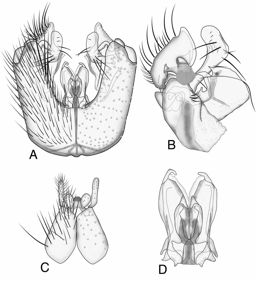
FIGURE 11. Pseudexechia monica Kjaerandsen & Chandler, 2006. — A. Male terminalia, ventral view. — B. Male gonostylus, internal view, enlarged. — C. Male tergite IX and cerci, dorsal view. — D. Hypandrial lobe, ventral view, enlarged.
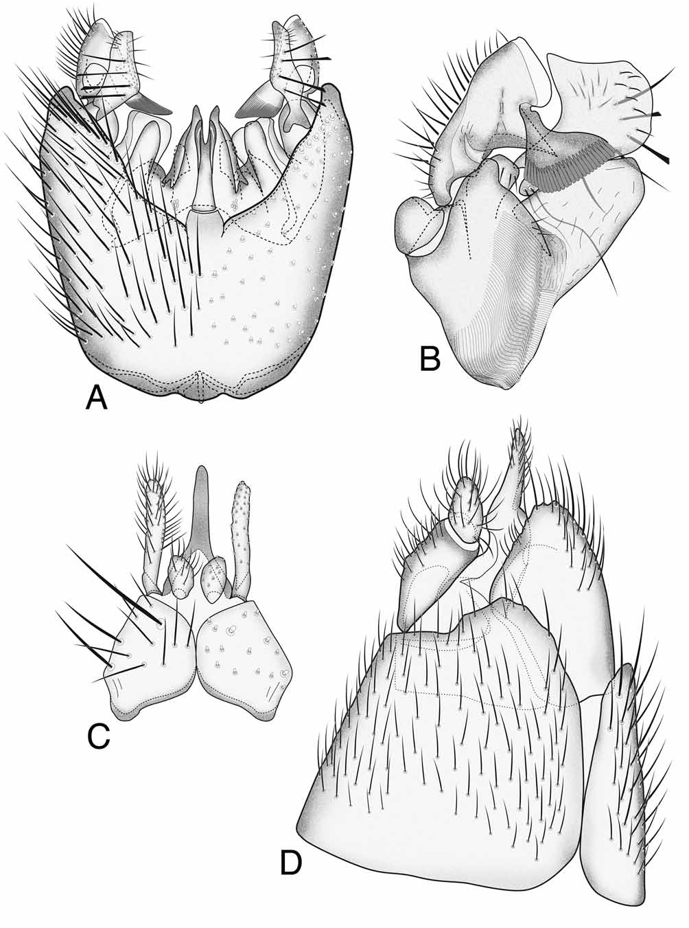
FIGURE 12. Pseudexechia parallela (Edwards, 1925). — A. Male terminalia, ventral view. — B. Male gonostylus, internal view, enlarged. — C. Male tergite IX and cerci, dorsal view. — D. Female terminalia, lateral view.

FIGURE 13. Pseudexechia latevittata Chandler & Blasco-Zumeta, 2001. — A. Male terminalia, ventral view. — B. Male gonostylus, internal view, enlarged. — C. Male tergite IX and cerci, dorsal view. — D. Female terminalia (redrawn from Chandler & Blasco-Zumeta 2001), lateral view.
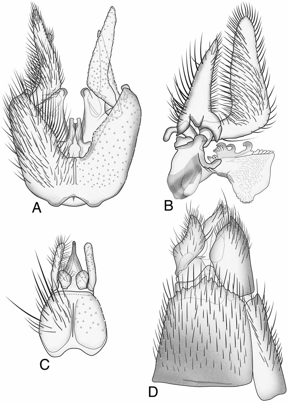
FIGURE 14. Pseudexechia tristriata (Stackelberg, 1969). — A. Male terminalia, ventral view. — B. Male gonostylus, internal view. — C. Male tergite IX and cerci, dorsal view. — D. Female terminalia, lateral view.
No known copyright restrictions apply. See Agosti, D., Egloff, W., 2009. Taxonomic information exchange and copyright: the Plazi approach. BMC Research Notes 2009, 2:53 for further explanation.
|
Kingdom |
|
|
Phylum |
|
|
Class |
|
|
Order |
|
|
Family |
1 (by plazi, 2016-04-19 08:51:10)
2 (by ImsDioSync, 2016-11-30 05:41:03)
3 (by ImsDioSync, 2016-11-30 05:42:29)
4 (by ImsDioSync, 2017-06-22 02:41:08)
5 (by ImsDioSync, 2017-06-22 03:11:14)
6 (by ExternalLinkService, 2019-09-26 12:25:13)
7 (by ExternalLinkService, 2022-01-30 17:42:25)
8 (by ExternalLinkService, 2022-02-22 00:43:21)
9 (by plazi, 2023-10-29 12:04:30)