
Euophrys
|
publication ID |
https://doi.org/10.5281/zenodo.194411 |
|
persistent identifier |
https://treatment.plazi.org/id/911E87A7-E408-FFCD-FF4B-FF5A3DB08F95 |
|
treatment provided by |
Plazi (2016-04-17 09:34:03, last updated 2022-01-30 16:33:35) |
|
scientific name |
Euophrys |
| status |
|
Key to Euophrys species
Females
1 More than 50 % of the spermatheca posterior to the fossa and guide (e.g. Fig. 59 View FIGURES 56 – 62 ) .................................................... 2
- Less than 50 % of the spermatheca posterior to the fossa and guide (e.g. Fig. 64 View FIGURES 63 – 65 )...................................................... 6
2 Spermatheca smaller than the fossa and inner edge of the fossa well beyond the outer edge of the spermatheca; L 4 patterning as in Fig. 3 View FIGURE 3 D ............................................................................................................ E. anotata ( Figs 49–55 View FIGURES 49 – 55 )
- Spermatheca equal to or larger than the fossa and inner edge of the fossa near or overlapping the outer edge of the spermatheca ................................................................................................................................................................. 3
3 Insemination duct anterior to the fossa and guide, fossa O-shaped, and wide apart so just overlapping spermatheca; L 4 patterning as in Fig. 3 View FIGURE 3 F ................................................................................................. E. patagonica ( Figs 73–79 View FIGURES 73 – 79 )
- Insemination duct not passing anterior to the fossa and guide, fossa C-shaped, inner edges of guide overlap the sper- matheca ........................................................................................................................................................................ 4
4 Fossa placed dorsolaterally to, and overlap, the spermatheca, guides open anteriorly; L 4 patterning as in Fig. 3 View FIGURE 3 B .... .............................................................................................................................................. E. rusticana ( Figs 89–95 View FIGURES 89 – 95 )
- Fossa dorsal to the spermatheca and guides open ventrolaterally .............................................................................. 5
5 Dark pattern on the abdomen forms two longitudinal bands; femur of L 4 a plain yellow colour (may occasionally have faint markings ( Fig. 3 View FIGURE 3 E) .......................................................................................... E. saitiformis ( Figs 96–102 View FIGURES 96 – 102 )
s- Dark pattern on the abdomen scattered, does not form two longitudinal bands; three dark bands on a yellow back- ground on the femur of L 4 ( Fig. 3 View FIGURE 3 H) ...................................................................... E. flordellago n. sp. ( Figs 56–62 View FIGURES 56 – 62 )
6 Inner edge of the fossa beyond the outer edge of the spermatheca ............................................................................ 7
- Inner edge of the fossa overlaps the spermatheca ....................................................................................................... 8
7 Entrance to the insemination duct close to the anterior edge of the fossa; L 4 patterning as in Fig. 3 View FIGURE 3 E ........................ .................................................................................................................................................. E. laetata ( Figs 63–65 View FIGURES 63 – 65 )
- Entrance to the insemination duct close to the posterior edge of the fossa; L 4 patterning as in Fig. 3 View FIGURE 3 G ...................... .......................................................................................................................................... E. tehuelche ( Figs 103–109 View FIGURES 103 – 109 )
8 Median edges of the fossae touching, guide opens posterolaterally; L 4 patterning as in Fig. 3 View FIGURE 3 C ................................. ................................................................................................................................................ E. mapuche ( Figs 66–72 View FIGURES 66 – 72 )
- Median edges of the fossae not touching, guide opens dorsolaterally; L 4 patterning as in Fig. 3 View FIGURE 3 A .............................
.................................................................................................................................................. E. rapida ( Figs 80–88 View FIGURES 80 – 88 )
Males
As the males are not known for E. laetata , E. saitiformis and E. flordellago no key is given. Compare specimens with Figs 2 and 3.
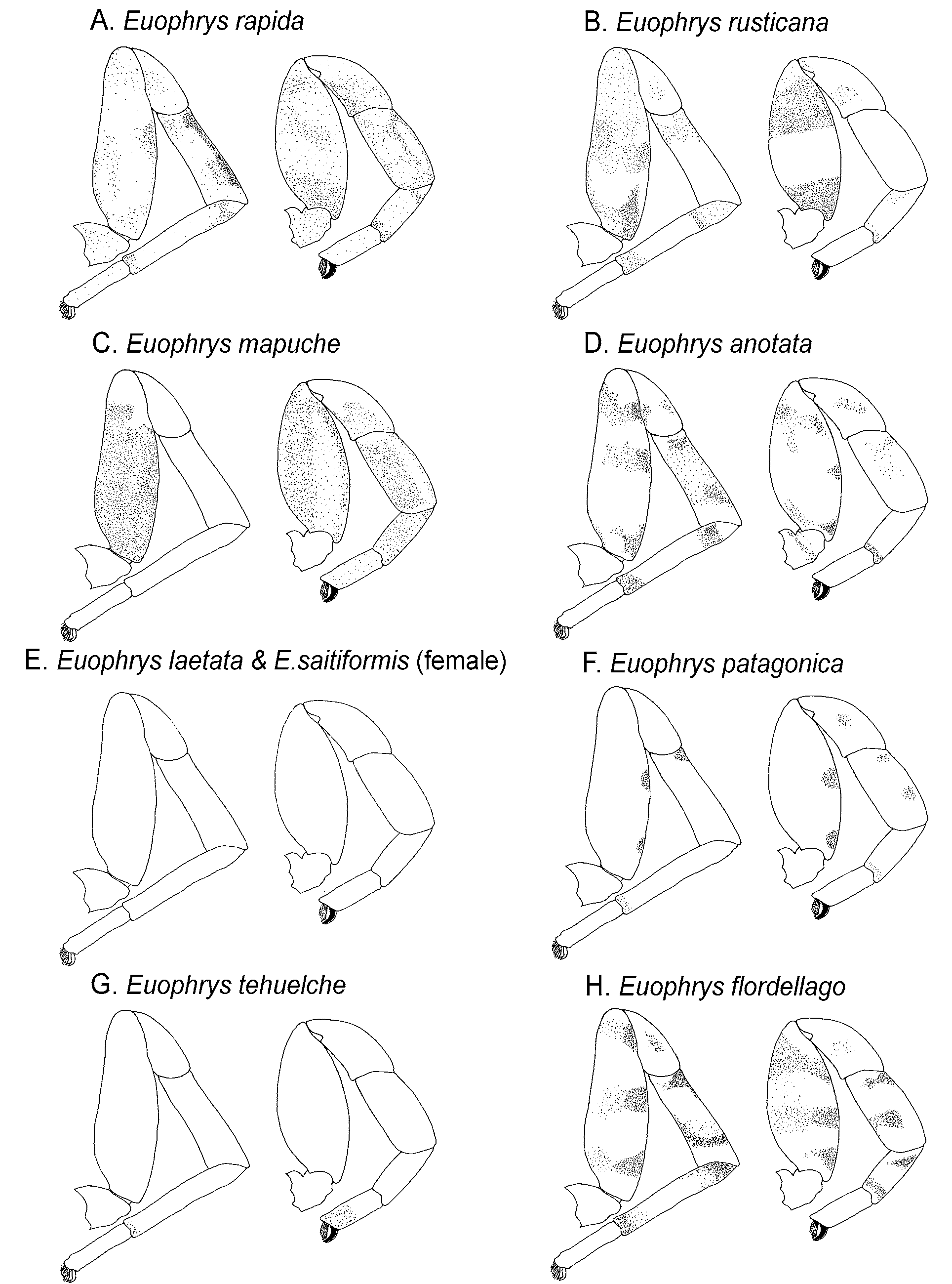
FIGURE 3. Semi-diagrammatic colour patterns of the first and fourth legs of males in the genus Euophrys. Light stippled areas are orange / mid-brown in colour, plain areas yellow, dark stipples areas are black or very dark brown. In each species there is some variation in the intensity of the colouring. L 4, left; L 1 right.
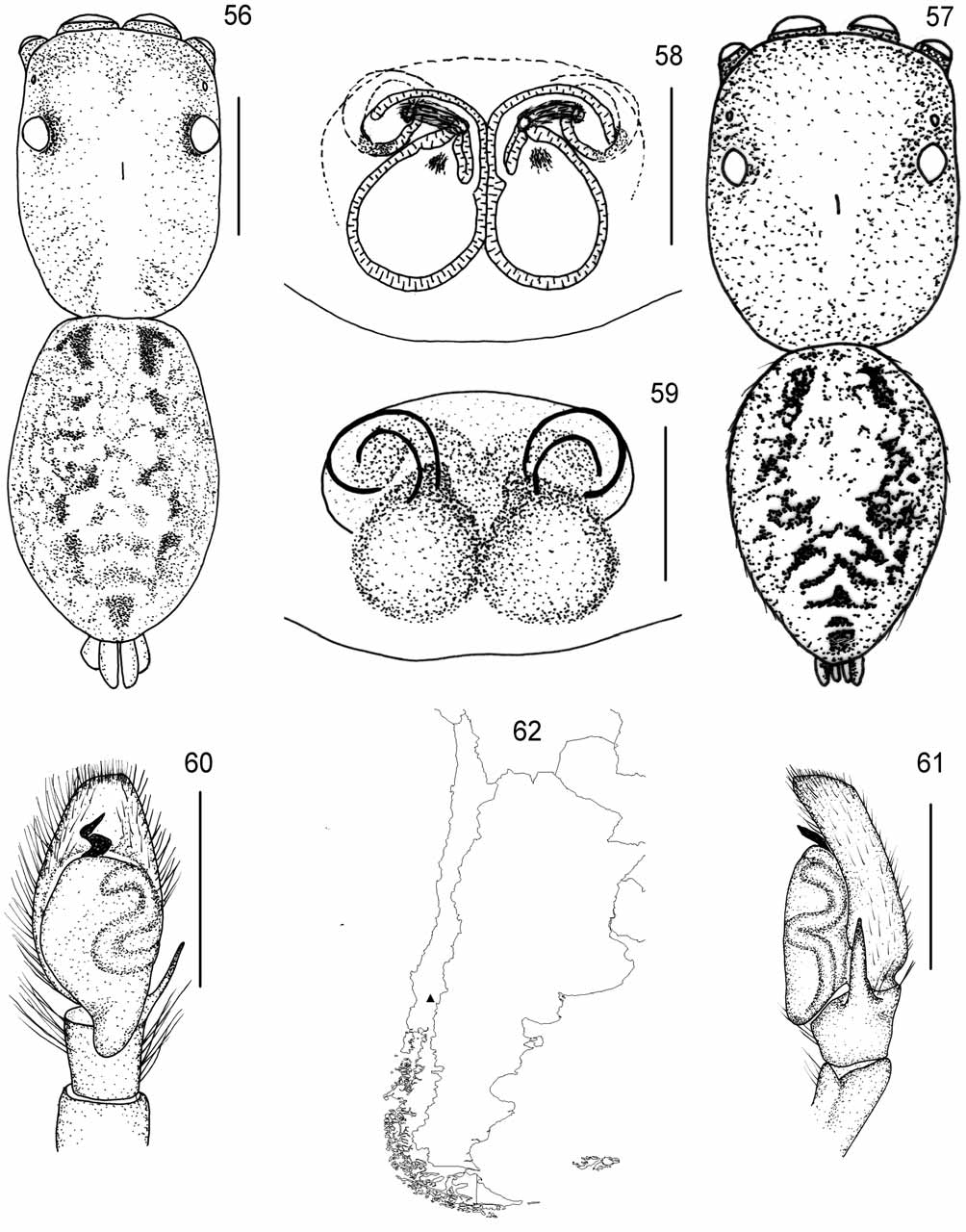
FIGURES 56 – 62. Euophrys flordellago. 56 – 57 dorsal view (56 female, 57 possible male, redrawn after Braul et al. 1997, see text); 58 – 59 female genitalia (58 dorsal view of cleared specimen, 59 ventral view of external characteristics); 60 – 61 possible male palp (60 ventral view, 61 lateral view, redrawn after Braul et al. 1997, see text); 62 known distribution. Scales: total body 1 mm; remainder 0.2 mm

FIGURES 63 – 65. Euophrys laetata. 63 female dorsal view; 64 – 65 female genitalia, (64 dorsal view of cleared specimen, 65 ventral view of external characteristics). Scales: total body 1 mm; remainder 0.2 mm

FIGURES 49 – 55. Euophrys anotata. 49 – 50 dorsal view (49 female, 50 male); 51 – 52 female genitalia (51 dorsal view of cleared specimen, 52 ventral view of external characteristics); 53 – 54 male palp (53 ventral view, 54 lateral view); 55 known distribution. Scales: total body 1 mm; remainder 0.2 mm

FIGURES 73 – 79. Euophrys patagonica. 73 – 74 dorsal view (73 female, 74 male); 75 – 76 female genitalia (75 dorsal view of cleared specimen, 76 ventral view of external characteristics); 77 – 78 male palp (77 ventral view, 78 lateral view); 79 known distribution. Scales: total body 1 mm; remainder 0.2 mm
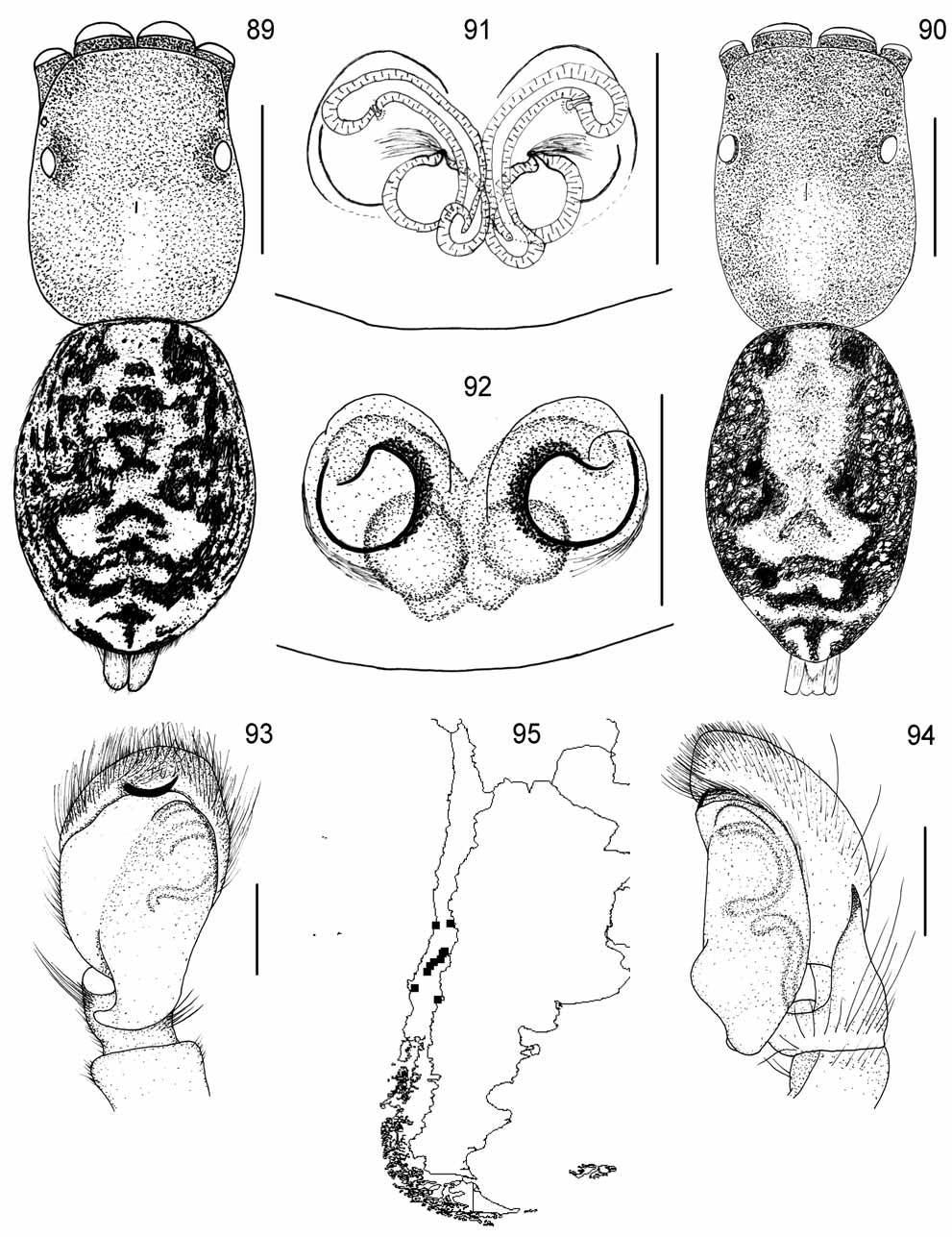
FIGURES 89 – 95. Euophrys rusticana. 89 – 90 dorsal view (89 female, 90 male); 91 – 92 female genitalia (91 dorsal view of cleared specimen, 92 ventral view of external characteristics); 93 – 94 male palp (93 ventral view, 94 lateral view); 95 known distribution. Scales: total body 1 mm; remainder 0.2 mm
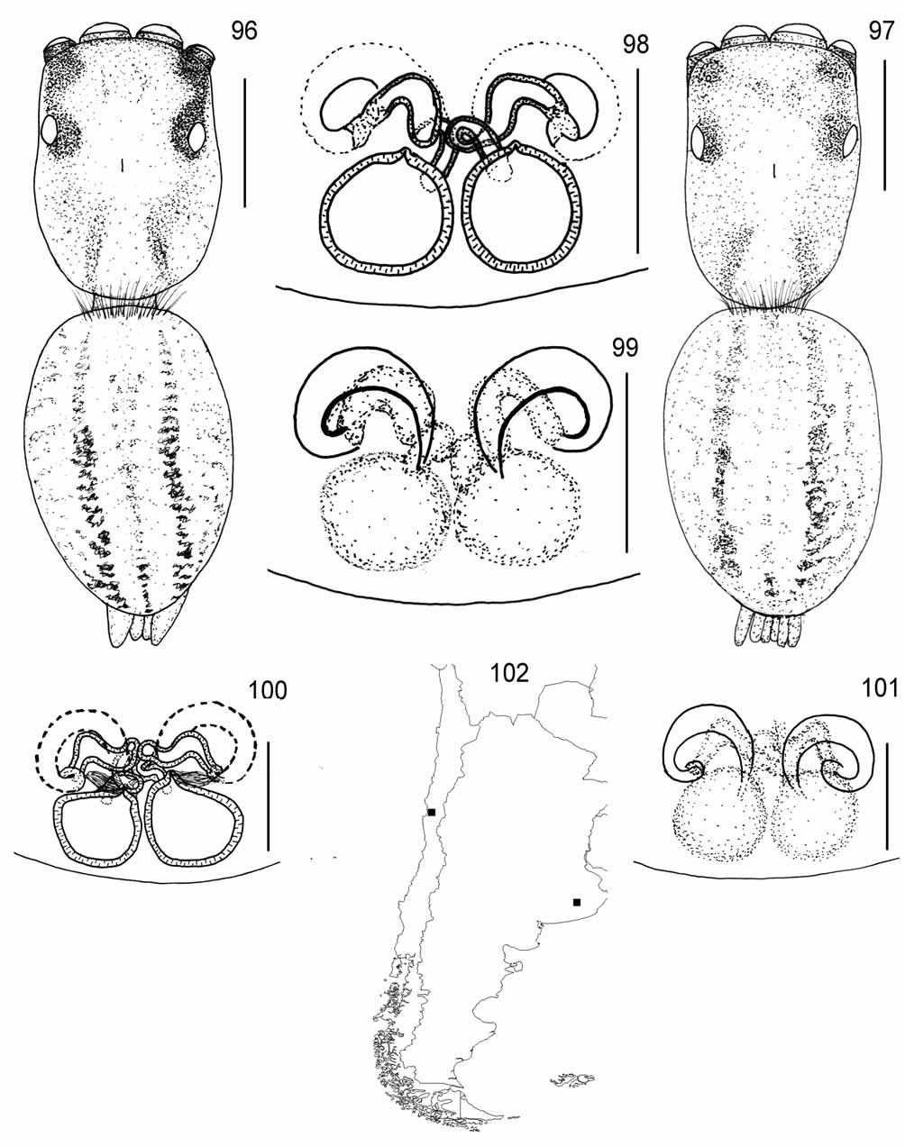
FIGURES 96 – 102. Euophrys saitiformis. 96 – 97 female dorsal view (96 E. saitiformis type, 97 E. cruziana type). 98 – 101 female genitalia (98 dorsal view of cleared type specimen of E. saitiformis. 99 ventral view of external characteristics of type specimen of E. saitiformis, 100 dorsal view of cleared type specimen of E. cruziana, 101 ventral view of external characteristics of type specimen of E. cruziana); 102 known distribution. Scales: total body 1 mm; remainder 0.2 mm
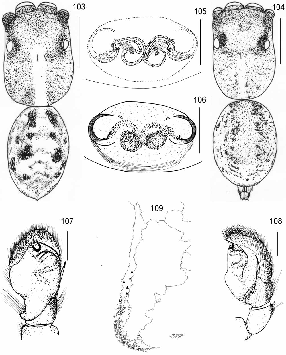
FIGURES 103 – 109. Euophrys tehuelche. 103 – 104 dorsal view (103 female, 104 male); 105 – 106 female genitalia (105 dorsal view of cleared specimen, 106 ventral view of external characteristics); 107 – 108 male palp (107 ventral view, 108 lateral view); 109 known distribution. Scales: total body 1 mm; remainder 0.2 mm
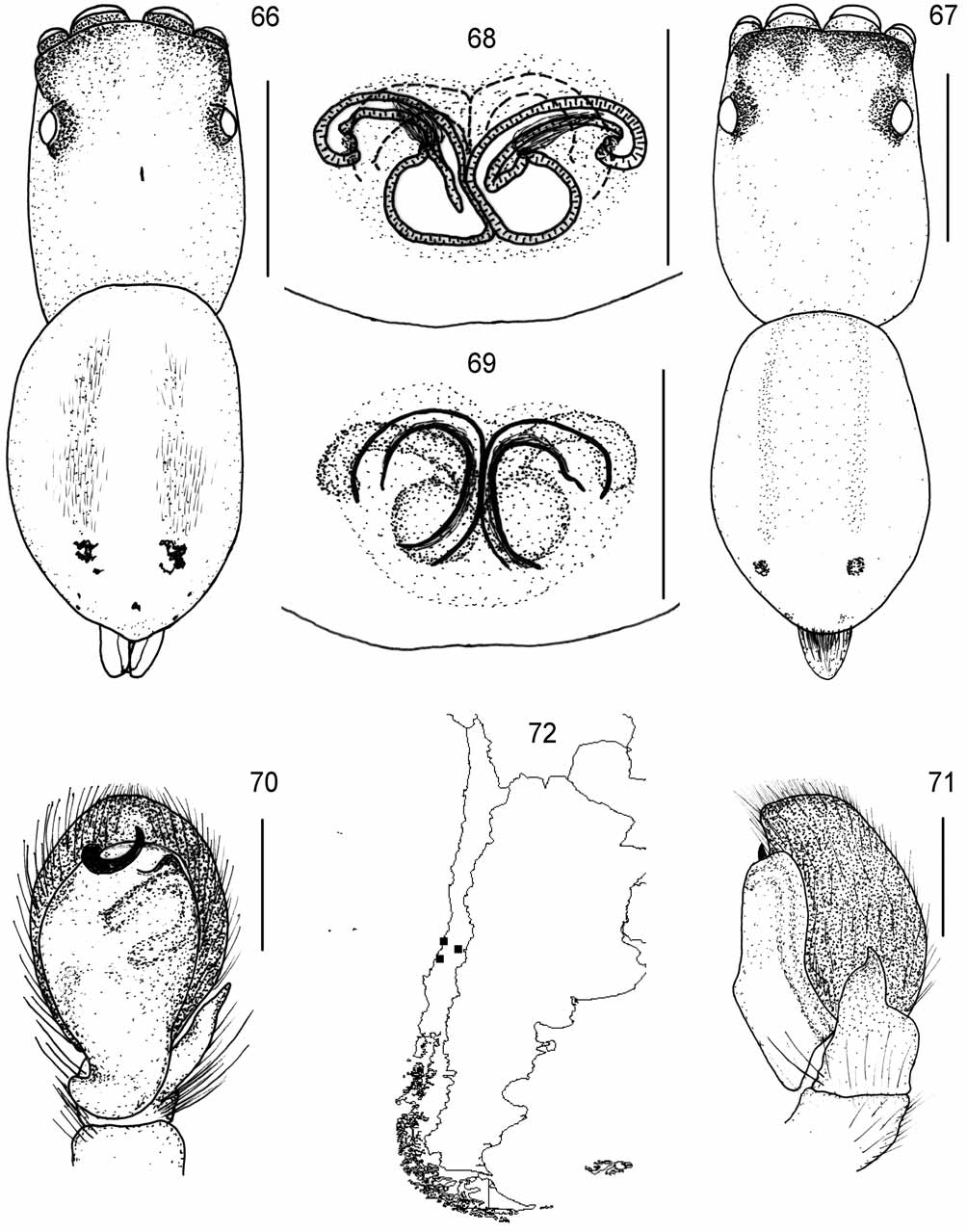
FIGURES 66 – 72. Euophrys mapuche. 66 – 67 dorsal view (66 female, 67 male); 68 – 69 female genitalia (68 dorsal view of cleared specimen, 69 ventral view of external characteristics); 70 – 71 male palp (70 ventral view, 71 lateral view); 72 known distribution. Scales: total body 1 mm; remainder 0.2 mm
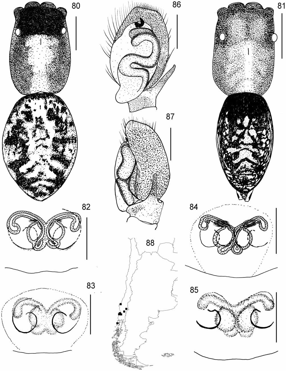
FIGURES 80 – 88. Euophrys rapida. 80 – 81 dorsal view (80 female, 81 male); 82 – 85 female genitalia (82 dorsal view of cleared E. rapida holotype, 83 ventral view of external characteristics of E. rapida holotype, 84 dorsal view of cleared E. pehuenche paratype, 85 ventral view of external characteristics of E. pehuenche paratype); 86 – 87 male palp (86 ventral view, 87 lateral view); 88 known distribution. Scales: total body 1 mm; remainder 0.2 mm
No known copyright restrictions apply. See Agosti, D., Egloff, W., 2009. Taxonomic information exchange and copyright: the Plazi approach. BMC Research Notes 2009, 2:53 for further explanation.
|
Kingdom |
|
|
Phylum |
|
|
Class |
|
|
Order |
|
|
Family |
1 (by plazi, 2016-04-17 09:34:03)
2 (by ImsDioSync, 2016-04-17 09:50:43)
3 (by ImsDioSync, 2016-04-17 10:11:16)
4 (by ImsDioSync, 2016-04-17 10:14:20)
5 (by ImsDioSync, 2016-04-24 09:11:16)
6 (by ImsDioSync, 2016-12-07 08:31:26)
7 (by ImsDioSync, 2016-12-07 08:33:04)
8 (by ExternalLinkService, 2019-09-26 14:08:17)
9 (by ExternalLinkService, 2022-01-30 16:33:35)
10 (by ExternalLinkService, 2022-02-21 14:53:29)
11 (by plazi, 2023-10-26 22:23:19)