
Neoperla
|
publication ID |
https://dx.doi.org/10.11646/zootaxa.3838.2.2 |
|
publication LSID |
lsid:zoobank.org:pub:18E650FD-3035-4C1B-967C-E72E4AE9AADF |
|
DOI |
https://doi.org/10.5281/zenodo.6133202 |
|
persistent identifier |
https://treatment.plazi.org/id/3918572F-FFC6-7369-FF68-FF2A5AB5FD21 |
|
treatment provided by |
Plazi (2016-04-12 10:58:55, last updated 2022-02-18 06:26:14) |
|
scientific name |
Neoperla |
| status |
|
Key to species of Neoperla (males) known from Henan
1. Aedeagal tube completely sclerotized...................................................................... 2
-. Aedeagal tube incompletely sclerotized.................................................................... 4
2. Aedeagal tube with ventral lobes (Fig. 5, Li et al. 2011)............................................. N. flavescens
-. Aedeagal tube without ventral lobes....................................................................... 3
3. Aedeagal sac about as long as tube, with small spines ( Fig. 4 View FIGURE 4 , Li et al. 2014).............................. N. cavaleriei
-. Aedeagal sac ca. 1.5x tube length, with linear large spines ( Fig. 1 View FIGURE 1 d).................................. N. baotianmana
4. Aedeagal tube with single ventral lobes.................................................................... 5
-. Aedeagal tube with one pair of ventral lobes................................................................ 6
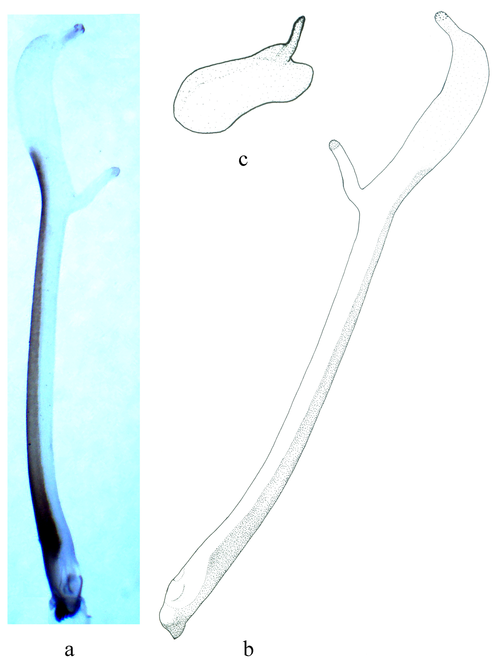
5. Aedeagal lobe finger like ( Figs. 4 View FIGURE 4 a & b)................................................. N. jigongshana sp. nov.
-. Aedeagal lobe bifurcate........................................................................ N. forcipata
6. Aedeagal tube with a pair of ventral projections in addition to lateral lobes apically.................... N. magisterchoui
-. Aedeagal tube only with a pair of ventral projections apically................................................... 7
7. Aedeagal sac lacking ventral spines (Fig. 15, Li et al. 2011); apical half with a field of fine spinules separated by a lateral membranous area (Fig. 17, Li et al. 2011)........................................................ N. yaoshana
-. Aedeagal sac with ventral spines medially; apical half evenly covered by fine lateral spinules with no membranous area ( Fig. 3 View FIGURE 3 , Li et al. 2011)................................................................................ N. henana
Li, W. H., Muranyi, D. & Wang, R. F. (2014) Species of Neoperla (Plecoptera: Perlidae) from Yunnan Province, China. Zootaxa, 3753 (1), 1 - 9. http: // dx. doi. org / 10.11646 / zootaxa. 3753.1.1
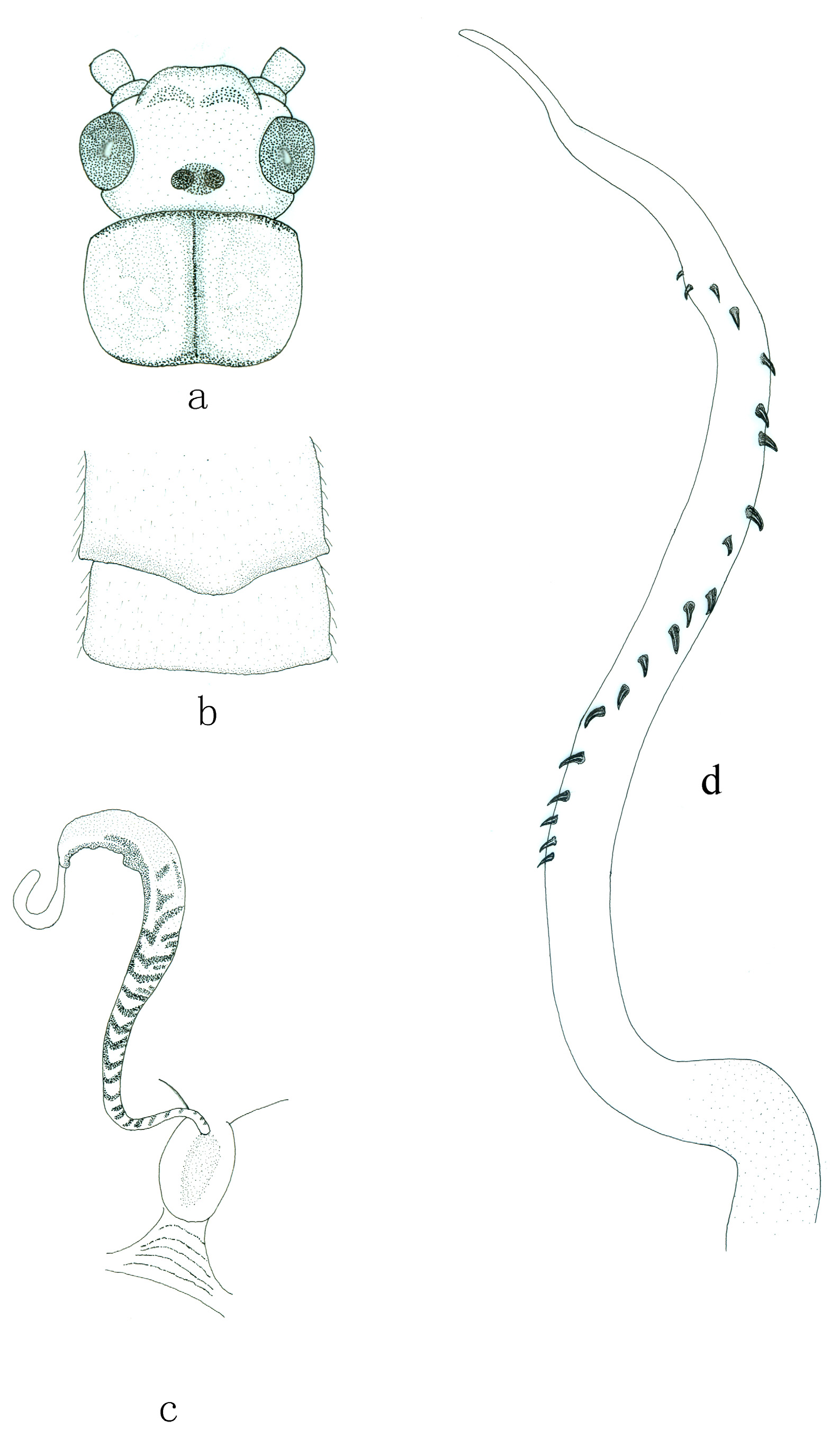
FIGURE 1. Neoperla baotianmana Li & Wang. a. Female head and pronotum, dorsal view. b. Female terminalia, ventral view. c. Vagina and spermatheca inside female terminalia. d. Male aedeagal sac, lateral view.
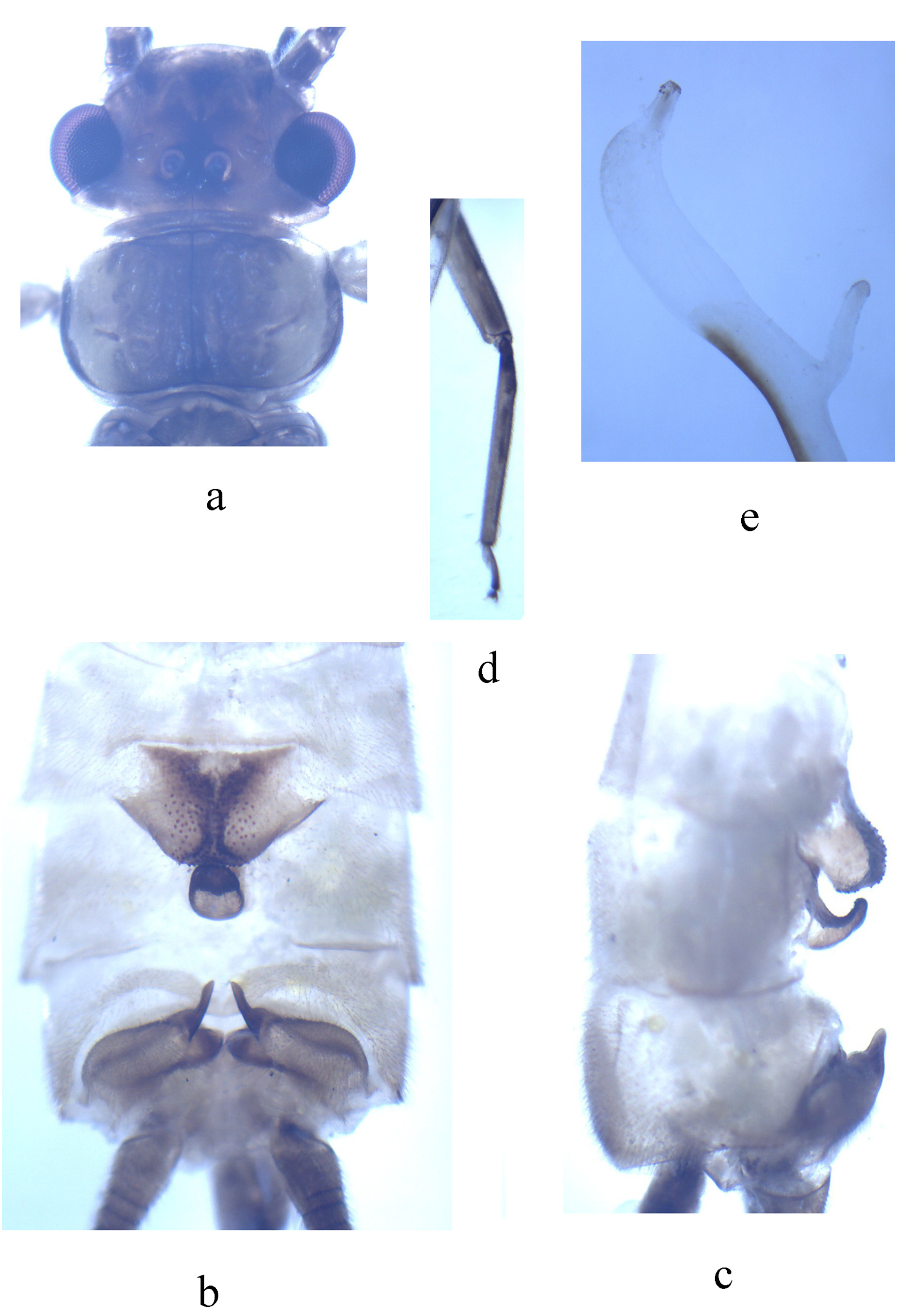
FIGURE 3. Neoperla jigongshana Li & Li, sp. nov., male. a. Head and pronotum, dorsal view. b. Terminalia, dorsal view. c. Terminalia, lateral view. d. Right midleg, dorsal view. d. Aedeagal sac, lateral view.
No known copyright restrictions apply. See Agosti, D., Egloff, W., 2009. Taxonomic information exchange and copyright: the Plazi approach. BMC Research Notes 2009, 2:53 for further explanation.
|
Kingdom |
|
|
Phylum |
|
|
Class |
|
|
Order |
|
|
Family |
1 (by plazi, 2016-04-12 10:58:55)
2 (by ImsDioSync, 2017-01-01 06:35:27)
3 (by ImsDioSync, 2017-01-01 06:36:09)
4 (by ImsDioSync, 2017-06-25 19:51:07)
5 (by ExternalLinkService, 2019-09-26 19:45:25)
6 (by ExternalLinkService, 2022-01-30 05:49:51)
7 (by ExternalLinkService, 2022-02-18 06:26:14)