
Psolidium multipes, Thandar, Ahmed S., 2006
|
publication ID |
https://doi.org/ 10.5281/zenodo.172917 |
|
DOI |
https://doi.org/10.5281/zenodo.5674068 |
|
persistent identifier |
https://treatment.plazi.org/id/1E5A87CB-0A58-577F-FF37-91E5F366F933 |
|
treatment provided by |
Plazi |
|
scientific name |
Psolidium multipes |
| status |
sp. nov. |
Psolidium multipes View in CoL sp. nov.
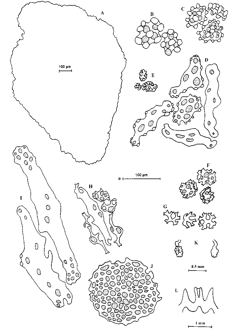
Figure 12 View FIGURE 12
Diagnosis
Small species, holotype 11 mm long; colour in alcohol white. Sole not well defined; oral valves indistinct. Tentacles?10, ventral two reduced. Anal teeth present. About 10 scales between mouth and anus, 8–9 laterally. Dorsal podia distinct, emitting through and between scales; ventral podia crowded, 4–5 rows in each ventrolateral ambulacrum and 3–4 rows in midventral ambulacrum. Dorsal scales imbricating, complex, multilayered, up to 1.5 mm long; other dorsal deposits include buttons, baskets and rosettes. Buttons knobbed, 68–84 µm, with 4–6 holes, knobs of various sizes, central ones often united by a halfring on one side; baskets mostly incomplete; rosettes minute, mulberrylike. Ventral scales also complex but smaller than dorsal ones, up to 660 µm long; other ventral deposits include knobbed buttons, up to 94 µm, with three or more holes and mostly complete baskets and rosettes.
Type
SAMA23175.
Type locality
Coconut Bay, Mozambique.
Material examined
Holotype only, collected 17.v.1973.
Description
Specimen small, length approximately 11 mm, width of midbody about 5 mm. Colour beigewhite middorsally, white elsewhere, including oral and anal ends. Dorsal surface arched, ventral flattened, but sole not clearly defined. Mouth anterior, oral valves indistinct but mouth surrounded by scales appearing as pairs in the five radii; buccal membrane distinct. Tentacles nine, bushy, white, few protruding through mouth, seven well developed, midventral two reduced. Anus situated on a dorsally directed cone, encircled by five calcareous teeth. Dorsal and lateral surfaces invested in large, imbricating scales covered with granules. About 10 scales middorsally, between mouth and anal cone, 8–9 scales laterally. Dorsal podia small, distinct, numerous, scattered, with about 2–3 emitting from each scale but most between scales, spreading to the dorsolateral radii where they are smaller and fewer. Ventral podia retracted, crowded, in 4–5 rows on each ventrolateral radius and 3–4 rows in midventral radius, increasing to five midventrally. Ventral suckers well developed, dorsal less so.
Calcareous ring ( Figure 12 View FIGURE 12 L) well calcified with radial and interradial plates simple, of more or less the same length, the former anteriorly bifid with a concavity for insertion of radial muscle; all plates with a posterior concavity but no posterior processes. Polian vesicle single, bulbous, arising ventrally; stone canal short, only slightly coiled; madreporite ( Figure 12 View FIGURE 12 K) poorly calcified, lodged in the posterior notch of middorsal interradial plate. Respiratory trees delicate, well branched, both trees extending to anterior end. Gonad (testis) as two tufts of unbranched, white, sausagelike tubules, filling most of body cavity; gonadal stolon short, lodged in dorsal mesentery. Longitudinal muscles unpaired, retractor muscles arise from anterior third of longitudinal bands.
Dorsal scales ( Figure 12 View FIGURE 12 A) large, up to 1.5 mm long, complex, multilayered, those surrounding mouth elongated, specialised, up to 1.3 mm long; other dorsal deposits include buttons, baskets and rosettes. Buttons of three types: robust ones, 84–113 µm (mean 95 µm), pierced by 4–6 holes, with large knobs and with central knobs developed as a halfring; other robust buttons, 68–84 µm (mean 76 µm), perforated by usually four (sometimes 2–5) holes, also with large knobs but without halfring; remaining buttons small, ca. 70 µm, fewer, small knobbed and usually perforated by four holes and with varying number of knobs. Baskets in the form of spinose, often cuplike crosses ( Figure 12 View FIGURE 12 G), with spines usually restricted to the branches; complete baskets not detected amongst dorsal deposits. Rosettes minute, mulberrylike. Ventral deposits also comprise complex, multilayered scales, much smaller (194–660 µm, mean 395 µm) than the dorsal ones, and buttons, baskets and rosettes. Buttons usually smallknobbed ( Figure 12 View FIGURE 12 C), 45–74 µm long (mean 57 µm), with four or more holes and a varying number of knobs; other knobbed buttons fewer but slightly larger (58–94 µm, mean 73 µm), with large knobs and three or more holes ( Figure 12 View FIGURE 12 B). Ventral baskets (26–48 µm, mean 38 µm) mostly complete ( Figure 12 View FIGURE 12 F), with spinose margins and surface, few may be incomplete and similar to those of dorsal body wall. Ventral rosettes ( Figure 12 View FIGURE 12 E) 16–39 µm (mean 25 µm), similar to those of dorsal body wall but commoner. Podial deposits ( Figure 12 View FIGURE 12 D) comprise marginallyknobbed, perforated plates and smooth rods (ventrally 87–184 µm, mean 130 µm; dorsally 74–110 µm, mean 91 µm); dorsal endplates reduced, ventral endplates ( Figure 12 View FIGURE 12 J) larger (77 µm cf. 187 µm). Tentacle branches with minute plates and perforated rods of varied shapes and sizes (36–187 µm, mean 93 µm) ( Figure 12 View FIGURE 12 H), as well as both open and closed rosettes (10–29 µm, mean 17 µm). Tentacle stalks supported by larger rods (265–426 µm, mean 349 µm)( Figure 12 View FIGURE 12 I). Deposits of the buccal membrane comprise largeknobbed rods (ca 185 µm), perforated with several holes, baskets similar to those of the dorsal body wall but smaller (26–29 µm, mean 27 µm), and closed rosettes (13–26 µm, mean 20 µm). No spicules detected in gonad.
Distribution
Type locality only.
Remarks
In the presence of dorsal podia emitting through or between scales, the distribution of the ventral podia and the presence of four distinct types of body wall deposits besides the scales, this species appears unique. Its body wall deposits come quite close to Psolidium ornatum (Ed. Perrier, 1893) , redescribed by Cherbonnier (1988) from designated neotype, but the new species differs in its distribution of podia. Cherbonnier (1988) describes ventrolateral ambulacra with two rows of podia and the midventral ambulacrum with three rows. Further, in P. o r n a t u m the plates of the calcareous ring appear separate while in the new species, they are partially fused. Hence, the new species appears to be very close to P. ornatum but not identical with it. The only other species where the podia are arranged in crowded bands is P. nigrescens Clark, 1938 from southeast Australia and P. dorsipes Ludwig (1887) from Chile and Patagonia. However, the dorsal deposits of the former species are stated to be scarce and also include numerous triradiate bodies. Such bodies are also found in the sole together with knobbed, perforated plates unlike those found in the new species. In P. dorsipes , on the other hand, there are towerlike deposits in the dorsal body wall and the baskets are of a different kind.
No known copyright restrictions apply. See Agosti, D., Egloff, W., 2009. Taxonomic information exchange and copyright: the Plazi approach. BMC Research Notes 2009, 2:53 for further explanation.