
Zoothamnium hiketes Precht, 1935
|
publication ID |
https://doi.org/ 10.1080/00222930500239785 |
|
persistent identifier |
https://treatment.plazi.org/id/03DD1129-F01D-FFDA-2FCD-23165E9AFCF3 |
|
treatment provided by |
Felipe |
|
scientific name |
Zoothamnium hiketes Precht, 1935 |
| status |
|
Zoothamnium hiketes Precht, 1935 View in CoL
( Figures 19–38 View Figures 19–26 View Figures 27–38 ; Table I) Zoothamnium hiketes was originally described by Precht (1935) from the North Sea, Germany. No further studies have been carried out since then and hence neither the infraciliature nor silverline system is clear. Based on the Qingdao population and previous studies, an updated diagnosis and a detailed redescription are presented here.
New diagnosis
Marine Zoothamnium with dichotomously branched stalk. Zooids elongate, measuring about 40–80× 30–40 mm, with fine pellicular striations. Peristomial lip double-layered. Single contractile vacuole apically located. Macronucleus generally C-shaped and transversely orientated. Number of transverse lines from oral area to aboral ciliary wreath 89–109, from aboral ciliary wreath to scopula, 35–43.
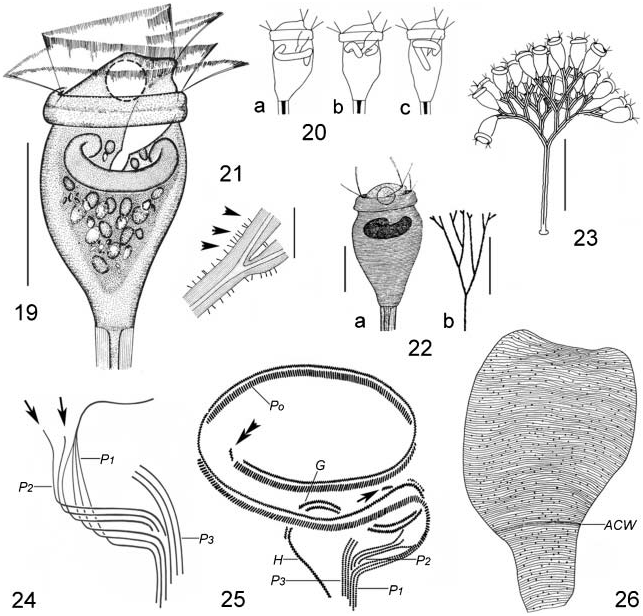
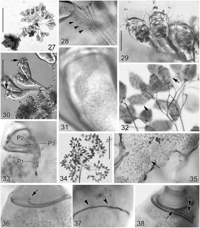
Description of Qingdao population Zooids usually elongated vase-shaped, in vivo about 60–80× 30–40 mm, and only slightly constricted below the thick peristomial lip, which is inconspicuously double-layered; large peristomial disc highly elevated ( Figures 19 View Figures 19–26 , 29 View Figures 27–38 ).
Pellicle smooth when observed at low magnification, fine striations recognizable under high magnification (×400 or higher) ( Figure 31 View Figures 27–38 ).
Cytoplasm colourless or slightly greyish, usually contains several to many large granules (4–8 mm in diameter) ( Figures 19 View Figures 19–26 , 29 View Figures 27–38 ). One large contractile vacuole apically located, contracting every 4 min ( Figure 19 View Figures 19–26 ; Figure 30 View Figures 27–38 , arrows). Macronucleus band-like, generally C-shaped, transversely orientated ( Figures 19, 20 View Figures 19–26 ); micronucleus not observed.
Colony dichotomously branched with zooids located regularly in pairs ( Figures 23 View Figures 19–26 , 27 View Figures 27–38 ), consisting of up to 100 zooids in a large colony ( Figure 34 View Figures 27–38 ). Stalk up to 400 mm long, diameter about 12 mm in main stalk and 6–8 mm in accessory branches. Surface of stalk smooth and often covered with rod-shaped bacteria (about 1.2–1.8 mm in length) ( Figures 21 View Figures 19–26 , 28 View Figures 27–38 , arrowheads). Spasmoneme about 2 mm in diameter, without visible mitochondria (thecoplasmic granules).
Oral infraciliature as shown in Figures 24, 25 View Figures 19–26 , 33, 36, 38 View Figures 27–38 , haplo- and polykinety circling about 1.5 turns around peristomial disc, and making a further turn after entering vestibulum. Near distal end of haplo- and polykinety, always one short kinety fragment recognizable ( Figure 25 View Figures 19–26 , double arrowhead; Figure 36 View Figures 27–38 , arrow).
At lower half of vestibulum, polykinety transforming into three peniculi (P1–3), each of which consists of three rows. P1 longer than other two, extending to cytostome. P2 interposes between P1 and P3 and ends at curvature of P1. Rows in P3 are parallel to each other ( Figure 33 View Figures 27–38 ). Germinal kinety parallel to haplokinety within upper half of vestibulum ( Figure 25 View Figures 19–26 ; Figure 38 View Figures 27–38 , double arrowhead). Epistomial membrane located at opening of oral cavity ( Figures 25 View Figures 19–26 , 38 View Figures 27–38 , arrow). Aboral ciliary wreath formed by probably double-rowed kineties ( Figure 26 View Figures 19–26 ; Figures 32, 35 View Figures 27–38 , arrow; Figure 37 View Figures 27–38 , arrowheads).
Silverline system genus typical, 89–109 striations between peristomial area and aboral ciliary wreath, 35–43 striations from aboral ciliary wreath to scopula, with many sparsely distributed pellicular pores ( Figures 26 View Figures 19–26 , 35 View Figures 27–38 ).
Comparison
Zoothamnium hiketes Precht, 1935 View in CoL was originally found on the seta of Gammarus species as epizoons, and the brief description concerned only the morphology of live cells ( Precht 1935; Figure 22a, b View Figures 19–26 ). Since then, no redescriptions have been made. In the absence of data concerning the infraciliature and silverline system, we identified our organism mainly on the basis of its body shape, appearance of the thick peristomial lip (inconspicuous doublelayered), position of contractile vacuole, branching form, and marine habitat. The only difference is the size of the zooid. According to the original description, the zooid of Z. hiketes View in CoL is smaller, about 40–55 mm in length, while in the Qingdao population the length is about 60–80 mm. Since the size of the organism, to the authors’ knowledge, is usually a population-dependent feature in most cases, we consider this difference as an intra-species variation.
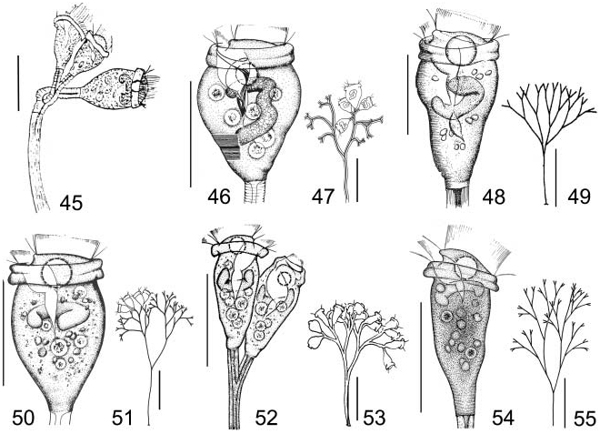
Morphologically, Zoothamnium cienkowskii Wrzesniowski, 1877 View in CoL ( Figure 45 View Figures 45–55 ; Table III) is similar to Z. hiketes View in CoL in vivo. Unfortunately, neither the infraciliature nor silverline system of the former has been described. However, Z. cienkowskii View in CoL can be distinguished at least by the appearance of the stalk (transversely wrinkled at accessory branch versus smooth) and differentiation of the zooids (having enlarged zooids versus all zooids about equal size) ( Kahl 1935).
Zoothamnium affine sensu Song, 1991 ( Figures 46, 47 View Figures 45–55 ) whose infraciliature also remains unknown, matches in some aspects Z. hiketes , e.g. in body size, in branching form and in some other morphometric features (Table III). However, the former can be identified by having fewer silverlines between oral area and aboral ciliary wreath (75–81 versus 89–109) and a cross-striated stalk (versus smooth) ( Song 1991b).
Another similar form, Zoothamnium maximum Song, 1986 ( Figures 48, 49 View Figures 45–55 ; Table III) differs from Z. hiketes in: (1) the larger size (80–120 versus 40–80 mm); (2) more silverlines between aboral ciliary wreath and scopula (45–58 versus 35–43); (3) the appearance of P3 (the outer row loosely ciliated, about two-thirds of the other two in length versus inner row slightly longer than the other two) ( Song 1986; Ji and Song 2004).
Zoothamnium duplicatum Kahl, 1933 ( Figures 50, 51 View Figures 45–55 ; Table III) also exhibits a similar size and dichotomous branching pattern, but differs from Z. hiketes by the following features: (1) significantly fewer silverlines between peristomial area and aboral ciliary wreath (48–53 versus 89–109); (2) the form of P3 (outer two rows close set, with their upper halves separated widely from the inner one versus three rows parallel to each other) ( Ji et al., 2005).
Zoothamnopsis mengi Song, 1997 View in CoL ( Figures 52, 53 View Figures 45–55 ) and Zoothamnopsis sinica Ji and Song, 2004 View in CoL ( Figures 54, 55 View Figures 45–55 ) might also be similar to the present form (Table III). However, they differ from Z. hiketes View in CoL by the different silverline system ( Pseudovorticella View in CoL - type versus Vorticella View in CoL - type) and the structure of P3 ( Song 1997; Ji and Song 2004).
No known copyright restrictions apply. See Agosti, D., Egloff, W., 2009. Taxonomic information exchange and copyright: the Plazi approach. BMC Research Notes 2009, 2:53 for further explanation.
|
Kingdom |
|
|
Phylum |
|
|
Class |
|
|
Order |
|
|
Family |
|
|
Genus |
Zoothamnium hiketes Precht, 1935
| Sun, Ping, Song, Weibo, Ji, Daode & Hu, Xiaozhong 2005 |
Zoothamnopsis sinica
| Ji and Song 2004 |
Zoothamnopsis mengi
| Song 1997 |
Pseudovorticella
| Foissner and Schiffmann 1974 |
Zoothamnium hiketes
| Precht 1935 |
Z. hiketes
| Precht 1935 |
Z. hiketes
| Precht 1935 |
Z. hiketes
| Precht 1935 |
Zoothamnium cienkowskii
| Wrzesniowski 1877 |
Z. cienkowskii
| Wrzesniowski 1877 |