
Trachycephalus mambaiensis, Cintra, Carlos Eduardo Domingos, Silva, Hélder Lúcio Rodrigues, Jr, Nelson Jorge Da Silva, Garcia, Paulo Christiano De Anchietta & Zaher, Hussam, 2009
|
publication ID |
https://doi.org/ 10.5281/zenodo.185123 |
|
DOI |
https://doi.org/10.5281/zenodo.5670654 |
|
persistent identifier |
https://treatment.plazi.org/id/570887B4-BC24-FF86-A1F2-FADAFA74EEE8 |
|
treatment provided by |
Plazi |
|
scientific name |
Trachycephalus mambaiensis |
| status |
sp. nov. |
Trachycephalus mambaiensis View in CoL sp. nov.
Figs. 1–4 View FIGURE 1 View FIGURE 2 View FIGURE 3 View FIGURE 4
Holotype. MZUSP 135715, an adult male ( Fig. 1 View FIGURE 1 ) from the vicinity of Santa Edwiges I hydroelectric power plant dam, Municipality of Mambaí, State of Goiás, Brazil (14°17'24''S 46°11'37''W or UTM 0 371233 and 8419860) ( Fig. 2 View FIGURE 2 ), collected by Carlos Eduardo Domingos Cintra on 2 August 2006.
Paratypes. Two adult females ( MZUSP 135713, 135716) and two adult males ( MZUSP 135714, 135717) ( Fig. 3 View FIGURE 3 ) from the vicinity of the Santa Edwiges I hydroelectric power plant, Municipality of Mambaí, State of Goiás, collected by Carlos Eduardo Domingos Cintra on 31 July and 17 August 2006 ( Fig. 2 View FIGURE 2 ); two adult males ( CHUNB 21868, 35471) from the municipality of São Domingos, State of Goiás, collected by Laurie J. Vitt and Janalee P. Caldwell between 19 November and 15 December 2003.
Diagnosis. The species is included in the genus Trachycephalus which present paired vocal sacs protruding posterior to the angles of the jaws when inflated as a unique synapomorphy (fide Trueb and Duellman, 1971; Tyler, 1971; Faivovich et al., 2005). A medium-sized species of Trachycephalus (SVL in males 76.4–82.0 mm; females 83.67–86.71 mm) characterized by the combination of the following traits: presence of paired vocal sacs protruding posterior to the angles of the jaws when inflated; a rounded snout without a medial (premaxillary) notch in dorsal view and truncate in lateral view; skin co-ossified with the skull; frontoparietals converge medially throughout their lengths, covering the fontanelle; frontoparietals not covering the prootics posteriorly; frontoparietals not articulating with squamosals; presence in life of small reddish spots sparsely disposed on the dorsum.
The new species differs from T. coriaceus , T. hadroceps , T. imitatrix , T. lepidus , T. mesophaeus , T. resinifictrix , and T. venulosus by the presence of a well ossified skull in which the skin is adhered, and from T. atlas , T. jordani , and T. nigromaculatus by frontoparietals that fail to cover the prootics posteriorly and by the lack of an occipital crest and medial premaxillary notch. Further, the new species differs from all ten species of Trachycephalus (except T. nigromaculatus ) by the presence of small reddish spots on the dorsum.
Description of the holotype. Body robust; head narrower than the body, as wide as long ( Fig. 1 View FIGURE 1 ); top of the head flat, skin visibly adhered to the skul; snout short, rounded in dorsal profile and nearly truncate in lateral profile; canthus rostralis rounded distinct; loreal region barely concave; lips moderately thin and flared; nostrils protuberant, directed laterally; distance of nostril to tip of snout smaller than internarial and eye-nostril distances; internarial region slightly depressed; eyes lateral, directed anteriorly, moderate size; eyes diameter being close to 1/4 of head length; pupil horizontal; tympanum distinct, large, its diameter approximately four-fifth of the eye diameter; supratympanic fold extends posteriorly from the posterior corner of the eye, above the tympanum. Vocal sacs paired and lateral, behind the angle of the jaw; vocal slits large, located laterally under the tongue; tongue large, cordiform, covering approximately 2/3 of the mouth floor, free and notched posteriorly; dentigerous processes of prevomer slightly curved in a transverse plane, each one bearing eigth and seven teeth. Arms short, moderately robust; without axillary membrane and distinct ulnar fold; hands large, length slightly smaller than head length ( Fig. 4 View FIGURE 4 A); fingers medium sized and moderately robust; finger discs large; width of the disc on the third finger smaller than the tympanum diameter; subarticular tubercles single, large and oval on finger I-IV; outer palmar tubercle large, bifid; inner palmar tubercle large, elliptical; numerous palmar supranumerary tubercles; finger disks large, nearly rounded; relative finger lengths: I <II <IV <III; fingers slightly webbed; webbing formula: I – II 2 - - 3+ - 2+ IV. Legs short, robust; thighs slightly smaller than mead of SVL; feet relatively small, about 1/3 of SVL; robust toes; toe discs large ( Fig. 4 View FIGURE 4 B); relative toe lengths: I <II <V ˜ III <IV; foot webbing formula: I 1 1/2 – 2 + II 1 1/3 – 2 1/2 III 1 1/ 2– 3- IV 2 1/2 – 1+ V; inner metatarsal tubercle larger, elliptic, visible from dorsal view; outer metatarsal tubercle large, bifid. Toes moderately short; toe discs slightly smaller than those on fingers; subarticular tubercles moderately large, subconical; small supernumerary metatarsal tubercles; inner tarsal fold absent. The dorsal skin texture is finely granular; lateral and undersurface skin texture smooth.
Variation. Individuals of Trachycephalus mambaiensis show some variation in size ( Table 1 View TABLE 1 ), development of toe, finger webbing, and pattern of dorsal blotches. The holotype is the largest male of the series. Females are slightly larger than males, have larger fingers and toes, more extensive webbing.
Color in life of the holotype. Dorsum dark brown with several irregular light brown or creamy blotches. There are two wide and long, dorsolateral creamy blotches that extend from the scapular to the sacral region; some small, round red-cooper blotches randomly distributed on the dorsum and flanks; several large, round creamy blotches on a brown-grayish background on the flanks; dorsal surfaces of the anterior and posterior limbs show creamy transversal bars on a dark brown background; dorsum of fingers and toes yellowish; ventral surface of the body is creamy, with irregular gray spots disposed mostly in the gular region and flanks. Iris golden and finely reticulated with black. If not inflated, vocal sacs vary from dark brown to black.
Color in preservative of the holotype. Dorsum brownish-gray with creamy blotches; lighter surfaces of the venter and flanks grayish white (small red blotches on the body in life are not noticeable). Dorsal surfaces of fingers and toes without any differential coloration related to the limbs.
Cranial osteology. (based in an adult female paratype MZUSP 135713; Fig. 5 View FIGURE 5 ) skull of T. mambaiensis slightly wider than long (cranium length 24.22mm; cranium width 25.05mm; cranium height 8.45mm). Snout in dorsal view truncate and bears a poor medial notch; all dorsal surfaces of the dermal bones sculptured into rugosities and irregular ridges; distal margins of the dermal bones rugose or slightly spinose; premaxillaries robust and lie anteromedially to the maxillaries; left and right pars dentalis of the premaxillae bear 11 and 13 teeth, respectively; alary processes of the premaxillaries slightly expanded and co-ossified anteriorly, slightly concave posteriorly and oriented vertically; it approach but not articulate with the anterior tip of the nasal anterodorsally; dorsomedially, the alar processes form part of the anterior margin of the external nares; laterally, the alary processes approach but not to articulate with the pars facialis of the maxillary, whereas medially articulate with one another; the pars palatina of the premaxillary developed and the posteromedial palatine process conspicuous. Maxillary moderately robust, but lacks a labial flange; the pars facialis well developed, it is larger anteriorly to the orbit, and articulates with the alary process of the premaxillary to form the ventral margin of the external naris; posterior to the external naris, the dorsal edge of the pars facialis is free, and articulates with the maxillary process of the nasal at the anterior margin of the orbit; the pars facialis diminishes in size ventrally to the orbit, but is conspicuous along the entire length of the maxillary; the pars palatina of the maxillary well developed and extends from the anterior end of the bone posteriorly to the level of termination of the pars dentalis; posteriorly, the maxillary firmly articulates with the quadratojugal which lies medially, adjacent to the maxillary; the outer surface of the maxillary is co-ossified and slightly rugose, principally in the anterior part; each maxillae bears approximately 70 pedicillate teeth. Quadratojugal increases in size posteriorly, whereas the maxillary diminishes in size; posteriorly, ossification of the quadratojugal invades the cartilage of the quadrate process.
Measurements Holotype Males including the Holotype (n = 7) Females
MZUSP 135715 ± SD (Range) (n = 2) In ventral view, the paired vomer lies lateral to the midline of the skull and ventral to the sphenethmoid; anterior end of vomer contacts the premaxillary-maxillary suture dorsolaterally; vomer bears well developed lateral wings which form the anterior and medial margins of the internal naris; vomers converge medially just anterior to the vomerine teeth; the dentigerous processes slightly curved in a transverse plane, each one bearing nine teeth; vomer not contact the neopalatines; paired neopalatine long, robust, and bears a ventral ridge without odontoids; the distal end of the neopalatine embedded in connective tissue and lies dorsally to the pars palatina of the maxillary and adjacent to the anterior maxillary process; proximal end of the neopalatine pointed and lies ventral to the sphenethmoid and posterior to the prevomer, without a medial contact; parasphenoid robust and tetraradiate; acuminate anterior end lies slightly posterior to the level of the palatines, ventrally to the sphenethmoid; lateral alae approximately half the length of the cultriform process; lateral alae invest medially to the prootic; posteriorly, parasphenoid extends to the edge of the foramen magnum. In dorsal view, nasals large; anterior end of the nasal blunt and articulates with the dorsal margin of the alary process of the premaxilla; laterally, the nasal arches over the external nares and articulates posteriorly and ventrolaterally with the pars facialis of the maxillary at the corner of the orbit; the nasal forms the anterior margin of the orbit; articulates with the frontoparietal dorsolaterally and the dorsally exposed dermal sphenethmoid dorsomedially; the nasals converge medially anterior to the dermal sphenethmoid; canthal ridge distinct, not pronounced; it extends from the anterior end of the nasal along the lateral margin to the anterodorsal corner of the orbit; entire nasal surface involved in the integumentary-cranial co-ossification; surface of the nasal rugose externally and marked by prominent spines internally; frontoparietals converge medially throughout their length, without exposing the fontanelle; anteriorly, the frontoparietal articulates with the nasal laterally and the dermal sphenethmoid medially and anterolaterally, forming a slight supraorbital shelf; posterolaterally frontoparietal poorly developed and not articulate with the squamosal; posteriorly frontoparietal not form an occipital crest covering the prootic; entire dorsal surface of the frontoparietal involved in integumentary cranial co-ossification. Medially, the frontoparietal is rugose; irregular bony ridges radiate out from the central rugosity toward the distal margins of the bone; dermal sphenethmoid triangular, centrally located at the anterior level of the orbit; sphenethmoid lies posteromedial to nasals and anteromedial to the frontoparietals; dermal sphenethmoid is completely involved in integumentary-cranial co-ossification and its dorsal surface slightly rugose. Exoccipital and prootic completely fused, lacking a visible suture; anterodorsally exoccipitals partially covered by frontoparietals; latter form the posterior border of the optic capsule; anterolaterally and laterally, its crista parotica is in contact with the otic plate of the squamosal; dorsal and lateral surfaces of the squamosal co-ossified. slightly rugose; zygomatic ramus robust and forms a part of the posterior margin of the orbit, it is connected to the maxillary and anterior rami of the pterygoid; optic ramus of squamosal poorly developed, smaller than the zygomatic ramus and not extend posteriorly to the crista parotica; zygomatic ramus articulates with the crista parotica, not with the frontoparietal; ventral arm of the squamosal moderately robust; ventrally, it terminates between the quadratojugal laterally and the pterygoid medially, and dorsal to the quadrate process. Pterygoid moderately robust; the anterior ramus lies adjacent to the posterior maxillary process at a level just posterior to the orbitonasal foramen; medial ramus short, poorly developed and not articulate with the prootic medially; posterior ramus robust and diverges medially from the maxillary to articulate with the quadrate process and ventral arm of the squamosal posteriorly. Quadratojugal well developed; anterior end lies medially, adjacent to the maxillary at the level of the optic foramen; columella appears between the levels of the prootic and anterior acoustic foramen.
Comparison with other species. The skull of T. mambaiensis is similar to T. nigromaculatus ( Trueb, 1970, plate 6a and 6b), differing from the latter by the less ossified conditions of the nasals, premaxillaries, and especially frontoparietals. The small development of the frontoparietals, failing to cover the prootics posteriorly, is a characteristic that easily differentiates the new species from the other species of the genus with ossified skulls ( Bokermann, 1966; Trueb, 1970). Trachycephalus mambaiensis can be further distinguished from the the remaining species of Trachycephalus (all previously included in the genus Phrynohyas ) by the presence of a dermal sphenethmoid and by the less developed labial flange (see Trueb, 1970, Table 1 View TABLE 1 ). The overall characteristic of the skull of T. mambaiensis is intermediary between the condition shown by the species of Trachycephalus with an ossified skull and the ones without dermal ossifications ( Fig. 6 View FIGURE 6 ).
Distribution. The species is currently known only from the type-locality. However, it is possible that its distribution includes the upper rio Corrente region and its tributaries where several large fragments of natural Cerrado and gallery forests are still well preserved.
Etymology. A specific epithet in allusion to the municipality of Mambaí, where the species was collected.


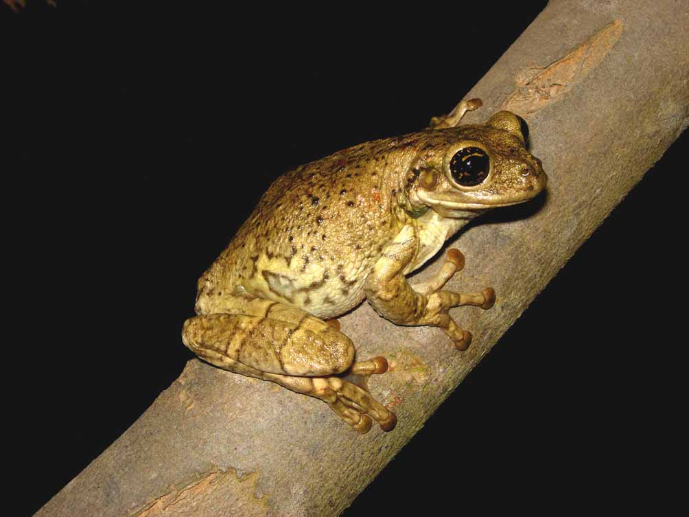

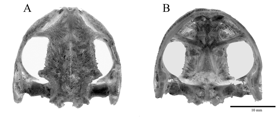
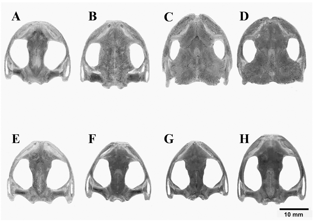
TABLE 1. Measurements (mm) and body ratios (%) of Trachycephalus mambaiensis:, arithmetic mean; SD, standard deviation.
| SVL HL | 82.03 24.84 | 78.64 ± 2.43 (76.4–82.0) 24.52 ± 0.86 (23.1–25.4) | 83.67–86.71 28.17–29.19 |
|---|---|---|---|
| HW | 24.8 | 24.47 ± 0.95 (23.1–25.7) | 26.83–27.8 |
| ED TD | 6.43 5.35 | 6.55 ± 0.46 (6.0–7.1) 5.25 ± 0.15 (5.0–5.4) | 7.21–7.47 5.92–6.14 |
| END | 6.98 | 6.84 ± 0.61 (6.1–7.8) | 8.50–8.81 |
| NSD IND | 3.32 5.13 | 3.16 ± 0.18 (2.9–3.3) 4.96 ± 0.20 (4.7–5.1) | 3.47–3.35 5.13–5.32 |
| IOD | 10.39 | 10.43 ± 0.56 (9.7–11.1) | 11.22–11.63 |
| FAL HAL | 12.19 23.7 | 12.35 ± 0.80 (11.4–13.4) 23.43 ± 0.84 (22.1–24.2) | 14.05–16.87 25.35–26.27 |
| THL | 35.76 | 35.30 ± 1.36 (33.3–37.1) | 40.04–41.68 |
| TL TAL | 35.61 19.45 | 35.22 ± 1.39 (33.2–37.0) 18.88 ± 0.61 (18.1–19.5) | 39.9–42.22 20.25–21.01 |
| FL | 30.65 | 30.44 ± 0.58 (29.5–31.0) | 33.93–34.33 |
No known copyright restrictions apply. See Agosti, D., Egloff, W., 2009. Taxonomic information exchange and copyright: the Plazi approach. BMC Research Notes 2009, 2:53 for further explanation.