
Tomocerus similis Chen & Ma, 1997
|
publication ID |
https://doi.org/ 10.11646/zootaxa.4268.3.5 |
|
publication LSID |
lsid:zoobank.org:pub:517459CE-D20D-48CB-9F1B-3FDEA30F31E3 |
|
DOI |
https://doi.org/10.5281/zenodo.6023029 |
|
persistent identifier |
https://treatment.plazi.org/id/03DEBC14-AA6C-4C41-27CB-4466E42D2761 |
|
treatment provided by |
Plazi |
|
scientific name |
Tomocerus similis Chen & Ma, 1997 |
| status |
|
Tomocerus similis Chen & Ma, 1997
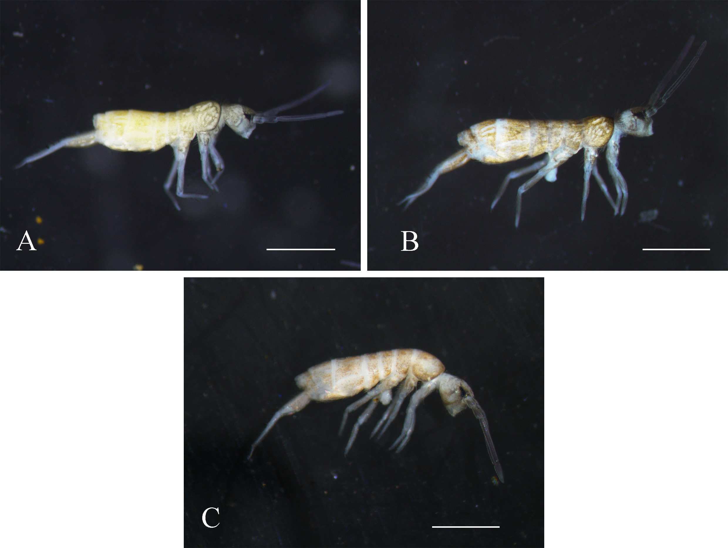
Figs 1 View FIGURE 1 A, 2, 3
Material examined. Holotype female adult on slide. Langya Mountain , Chuzhou (previously Chuxian County), Anhui Province, China, 8.iv.1990 (No. 8040). Other material: topotypes, one female and two male adults on slides, 2 in alcohol . Langya Mountain , Chuzhou, Anhui Province, China, 118°16'34.66"E, 32°17'5.17''N, alt. 110m, 30.viii.2015, by Daoyuan YU (No. 2016LY). One topotype in alcohol in NTU, other types in NJAU GoogleMaps .
Description. Body length 2.2–2.5mm. Body ground colour light yellow to orange, with diffuse dark pigment on head, thorax and legs. Anterior area of head and Th. II darker. Antennae bluish purple. Eye patches black. ( Fig. 1 View FIGURE 1 A).
Antennae short, 0.45–0.5 times as long as body. Length ratio of Ant. I:II:III:IV= 1.0:1.4–1.7:4.8–6.1:1.5–1.9. Dorsal side of Ant. I and Ant. II scaled, Ant. III and Ant. IV unscaled. PAO not seen. Eyes 6+6. Labral formula 4/ 5, 5, 4. Distal edge of labrum with four curved spines. Mandibular head asymmetrical, left one with 4 teeth and the right one with 5, left molar plate distally with a tapered tooth ( Fig. 2 View FIGURE 2 A). Maxillary lamella 5 without beard-like appendage. Maxillary outer lobe with trifurcate palp, one basal chaeta and 4 sublobal hairs. Both dorsal and ventral sides of head scaled. Cephalic dorsal macrochaetotaxy: anterior area: 2, 2; interocular area: 2, 6, central unpaired macrochaeta absent; postocular area: 2+2; posterior area: 0. Posterior margin of head with approximately 30+30 small chaetae ( Fig. 2 View FIGURE 2 B). Mentum with 5 chaetae, submentum with numerous chaetae.
Pattern of body chaetotaxy as in Fig. 2 View FIGURE 2 C. Bothriotricha 1, 1/ 0, 0, 1, 2, 0, 0 on Th. II–Abd. VI. Macrochaetae densely arranged along anterior margin of Th. II (not shown in figure). Th. II with an irregular row of macrochaetae behind anterior margin. Number of macrochaetae or large mesochaetae in the posterior row as 3, 3/ 3, 3, 4, 3, 4 from Th. II to Abd. V. Th. II with four central and one lateral macrochaetae, postero-central one near pseudopore; Th. III with anterior macrochaeta; Abd. III with two anterior macrochaetae; Abd. IV with one antero-lateral macrochaeta; Abd. VI with numerous chaetae of different sizes. Mesochaetae most abundant laterally and posteriorly on terga. Pseudopores near the axis of terga, 1, 1/ 1, 1, 1, 1, 0, 0 from Th. II to Abd. VI.
Trochantero-femoral organ with 1, 1 small slender chaetae ( Fig. 2 View FIGURE 2 D). Anterior, mid and hind tibiotarsi ventrally with 0, 0, 2–3 spine-like chaetae ( Fig. 2 View FIGURE 2 E). Each tibiotarsus with a distal whorl of 11 chaetae, ventral 6 as ordinary chaetae, dorsal 5 modified: tenent hair clavate on all legs, fine and short, about 0.6–0.75 times as long as inner edge of unguis; two accessory chaetae small, subequal to or slightly longer than pretarsal chaetae; two guard chaetae strong, subequal to or longer than tenent hair, about 0.85 times as long as unguis. Unguis slender, with baso-internal ridges about 1/3 distance from base; lateral teeth pointed, relatively small. Inner edge of unguis with distinct basal tooth, large sub-basal tooth, and 2–3 obscure distal teeth. Unguiculus lanceolate, about 0.5–0.65 times as long as unguis, inner edge with 0–2 small tooth. Pretarsus chaetae 1+1 ( Fig. 2 View FIGURE 2 F).
Ventral tube scaled only on anterior face. Anterior face with 15–20 chaetae on each side, posterior face with 40–65 chaetae, each lateral flap with 35–50 chaetae. Rami of tenaculum with 4+4 teeth, anterior face with 1 small chaeta and without scales ( Fig. 3 View FIGURE 3 A). Ratio manubrium:dens:mucro=2.2–2.8:3.0–3.3:1.0. Manubrium ventrally scaled and normally without chaetae, one specimen with two basal small chaetae; laterally with large round scales and 8–10 chaetae on each side, proximal 1–3 chaetae small, distal ones strong; dorsally with two longitudinal areas each with approximately 100 chaetae of different sizes; each chaetal area with a inner longitudinal row of scales, running from base to the middle of manubrium; prominent chaetae 2+2, slender and pointed; pseudopores 5–9 on each side ( Fig 3 View FIGURE 3 B); disto-external corner of manubrium with a microchaeta (manubrial distal corner chaeta, Fig. 3 View FIGURE 3 C). Dens basally with a pointed prominent dorsal chaeta, without large modified inner scale or strong outer chaetae. Dental spine formula as 3–4/1, II; all spines basally with several moderate to large sized denticles. Dens dorsally with ordinary chaetae and feather-like chaetae, ventrally with dense scales and several apical chaetae; distal subsegment with inner longitudinal row of short slender chaetae ( Fig. 3 View FIGURE 3 D). Mucro elongated and distally curved, bearing numerous smooth chaetae with elongated sockets (not shown in figure); both basal teeth with proximal lamellae, outer basal tooth with toothlet; apical tooth longer than subapical one; structure of dorsal lamellae of Tomocerus type, two dorsal lamellae running from subapical tooth, outer lamella ending in inner basal tooth, inner lamella ending at base of mucro; outer lamella with 1–2 moderate sized intermediate teeth ( Fig. 3 View FIGURE 3 E).
Ecology. Under stones in a mixed forest.
Remarks. The new material complies with most characters in the original description, but there are some differences. The main one is in the cephalic dorsal chaetotaxy. In the original description, there are 5 macrochaetae along the mid row, which was proposed as a main diagnostic character for distinguishing between T. similis and T. kinoshitai . But in the new material of T. similis , T. persimilis sp. nov. and T. dissimilis sp. nov. there are 6 or 7 macrochaetae along the mid row of head, with the most lateral ones close to the eye patches. As we have checked, all the previous types mounted on slides were insufficiently depigmented, so the pigment of the eye patches remained dark and diffused around, and the sockets of the lateral macrochaetae on one or both sides were obscured and unclear, probably leading to the miscounting. The other difference is in the number of ungual teeth. In the original description there are mostly 2, seldom 3 inner teeth on unguis, while more distal teeth are present in the new material. However, the number of ungual distal teeth is not always reliable for diagnosis. Despite the possibility of variability, the distal teeth in T. similis are minute, and their visibility may be affected by the mounting angle of the unguis, so they are easily not seen under low magnification compared to the large sub-basal tooth.
In the new diagnosis T. similis is still similar to T. kinoshitai , but central macrochaeta on the head and spinelike chaetae on the distal part of dens are present in T. kinoshitai and absent in T. similis .


No known copyright restrictions apply. See Agosti, D., Egloff, W., 2009. Taxonomic information exchange and copyright: the Plazi approach. BMC Research Notes 2009, 2:53 for further explanation.
|
Kingdom |
|
|
Phylum |
|
|
Class |
|
|
Order |
|
|
Family |
|
|
Genus |