
Tomocerus pseudospinulus, Gong & Qin & Yu, 2018
|
publication ID |
https://doi.org/ 10.11646/zootaxa.4514.2.10 |
|
publication LSID |
lsid:zoobank.org:pub:8D778B7C-07A2-48EB-8E80-F197A508EBA3 |
|
DOI |
https://doi.org/10.5281/zenodo.5984640 |
|
persistent identifier |
https://treatment.plazi.org/id/7F4F87E6-7620-FFE9-BFFD-FC348DE4186B |
|
treatment provided by |
Plazi |
|
scientific name |
Tomocerus pseudospinulus |
| status |
sp. nov. |
Tomocerus pseudospinulus View in CoL sp. nov.
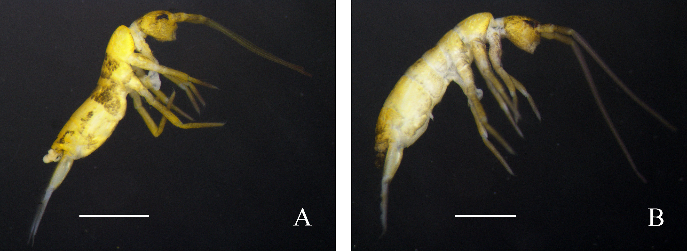
Figs 1A View FIGURE 1 , 2 View FIGURE 2 , 3 View FIGURE 3
Type material. Holotype: male on slide, near group of waterfalls, Tiantangzhai National Nature Reserve , Jinzhai County, Anhui Province, China, 115°46′40″E, 31°07′49″N ( WGS84 ), alt. 995m, 27.iii.2016, leg. Daoyuan Yu and Chunyan Qin (16 TTZ10 ) GoogleMaps . Paratypes: 2 females and 1 male on slides, 4 in alcohol, same data as holotype. All types deposited in NJAU GoogleMaps .
Description. Body length 2.8–3.3 mm (average 3.0, 4 specimens). Background body colour light yellow. Ant. I and Ant. II antero-laterally with diffuse purple pigment; ground colour of Ant. III grey, basally and apically with purple pigment, gradually darker towards apex; Ant. IV dark purple. Eye patch black, small purple patch behind eye. Clypeus with diffuse light purple pigment. Tibiotarsi usually with purple pigment ( Fig. 1A View FIGURE 1 ).
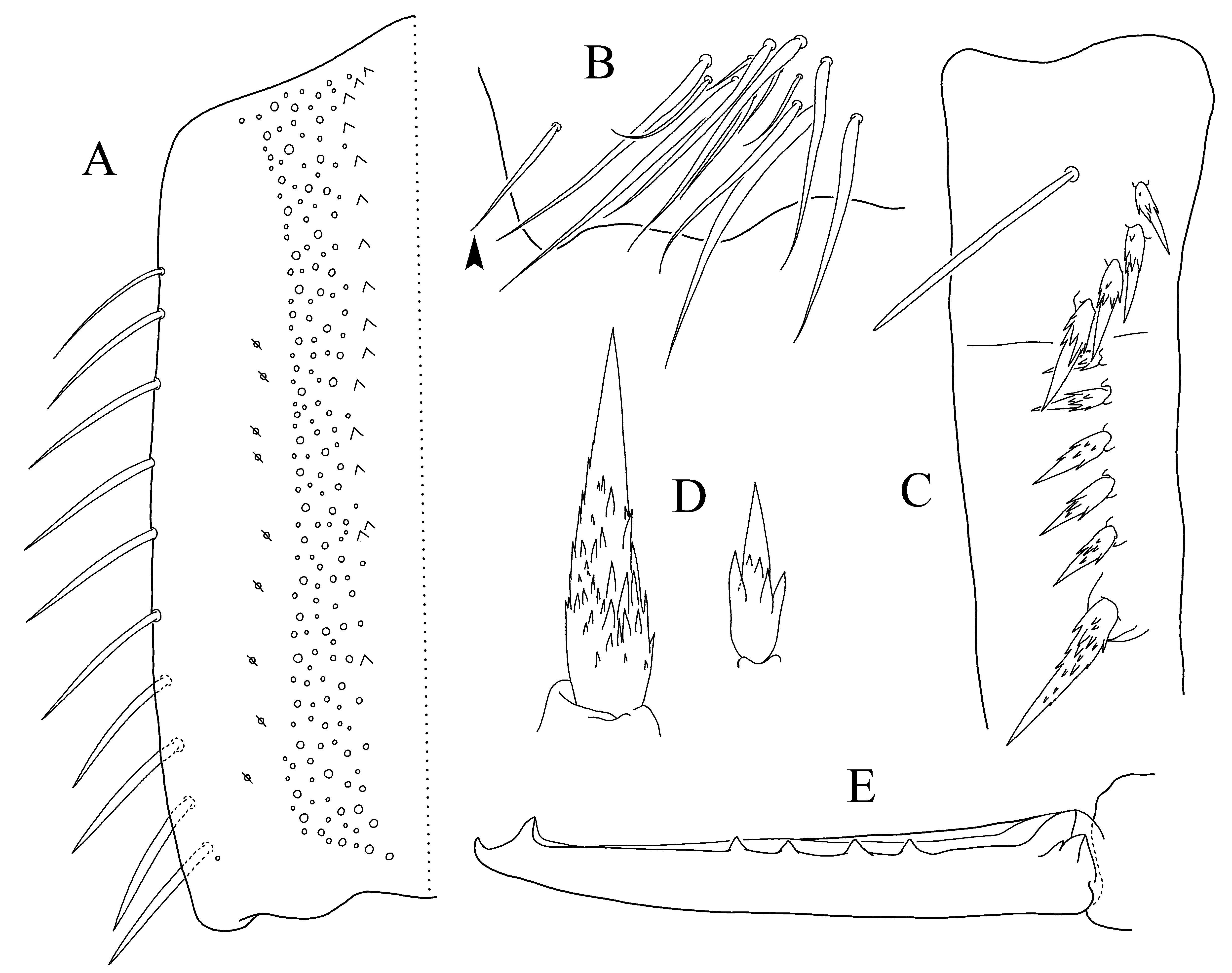
Intact four-segmented antennae 0.6–0.75× length of body (average 0.72, 3 specimens). Length ratio of Ant I:II:III:IV = 1.0:1.2–1.4:6.9–7.5:1.3–1.4. Ant. III unscaled. Cephalic dorsal macrochaetotaxy: anterior area: 2 (A2), 4 (A3, A5); interocular area: 2 (M2), 7 (S0, S2, S5, S 5i); postocular area: 2+2 (Pa5, Pa6); posterior area: 3+3 (Pa2, Pp3, Pe3). Posterior margin of head with 30–40 chaetae on each side ( Fig. 2A View FIGURE 2 ).
Pattern of body chaetotaxy as in Fig. 2B View FIGURE 2 . Number of macrochaetae or large mesochaetae in posterior row as 3 (p2, p3, p4), 3 (p1, p3, p5)/ 3 (m2, m3, m4), 3 (m2, m3, m4), 4 (p1, p3, p6, p7), 2 (p6, p7), 4 (m2, m3, m5, m6) from Th. II to Abd. V respectively. Th. II with macrochaetae a3, a4, a4a and a5a behind anterior marginal macrochaetae cluster; central macrochaetae a2, a5, m1, m2 and m3 arranged in triangle, m4 lateral to m2; Th. III with anterior macrochaeta a4; Abd. III with two anterior macrochaetae m3 and m6; Abd. IV with antero-lateral macrochaeta m6; Abd. VI with numerous chaetae of different sizes.
Trochantero-femoral organ with 1, 1 slender chaetae subequal in length ( Fig. 2C View FIGURE 2 ). Tibiotarsi I, II, III ventrally with 8–9, 8, 8 strong chaetae, 4–6, 6, 8 of them blunt ( Fig. 2D View FIGURE 2 ). Tenent hair 1.0–1.2× length of inner edge of unguis (average 1.1, 2 specimens); accessory chaetae weaker than pretarsal chaetae; guard chaetae 0.8–0.9× length of tenent hair (average 0.85, 2 specimens). Unguis slender, with baso-internal ridges about 1/4 – 1/3 distance from base; lateral teeth pointed, of moderate size. Inner edge of unguis with basal tooth and 4–6 (average 5, 4 specimens) more distal teeth, sub-basal tooth larger. Unguiculus lanceolate, about 0.55–0.75× length of unguis (average 0.68, 4 specimens), its inner edge with one tooth ( Fig. 2E View FIGURE 2 ).
Ventral tube scaled on both faces. Anterior face with 25–36 (average 31, 4 specimens) chaetae on each side, posterior face with 75–106 (average 90, 4 specimens) chaetae, each lateral flap with 70–106 (average 82, 4 specimens) chaetae and occasionally 1–2 scales. Anterior face of tenaculum with 7–10 (average 8, 3 specimens) chaetae and without scales ( Fig. 2F View FIGURE 2 ). Ratio manubrium:dens:mucro = 3.1–3.5:4.1–4.5:1.0. Manubrium ventrally scaled without chaetae; laterally with large round scales and 9–10 chaetae, proximal 1–2 chaetae slender, distal chaetae strong; each dorsal chaetal strip with 130–180 (average 153, 4 specimens) chaetae of different sizes, an irregular row of scales from base to 2/3 – 3/4 (average 7/10, 4 specimens) length of manubrium along inner edge, and 9–12 (average 10, 4 specimens) pseudopores on lateral side; without distinct prominent chaetae ( Fig. 3A View FIGURE 3 ); external distal corner chaeta as large as small mesochaetae in chaetal strip ( Fig. 3B View FIGURE 3 ). Dens basally with prominent blunt dorsal chaeta. Dental spine formula as 3–4/5–6, 1, sizes of spines gradually increase on basal subsegment ( Fig. 3C View FIGURE 3 ); small spines with large denticles at basal half and a few small to moderate-size denticles, large spines with numerous small to moderate-size denticles ( Fig. 3D View FIGURE 3 ). Mucro with 2–5 (average 4, 4 specimens) intermediate teeth ( Fig. 3E View FIGURE 3 ).
Etymology. Combination of the Ancient Greek word pseudḗs: false, and the specific name of the similar species T. spinulus .
Habitat. Living in moss on rocks.
Remarks. Of other species in the T. ocreatus group, T. pseudospinulus sp. nov. most resembles T. spinulus in having short antenna, a similar chaetotaxy and a single large distal dental spine, but differs from the latter mainly in absence of the distinct prominent chaetae on the manubrium, presence of a blunt prominent chaeta on the dens and larger denticles on the dental spines ( Table 1). Also the body colour of the new species is bright yellow and, in some individuals, with very light greenish tinge, while T. spinulus is dirty greyish yellow in the adults and light yellow only in the subadults. The type localities of T. pseudospinulus sp. nov. and T. spinulus are about 250 km apart, one belonging to the Dabie Cordillera and the other the Yellow Mountain Cordillera, respectively, which are geographically divided by the Yangtze River.

No known copyright restrictions apply. See Agosti, D., Egloff, W., 2009. Taxonomic information exchange and copyright: the Plazi approach. BMC Research Notes 2009, 2:53 for further explanation.
|
Kingdom |
|
|
Phylum |
|
|
Class |
|
|
Order |
|
|
Family |
|
|
Genus |