
Thraulodes zonalis Traver & Edmunds 1967
|
publication ID |
https://doi.org/ 10.11646/zootaxa.4756.1.1 |
|
publication LSID |
urn:lsid:zoobank.org:pub:9FF62616-A7FA-4331-AC51-0F534400631D |
|
DOI |
https://doi.org/10.5281/zenodo.3811785 |
|
persistent identifier |
https://treatment.plazi.org/id/039787A6-FFD3-8436-8CFB-FCA3D88BF9CC |
|
treatment provided by |
Carolina |
|
scientific name |
Thraulodes zonalis Traver & Edmunds 1967 |
| status |
|
10. Thraulodes zonalis Traver & Edmunds 1967 View in CoL
( Figs 317–349 View FIGURES 317–336 View FIGURES 337–345 View FIGURES 346–347 View FIGURES 348–349 )
Thraulodes zonalis Traver & Edmunds 1967: 383 View in CoL , figs 25, 41 (♂ imago and presumably associated larva); Allen & Brusca 1978: 421, fig. 26 (presumably associated larva).
Material examined. PANAMA, Provincia de Coclé, El Valle de Anton, Rio Anton , (8°35’N, 80°07’W), 3–5.II.2018, coll. N. Kluge & L. Sheyko: 1 L-S-I ♀, 1 L /S ♂, 2 larvae. GoogleMaps
Descriptions.
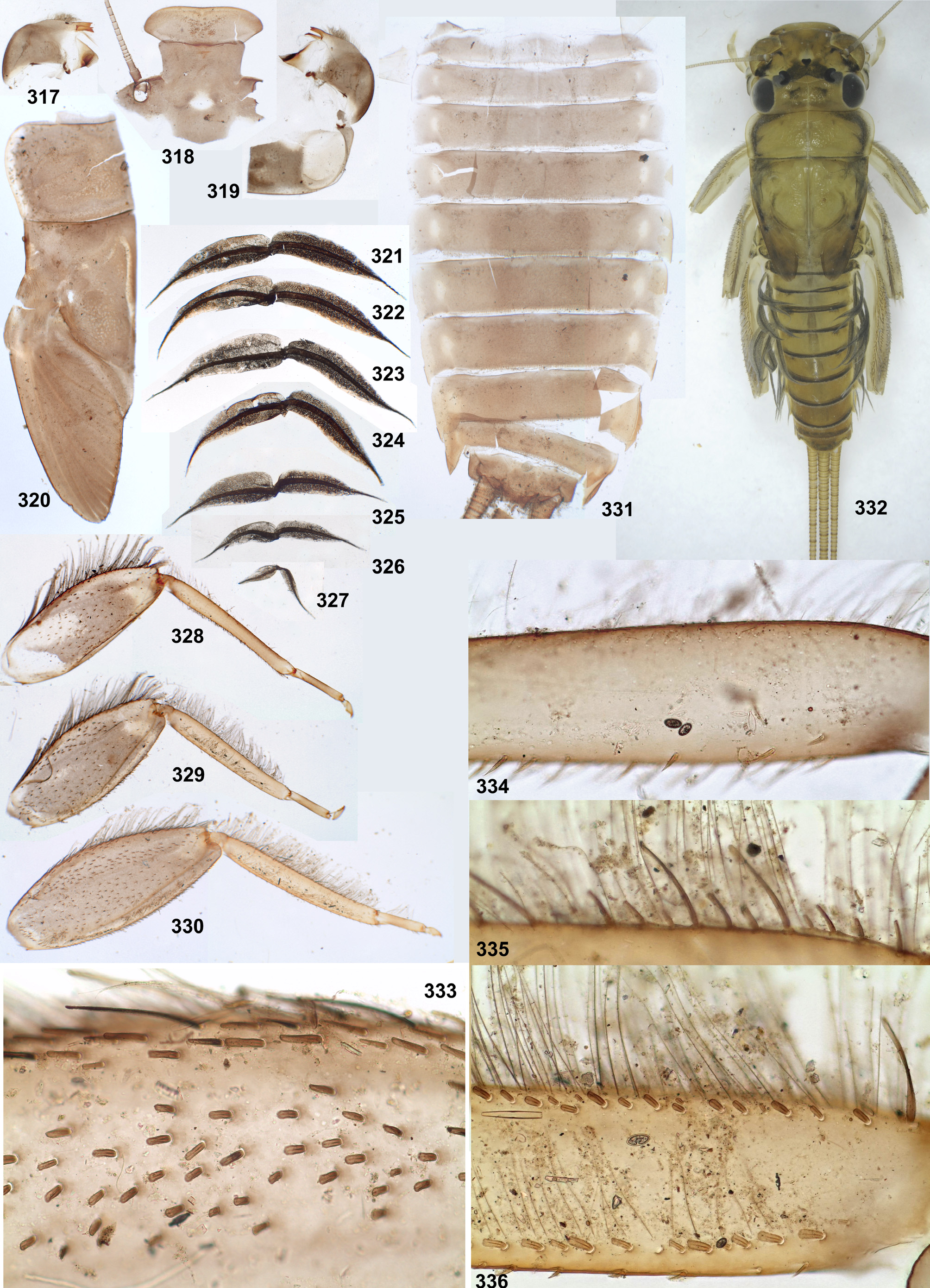
Larva. CUTICULAR COLORATION ( Figs 317–320, 328–331 View FIGURES 317–336 ). Dorsal side of head, thorax and abdomen mostly brown with few blanks: pronotum bordered by light laterally, without other blanks; abdominal terga with blanks on near tergalii bases, area mediad of them nearly unicolorous brown. Femora mostly brown with proximal and preapical blanks; tibia brown; tarsus brown with base and apex lighter.
HYPODERMAL COLORATION ( Fig. 331 View FIGURES 317–336 ). Abdominal terga I–IX with transverse band on posterior margins; terga II–V also with pair of brown spot. Tergalii gray with yellowish ( Figs 321–327 View FIGURES 317–336 ).
SHAPE AND SETATION. Clypeus widened distally; labrum 1.3–1.4 times wider than clypeus ( Fig. 318 View FIGURES 317–336 ). Labrum widest at 0.4 length from base; initial fore margin (turned ventrally) without median emargination, with all 5 denticles wide; anterior transverse setal row regular (as in Fig. 88 View FIGURES 86–93 ), as wide as all 5 denticles. Maxilla with 15–16 pectinate setae in apical-ventral row.
Femora: Stout setae on anterior surface parallel-sided and blunt ( Fig. 333 View FIGURES 317–336 ). Irregular row of hairs near inner margin absent on fore femur, present on middle and hind femora.
Fore tibia ( Fig. 334 View FIGURES 317–336 ): outer hairs form two irregular rows; inner-anterior row of recurved hairs absent; inneranterior row of stout setae absent or represented by single blunt stout seta near tibia base; inner field of stout pointed setae dense (i.e. setae longer than distances between them, about 3 setae in cross section), setae of most anterior row shorter than others, bipectinate or dentate.
Hind tibia ( Figs 335–336 View FIGURES 317–336 ): outer-anterior row of stout setae consists of two clearly different types of stout setae: numerous short rounded ones and several long spoon-like ones; outer-posterior row of stout setae consists of blunt and spoon-like stout setae of various lengths; hairs located between these rows, numerous and form more than one row (besides row of hairs posteriad of outer-posterior row of stout setae); stout setae of inner-anterior row short, widened distally and truncated.
Claws with 5–7 denticles on rigid portion, with several minute denticles on articulatory portion.
Tergalii ( Figs 321–327 View FIGURES 317–336 ): of moderate width; on both lamellae main trachea without branches; dorsal lamella with costal margin most convex in proximal part and anal margin most convex in distal part, gradually narrowed toward apex, with slender apical filament; ventral lamella widest near base, gradually narrowed toward apex, with slender apical filament.
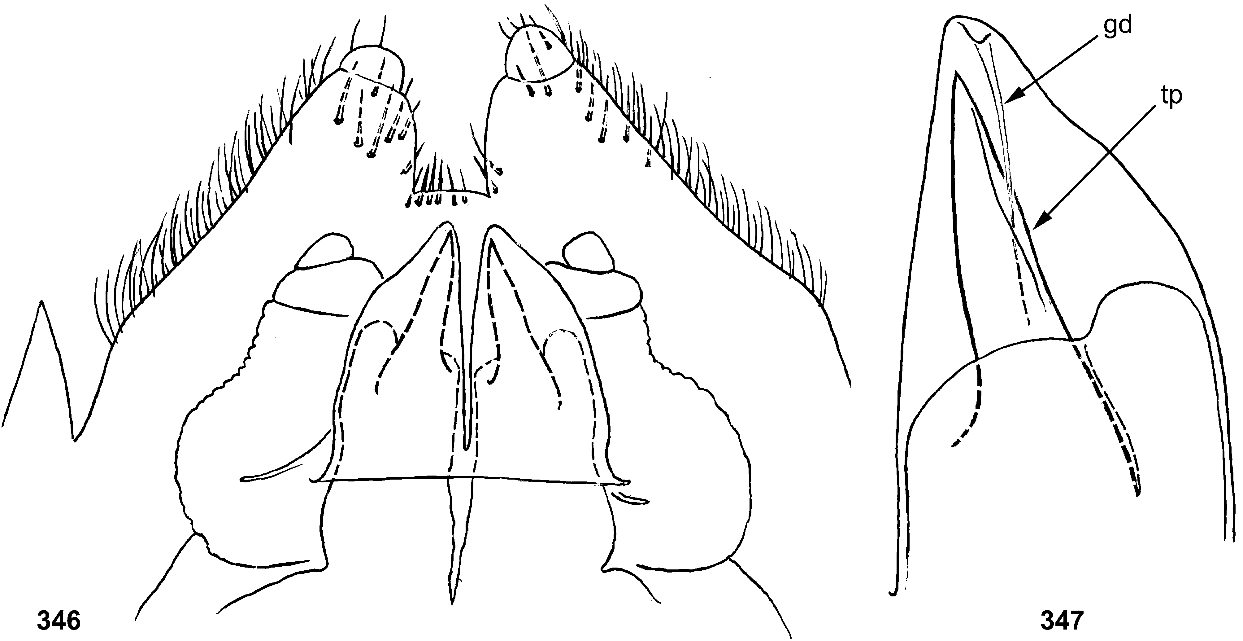
Male genitalia in last larval instar ( Figs 346–347 View FIGURES 346–347 ): protogonostyli separated one from another by deep emargination. Each protopenes lobe smoothly stretched to gonopore-bearing process located near median margin; gonopore opened caudally.
Subimago. CUTICULAR COLORATION ( Figs 337–338 View FIGURES 337–345 ). Cuticle brown with lighter brownish and colorless areas. Pronotum light brow. Mesonotum mostly brown, with darker brown antelateroparapsidal and lateroparapsidal sutures, with colorless oblique stripe on posterior scutal protuberance; chromozones of medioscutum and submedioscutum equally colored by brown; achromozones of medioscutum and submedioscutum contrastingly colorless. On all legs femur mostly light, bordered by brown on outer and inner margins, with apex brown; tibia brownish; tarsus lighter brownish. Wings look gray due to blackish microtrichia and small brown ring around base of each microtrichion. Abdominal terga, sterna and caudalii light brownish.
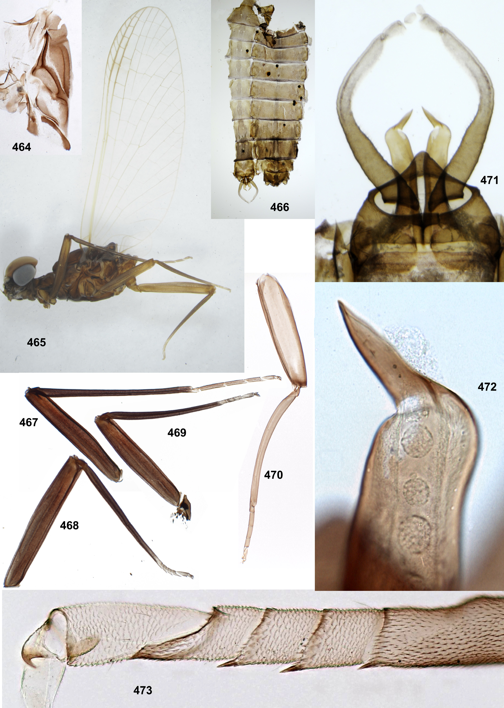
TEXTURE. On tarsi of all legs, 1st tarsomere with microtrichia (as tibia), 2nd–5th tarsomeres coved by blunt microlepides; pointed microlepides present near apical margins of 2nd–4th tarsomeres of middle and hind legs (as in Fig. 473 View FIGURES 464–473 ).
Male imago. Described by Traver & Edmunds (1967). Coloration as in female (see below).
Female imago ( Figs 338–342, 344–345 View FIGURES 337–345 ). Head and thorax dark brown.
Cuticle of legs mostly colorless; on fore leg apex of femur and base of tibia with cuticle brown; on middle and hind legs apex of femur with lighter brownish. Legs with following hypodermal pigmentation on whitish background: Fore femur entirely pigmented, with proximal part blackish-brown and distal part reddish-brown, with blackish stripe along inner margin. Fore tibia mostly reddish-brown, basally and apically darker brown. Fore tarsus light brown with non-pigmented joinings. Middle femur with gray-brown spot on posterior side close to base, larger brown spot on anterior side more distally and multicolored pre-apical band occupying distal half of femur both on anterior and posterior sides; pre-apical band mainly reddish-brown, proximally bordered by blackish brown. Hind femur with large basal brown spot on anterior side, occupying most part of proximal half of femur beginning from its base, and with multicolored pre-apical band as on middle femur. On middle and hind legs tibia mostly non-pigmented, with brown spots near base (close to patella-tibial suture) and on apex; tarsus with small brown spots on 3rd, 4th and 5th tarsomeres. In reared individual, relation of fore femur to fore wing length 80:350; proportions femur/tibia/tarsomeres on fore leg 80:80:4:7:7:5:10; on middle leg 80:75:4:5:4:3:9; on hind leg 95:90:4:5:4:3:9.
Fore wing with dark brown band crossing costal brace. Longitudinal veins ocher or light brown; cross veins light brown, in anterior part of wing narrowly bordered by light brown; costal cross veins proximad of bulla few and very thin, bordered by brown near subcostal vein ( Figs 344–345 View FIGURES 337–345 ). Pterostigmatic cross veins not dense, oblique, non-branched (as in male, Traver & Edmunds 1967: fig. 25). Hind wing short, with costal projection prominent; proximal part of hind wing brown ( Fig. 342 View FIGURES 337–345 ).
Abdomen brown with poorly expressed darker brown midway spots on terga III–V.
Eggs ( Figs 348–349 View FIGURES 348–349 ). Mostly barrel-shaped. Each KTC without ring-like cover, spirally coiled threads being exposed. Other chorion with evenly dispersed protuberances.
Dimension. Fore wing length (and approximate body length) 6–8 mm.
Distribution. Panama ( Traver & Edmunds 1967; this study).
Comments. The original description of Th. zonalis was based on male imagines ( Traver & Edmunds 1967). The male subimago, extracted from larva, has the same hypodermal coloration, especially characteristic coloration of legs (as in female—Figs 339–341) and abdomen ( Fig. 343 View FIGURES 337–345 ).
In the original description, coloration of proximal half of middle femur is characterized as «2 dark spots within this pale basal area, arranged in tandem», and coloration of hind femur—as «femur III twice-banded...» ( Traver & Edmunds 1967: 383). Actually, each of these femora has only one true band girdling the femur, this is the pre-apical one; on the proximal half of middle and hind femora dark pigment does not form bands girdling the femur, but forms separated spots, each of which is located on one side of the femur only. On the proximal half of middle femur, there are 2 dark spots located on different sides of the femur (more proximal spot—on posterior side, and more distal spot—on anterior side), so that they look as «tandem» only if the femur is translucent. On the hind femur the proximal large spot is located on the anterior side only.
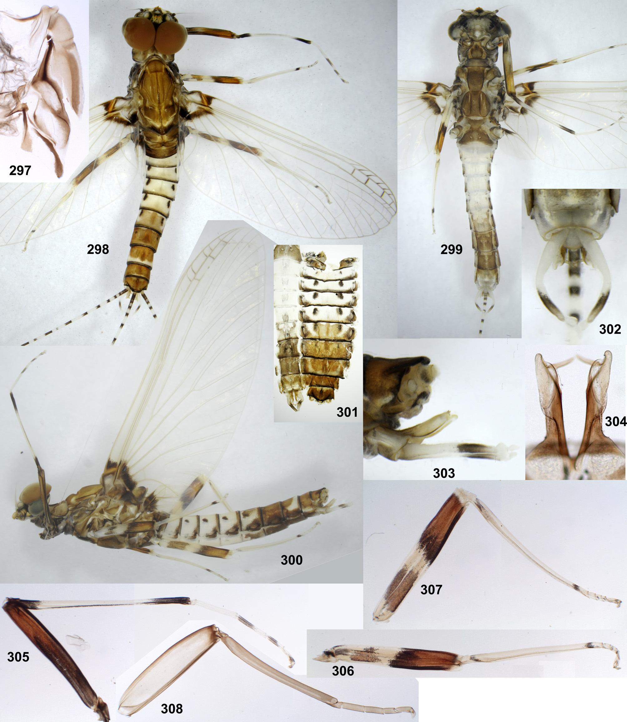
According to the original description, male imago of Th. zonalis has «most cross veins indistinct ... In apical area C, Sc, and R 1 somewhat thickened; 3–5 stigmatic veins, these on 2 or 3 in subcostal space also thickened» ( Traver & Edmunds 1967: 383, fig. 25). This is the same as in male imago of Th. fascipennis ( Fig. 298 View FIGURES 297–308 ). Female imago of Th. zonalis described here for the first time, has somewhat different coloration of fore wing, with all cross veins colored. Possibly, this is a sexual dimorphism in wing coloration.


No known copyright restrictions apply. See Agosti, D., Egloff, W., 2009. Taxonomic information exchange and copyright: the Plazi approach. BMC Research Notes 2009, 2:53 for further explanation.
|
Kingdom |
|
|
Phylum |
|
|
Class |
|
|
Order |
|
|
Family |
|
|
Genus |
Thraulodes zonalis Traver & Edmunds 1967
| Kluge, Nikita J. 2020 |
Thraulodes zonalis
| Allen, R. K. & Brusca, R. C. 1978: 421 |
| Traver, J. R. & Edmunds, G. F. Jr. 1967: 383 |