
Thornburghiella montana Ježek, Oboňa & Manko, 2021
|
publication ID |
https://doi.org/ 10.11646/zootaxa.4985.4.11 |
|
publication LSID |
lsid:zoobank.org:pub:E199BD57-5D29-4F5F-B611-52840883083F |
|
DOI |
https://doi.org/10.5281/zenodo.5075505 |
|
persistent identifier |
https://treatment.plazi.org/id/7C42675E-0000-B923-FF14-F951F0160845 |
|
treatment provided by |
Plazi |
|
scientific name |
Thornburghiella montana Ježek, Oboňa & Manko |
| status |
sp. nov. |
Thornburghiella montana Ježek, Oboňa & Manko View in CoL sp. nov.
( Figs 1–21 View FIGURES 1–11 View FIGURES 12–21 )
Description. Male. Head hardly as long as broad ( Fig. 1 View FIGURES 1–11 ), 1.2 times broader. Vertex conically a little inflated dorsally ( Figs 1 View FIGURES 1–11 ) with a cut top. Numerous setae alveoli are almost regularly spaced over the entire surface in spite of scar free areas above C-shaped compound eyes laterally. Eyes separated, interocular suture arcuate ( Figs 1 View FIGURES 1–11 , 12 View FIGURES 12–21 ), eye bridge formed by five facet rows, frontal marginal rows are reduced to four facets. Minimum distance between eyes corresponds roughly to six facet diameters; index of distance from tangential points of eye apices to minimum of frons 3.2. Setae alveoli of frontoclypeus arranged almost in a triangular centrally placed patch near the base of antennae, tapering to a dorsoventral stripe of hairs close below frontal suture ( Figs 1 View FIGURES 1–11 , 12 View FIGURES 12–21 ). Patagia cylindrical, bag-shaped, constricted and contracted in one third, bent, covered with microsetae, see Fig. 11 View FIGURES 1–11 . Antenna with 15 articles; scape club-shaped ( Fig. 2 View FIGURES 1–11 ), somewhat widened apically, 2.5 times as long as its maximum width, narrowed et base, 4.9 times as long as its minimum width. Pedicel pitcher-shaped, symmetrical. Flagellomere 1 (postpedicel) cylindrical, hardly as long as three following flagellomeres together ( Fig. 2 View FIGURES 1–11 ). Postpedicel with six conspicuous, strong bristles arranged in a row, sometimes is the longest distal bristle doubled (from the same insertion). Scape and pedicel with stiletto-shaped scales in contrast to needle-shaped macrosetae of flagellomeres. Flagellomeres 2–12 ovoid, with needle-shaped paired ascoids, a little bent, shorter than flagellomeres in which are inserted; apical flagellomere twice as long as the previous one including digital apiculus placed a little out of longitudinal axis ( Fig. 13 View FIGURES 12–21 ). Length ratio of maxillary palpus segments 1.0:1.2:1.5:2.1; apical segment annulated ( Fig. 3 View FIGURES 1–11 ). Terminal labial lobes ( Fig. 14 View FIGURES 12–21 ) with diverging rows of spines between them. Ratio of maximum length of cibarium ( Fig. 4 View FIGURES 1–11 ) to length of epipharynx 1.7:1.
Thorax. Anepisternum setae patch is almost trapezoid, anepimeron with triangular setose patch ( Fig. 15 View FIGURES 12–21 ). Spiracles set low on mesothorax. Wings ( Fig. 16 View FIGURES 12–21 ) lanceolate, 3.3 mm in holotype, 2.9–3.4 mm in paratypes, rounded distally, a little expanded at the posterior margin. The ending of R 5 beyond the tip of wing. Wing membrane slightly infuscated between Sc, R 1 and C and ends of all veins are a little strengthened distally with dark spots. Following veins or their parts strengthened: Sc with conspicuously marked origin and end, R 1 in distal three quarters, R 2, R 5, basal field, cross vein m 1 – m 2, CuA 1 and CuA 2 (conspicuously basally). Radial fork complete, medial fork in a form of a cross vein, their position see on Fig. 16 View FIGURES 12–21 . Both forks and the ending of CuA 2 are in one line (almost central area of wing). Wing index 2.4. Knob of halteres globular, with three close sensory microsetae ventrally, a prolonged stem as usually developed ( Fig. 5 View FIGURES 1–11 ). Ratio of maximum length of halteres to their maximum width approximately 2.8:1. Ratios of lengths of femora, tibiae and first tarsal segments P 1 2.0:2.3:1.0, P 2 2.1:2.8:1.1, P 3 2.4:3.2:1.2. Paired tarsal claws of P 1 gradually tapering, bent distad ( Fig. 6 View FIGURES 1–11 ).
Male genitalia. Ejaculatory apodeme almost straight, only inconspicuously bent proximally and contracted distally ( Figs 8 View FIGURES 1–11 , 21 View FIGURES 12–21 ), aedeagal complex with paired sclerotized boomerang-shaped ribs diverged laterally and converged caudally. The basis of distiphallus is braced by gonocoxal apodeme – a chitinized stripe with three prolonged lobes (arms) of different shape and length (triangular and pale-shaped) on both sides ( Fig. 8 View FIGURES 1–11 ). Gonocoxites almost hemisphaerical ( Figs 8–10 View FIGURES 1–11 ), gonostyli ovoid basally, with irregular margins, conspicuously scelrotized, distal parts V-shaped, forked in two protuberances: sickle-shaped thin longer arm and thicker shorter saw-shaped one with numerous teeth ( Figs 18–19 View FIGURES 12–21 ). Epandrium ( Figs 7 View FIGURES 1–11 , 17 View FIGURES 12–21 ) almost semicircular in dorsal view, hardly rectangular from lateral one, not bare, (see two divided areas of insertions of hairs distally), posterior margin conspicuously sclerotized, emarginate, with a deep cleft. Basal paired apertures conspicuous, crevice-shaped, connected. Ventral epandrial plate reduced ( Fig. 17 View FIGURES 12–21 ). Hypandrium narrow with a lobulus in the middle ( Fig. 8 View FIGURES 1–11 ). Epiproct inconspicuous, as a rounded fold, covered with microsetae and dark structures inside; hypoproct conspicuous, setose, tongueshaped, rounded apically from dorsal view ( Figs 7 View FIGURES 1–11 , 17 View FIGURES 12–21 ). Epandrial claspers (surstyli) strong, enlarged basally in contrast to the top, almost straight from dorsal view ( Fig. 17 View FIGURES 12–21 ), bent at about one-third from lateral view ( Fig. 7 View FIGURES 1–11 ). Tenacula are numerous (30–35), formed in longitudinal rows on inner sides of clasping lobes, apically frayed.
Female. Unknown.
Differential diagnosis. Thornburghiella montana sp. nov. resembles T. kovari Ježek, 1993 in body size, as well as wing venation. The new species have head vertex a little inflated dorsally ( Fig. 1 View FIGURES 1–11 ); frons with a dorsoventral stripe of hairs ( Figs 1 View FIGURES 1–11 , 12 View FIGURES 12–21 ); postpedicel not constricted subapically ( Fig. 2 View FIGURES 1–11 ); hypandrium narrow with a lobulus in the middle ( Fig. 8 View FIGURES 1–11 ); gonostyli with two protuberances. ( Figs 8–10 View FIGURES 1–11 , 18, 19 View FIGURES 12–21 ); aedeagal complex with paired sclerotized boomerang-shaped ribs diverged laterally and converged caudally ( Figs 8 View FIGURES 1–11 , 20, 21 View FIGURES 12–21 ). Thornburghiella kovari is readily distinguishable by vertex of head, conspicuously elevated dorsally; frons without dorsoventral stripe of hairs; postpedicel constricted subapically; hypandrium stripe-shaped of the same width; gonostyli with three quite different bizarre protuberances; aedeagal complex with two parallel almost spatula-shaped cut protuberances and inner two linear ribs diverged caudally by conspicuous sclerotized hooks protruded outline of distiphallus.
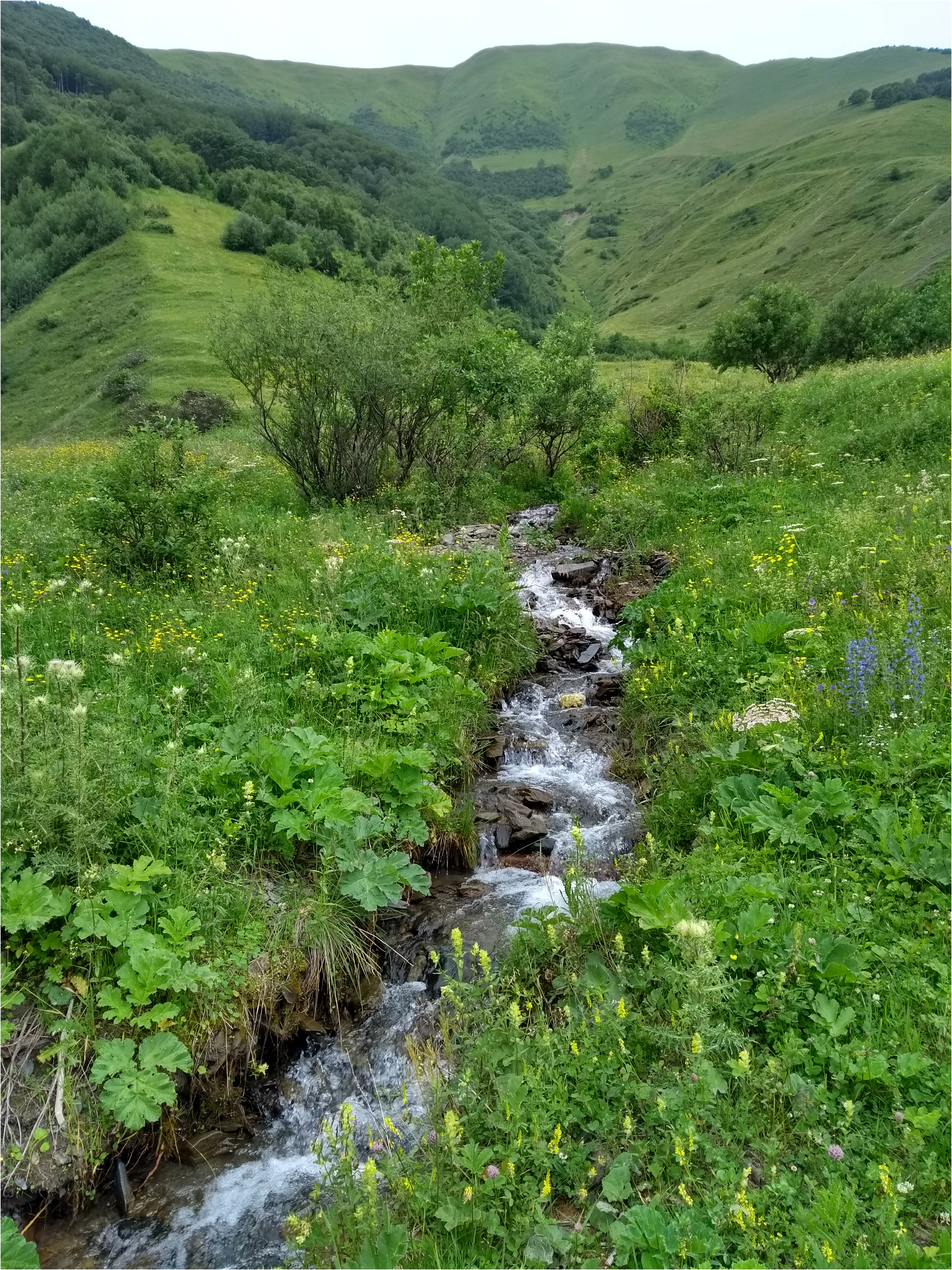
Type material. Holotype male: Transcaucasia , Georgia, Mtskheta – Mtianeti region , above the village Snotskali, a tributary of the Snotskali river, 1900 m a.s.l., 42°35’49.0”N 44°38’26.0”E ( Fig. 38 View FIGURE 38 ), 5.vii.2019, by sweep netting, Manko leg. Slide with a dissected specimen, Cat. No. 34899, Inv. No. 25956 ( NMPC). GoogleMaps
Paratypes of 15 males (slides, some specimens dissected): The same locality, method, collectors and date, Cat. No. 34900-34909, Inv. No. 25957-25966 ( NMPC) ; Gveleti , a stream beneath small waterfall, 1630 m a.s.l., 42°42’08.4”N 44°37’09.7”E, 12.vii.2019, by sweep netting, Kovács, Murányi and Vinçon leg., Cat. No. 34910- 34913, Inv. No. 25967-25970 ( NMPC) GoogleMaps ; Pansketi , the Snotskali River at its confluence with the Terek River, 1745 m a.s.l., 42°38’14.0”N 44°37’56.0”E, 12.vii.2019, by sweep netting, Kovács, Manko, Murányi and Vinçon leg., Cat. No. 34914, Inv. No. 25971 ( NMPC) GoogleMaps .
6. Tarsal claw of P 1, lateral view. 7. Epandrium and epandrial claspers, lateral view. 8. Aedeagal complex and gonopod, dorsal view. 9. Gonopod, lateral view. 10. Same, caudal view. 11. Patagium. [Scale: 1-5, 7-11 = 0.2 mm; 6 = 0.05 mm]
Type locality. Georgia, Mtskheta – Mtianeti region, Snotskali.
Etymology. The specific epithet is derived from the Latin word “montanus – a – um“ (adjective) = montane (mountain); it refers to the high elevation of the studied habitats of this species.
Bionomics. Unknown, males were collected near montane waterfalls and streams or confluences of rivers, 1630–1900 m a.s.l.
Distribution. Currently recorded only from Georgia.


| NMPC |
National Museum Prague |
No known copyright restrictions apply. See Agosti, D., Egloff, W., 2009. Taxonomic information exchange and copyright: the Plazi approach. BMC Research Notes 2009, 2:53 for further explanation.
|
Kingdom |
|
|
Phylum |
|
|
Class |
|
|
Order |
|
|
Family |
|
|
Genus |