
Tharybis kueishanensis, Wang & Hwang, 2022
|
publication ID |
https://doi.org/ 10.11646/zootaxa.5189.1.24 |
|
publication LSID |
lsid:zoobank.org:pub:944424E0-09D4-4D6B-B778-36172B1D86F9 |
|
DOI |
https://doi.org/10.5281/zenodo.7129639 |
|
persistent identifier |
https://treatment.plazi.org/id/114E87BD-FFCE-FFF6-FF70-A5D153587DE2 |
|
treatment provided by |
Plazi |
|
scientific name |
Tharybis kueishanensis |
| status |
sp. nov. |
Tharybis kueishanensis sp. n.
Holotype: Total body length, measured from anterior margin of rostrum to posterior margin of caudal rami. Dissected female 2.06 mm (KIC 22001), prosome 1.7 mm, urosome 0.36 mm . Paratypes: 5 undissected females 1.89 to 2.12 mm (KIC 22002 to KIC 22006) . Allotype: dissected male 1.69 mm (KIC 22007), prosome 1.29 mm, urosome 0.4 mm .
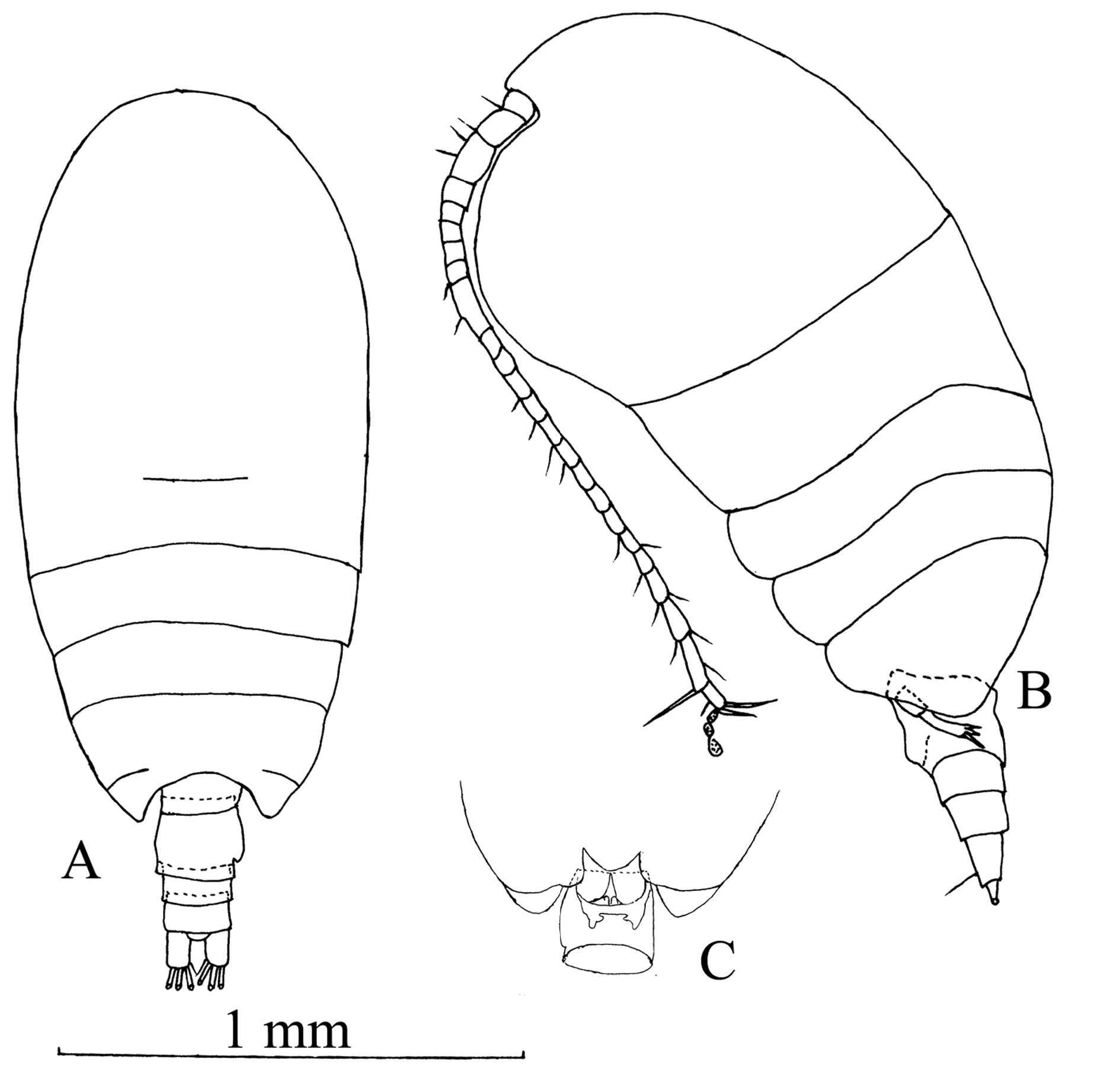
Female. Prosome ovoid in dorsal view ( Fig. 1A View FIG ). Rostrum absent. Cephalosome and first pedigerous somite partly fused. Fourth and fifth pedigerous somites partly fused. Posterior corner of prosome broadly rounded in lateral view, reaching almost first third of genital double-somite ( Fig. 1B View FIG ). Genital double-somite asymmetrical in dorsal view, right side slightly more produced laterally than left; ventral bulge close to distal margin; in ventral view, left side with small notch near distal margin ( Fig. 1C View FIG ). Posterior borders of urosomites unfurnished.
Antennule ( Fig. 2A View FIG ): Reaching slightly beyond posterior margin of third pedigerous somite, with 24 free segments, with the following armature: 3, 6+ae (=aesthetasc), 2+ae, 2, 2+ae, 2, 2+ae, 4+ae, 1, 1, 2+ae, 1, 2+ae, 1, 1, 1, 1, 1+ae, 1, 1, 2, 2, 2, 4+ae. Segments 8 and 9 completely separated.
Antenna ( Fig. 2B View FIG ): With short endopod; basis with 1 seta; endopod segment 1 with 2 setae, segment 2 with 14 (8+6) setae and outer border lined with small spinules; exopod 6-segmented, segment 1 without seta, segments 2-5 each with 1 seta, segment 6 with 3 terminal setae.
Mandible ( Fig. 2C View FIG ): Gnathobase with 8 ventral teeth and strong dorsal seta, anterior and posterior surfaces of cutting edge with rows of spinules; basis with 2 strong short setae and long more distally placed seta; endopod 2- segmented, proximally with 1 seta and distally with 7 setae.
Maxillule ( Fig. 2D View FIG ): Praecoxal arthrite very large, with 11 elements; coxal endite and coxal epipodite carrying 2 and 7 setae, respectively; basal endites with 4+3 setae; endopod indistinctly 2-segmented, bearing 2 and 5 setae, respectively. exopod fused to basis, and armed with 3 setae.
Maxilla ( Fig. 2E View FIG ): Praccoxa with an outer distal protrusion, endites 1 and 2 with 3 setae each; coxal endites 3 and 4 with 3 setae separately; basal endite bearing 3 setae; endopod with 3 long worm-like and 6 brush-like aesthetascs distally.
Maxilliped ( Fig. 2F View FIG ): Syncoxa and basis almost equal length; syncoxa with 2, 1, 3, 3 setae; basis with 3 setae; endopod half-length of basis, segment 1 apparently incorporated into basis with 2 setae, free segments 2-6 with 3, 4, 3, 2 + 1, and 4 setae respectively.
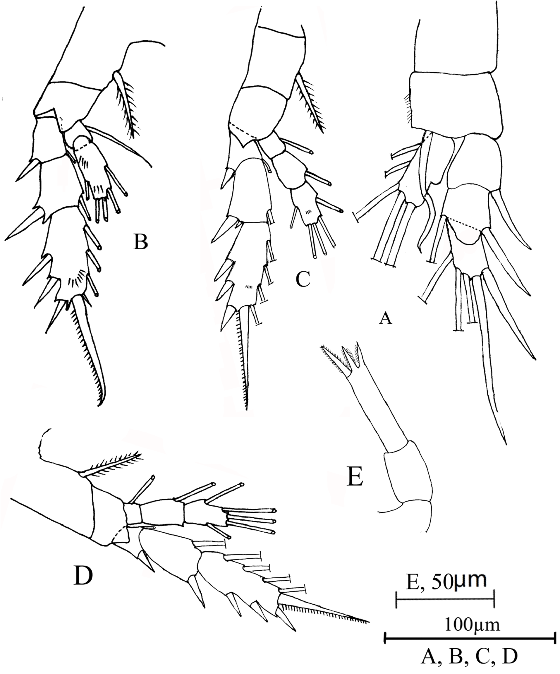
Swimming legs one to four ( Fig. 3 View FIG A-D): Segmentation and disposition of spines and setae as follows (Arabic and Roman numerals represent setae and spines, respectively; numbering proceeds from outer to inner edge of each segment):
Leg 1: Basis with curved distomedial seta. Tip of lateral seta on proximal exopod segment reaching midpoint of terminal segment.
Legs 2, 3: Posterior surface of distal segment of endopod and exopod with rows of small denticles.
Leg 5 ( Fig. 3E View FIG ): Uniramous, symmetrical and 3-segmented. Distal segment about 4 times longer than wide, with two spiniform projections of equal length distally, distolateral spine about 1.5 times longer than the projections.
Caudal rami ( Fig. 1 View FIG A-B): symmetrical and slightly longer than wide, with 4 large, terminal setae and small ventral seta; length about 1.4 times as long as width.
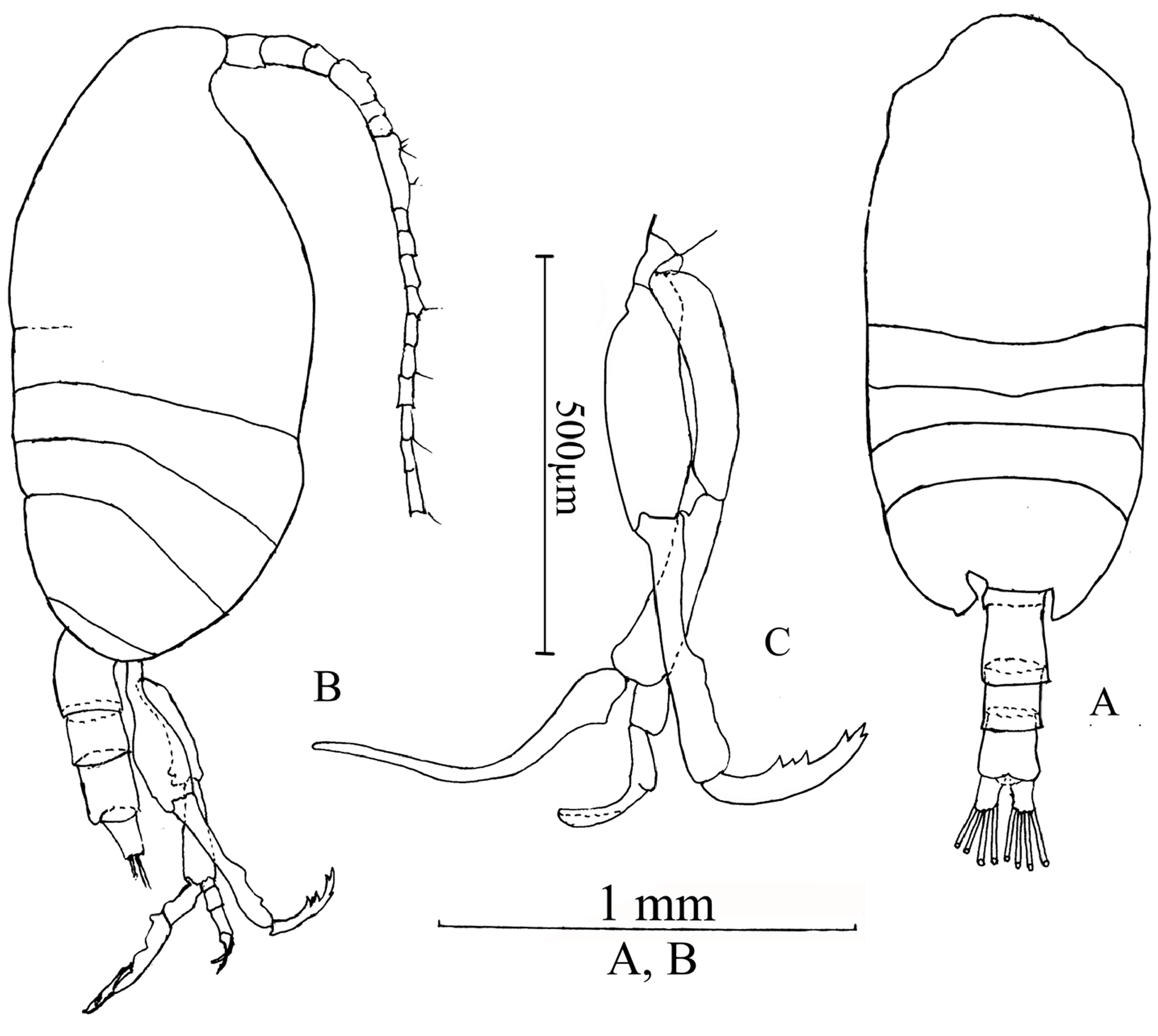
Male. Prosome 3.2 times longer than urosome. Rostrum absent. Cephalosome and first pedigerous somite partially fused laterally. Fourth and fifth pedigerous simites separated laterally. Posterior corners of prosome rounded in lateral view ( Fig. 4B View FIG ), asymmetrical in dorsal view with left posterior margin strongly modified as shown in Fig. 4A View FIG .
Leg 5 ( Fig. 4C View FIG ): right leg uniramous with 4 segments. Distal segment about half length of proximal segment, with 3 triangular projections at inner middle margin and tip. Left leg biramous. Endopod 1-segmented falciform, thin, narrowing distally. Exopod 3-segmented.
Etymology The species name kueishanensis refers to Kueishan Island where the species was collected.
Remarks
Females of Tharybis kueishanensis n. sp. can easily be distinguished from Tharybis shuheiella Ferrari & Markhaseva, 2005 ; Tharybis inflata Andronov, 2002 ; Tharybis scaura Andronov, 2002 and Tharybis inaequalis Bradford-Grieve, 2001 by symmetrical or asymmetrical leg 5. Leg 5 of T. kueishanensis was symmetrical. Genital complex of T. kueishanensis was asymmetrical which was obviously different to Tharybis juhlae Ferrari & Markhaseva, 2005 ; Tharybis fultoni Park, 1967 ; Tharybis macrophthalma Sars, 1902 ; Tharybis neptuni (Cleve, 1904) ; Tharybis tuberosa Andronov, 2002 ; Tharybis macrophthalmoida Andronov, 2002 ; Tharybis magna Bradford & Wells, 1983 ; Tharybis asymmetrica Andronov, 1976 ; Tharybis angularis Schulz, 1995 ; Tharybis tumidula Andronov, 2002 ; Tharybis megalodactyla Andronov, 1976 and Tharybis compacta ( Grice & Hulsemann, 1970) . The cephalon and pedigerous somite 1 fused, rostrum with 2 thin filaments in Tharybis sagamiensis Tanaka, 1960 and Tharybis pseudomegalodactyla Ferrari & Markhaseva, 2005 . In Tharybis lauta Andronov, 2002 , cephalon and pedigerous somite 1 separated and rostrum with 2 thin filaments. However, in T. kueishanensis , cephalon and pedigerous somite 1 partly fused and rostrum without filaments. The differences between Tharybis groenlandicus (Tupitzky, 1982) and T. kueishanensis as follows: pedigerous somites 4 and 5 fused in T. groenlandicus (Tupitzky, 1982) but partly fused in T. kueishanensis ; posterolateral corners of pedigerous somite 5 rounded and covering almost half of genital complex in T. groenlandicus (Tupitzky, 1982) , the posterolateral border of cephalosome only reaches the first third of the genital complex in T. kueishanensis .
Females of T. kueishanensis share several features with females of T. angularis Schulz, 1995 except the asymmetrical or symmetrical genital complex. Tharybis angularis Schulz, 1995 was collected in the south of Iceland, North Atlantic on 28 th July 1989 about 20 m above the bottom at a water depth of 2860 m ( Schulz & Beckmann, 1995). T. kueishanensis is very similar to T. angularis in the fifth pair of legs. Both of them have symmetrical fifth legs and distal segment of fifth legs are very long. The length of the distal segment is about 4 times longer than its width and the length of the inner terminal spine is about 1.5 times of the terminal projection. However, the main differences between these two species are as following: firstly, the body length of female T. angularis was 1.21 to 1.27 mm, the female body length of T. kueishanensis ranges from 1.89 to 2.12 mm which was longer than T. angularis ; secondly, cephalon and pedigerous somite 1 are almost separate in T. angularis and partly separate in T. kueishanensis ; thirdly, the pedigerous somites 4 and 5 are completely fused in T. angularis , however, in T. kueishanensis only partly fused dorsally; fourthly, the genital complex of T. angularis is symmetrical and somewhat spherical in dorsal view, the genital field is swollen in the middle part in lateral view, but in T. kueishanensis , there is a notch at the right side posterior margin in dorsal view and the genital field swelling is close to the posterior margin in lateral view; fifthly, the length to width ratio of the penultimate segment of the fifth swimming leg is about 1.5 in T. kueishanensis , whereas the ratio is only about 1 in T. angularis .
Female and male specimens have been assigned to the same species based on their co-occurrence and morphological similarity. The body shape of male T. kueishanensis is very similar with the female individual. The antennule was not modified as a grasping appendage on both sides in male. The significant character of the male is the 3 triangular projections at the inner middle margin and tip of the right leg 5, which is the main difference to previously described male species.

No known copyright restrictions apply. See Agosti, D., Egloff, W., 2009. Taxonomic information exchange and copyright: the Plazi approach. BMC Research Notes 2009, 2:53 for further explanation.