
Stereoderma mohammedi, Thandar, 2018
|
publication ID |
https://doi.org/ 10.11646/zootaxa.4532.1.3 |
|
publication LSID |
lsid:zoobank.org:pub:A6128B92-0B20-4D4D-AE8B-483D39BB2C04 |
|
DOI |
https://doi.org/10.5281/zenodo.5107304 |
|
persistent identifier |
https://treatment.plazi.org/id/2A0887A5-9915-5E29-BCF8-FEBAFD3CFE23 |
|
treatment provided by |
Plazi |
|
scientific name |
Stereoderma mohammedi |
| status |
sp. nov. |
Stereoderma mohammedi View in CoL n. sp.
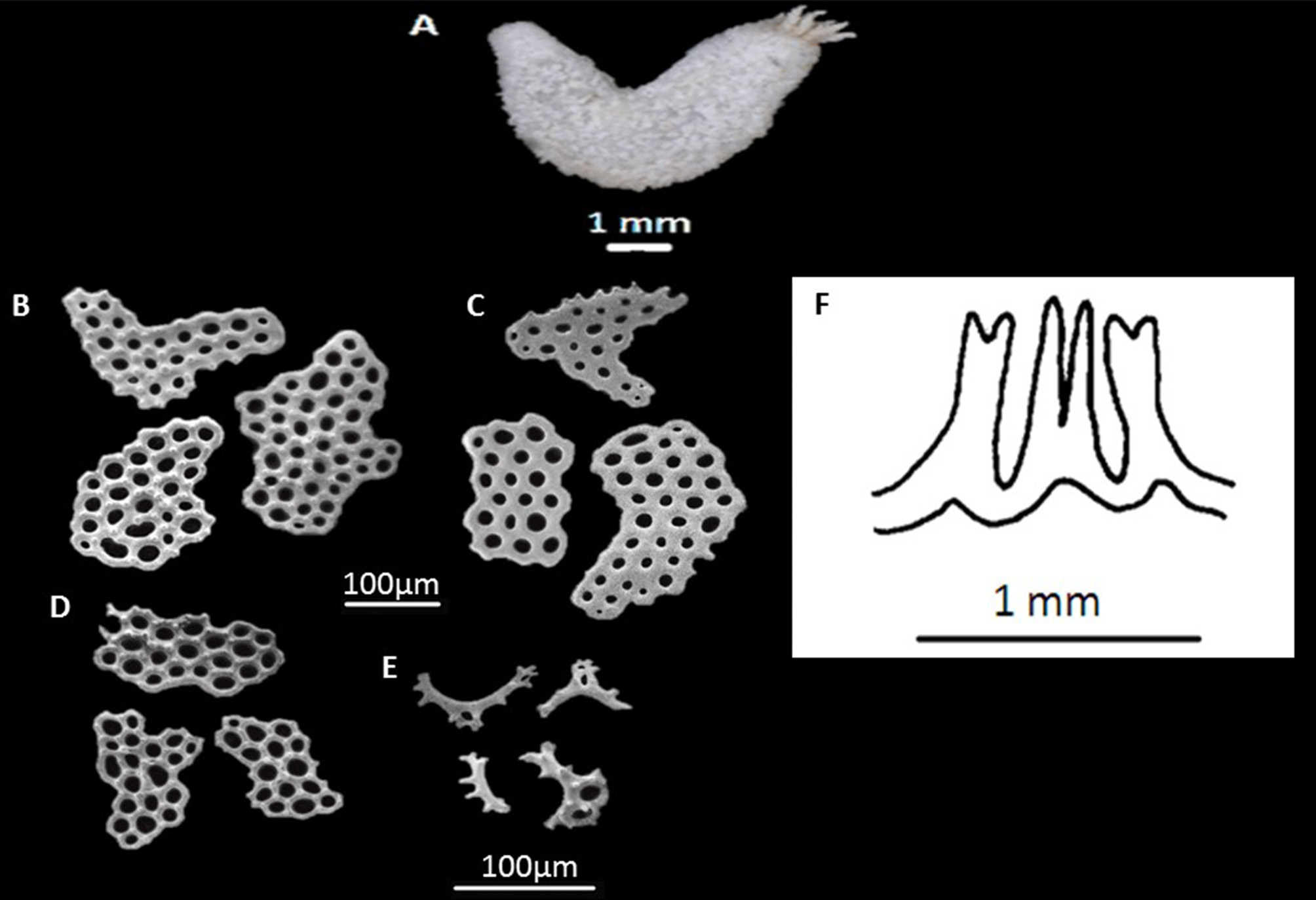
( Figure 8 View FIGURE 8 )
Material examined. Holotype: SAMC-A 090915 , SM232 , off coast of Transkei , south of Port St. Johns, 32° 10.9’ S, 29°10.4’ E, heavy dredge, 560–620 m, 26.VI.1979, 1 spec. GoogleMaps
Diagnosis. Holotype minute, U-shaped, length along ventrum 15 mm. Tentacles 10, of more or less equal size, not dendritic but with few sparse branches. Anal teeth present. Tube feet short, scattered. Calcareous ring simple. Stone canal short; Polian vesicles two. Respiratory trees elongate, unbranched. Body wall ossicles as simple, smooth or slightly knobbed imbricating, multilocular plates of a variety of shapes, those of ventral surface slightly elongate; nodules, when present, mostly minute, holes 7–16 in number, some plates polygonal. Tube feet rods curved, with characteristic short marginal projections; end-plates present. Tentacles with minute, smooth to slightly nodular, perforated plates.
Etymology. This species is named after my son, Mohammed, in appreciation of him providing us with a very comfortable retirement home in our twilight years.
Description. Holotype minute, U-shaped, oral and anal ends sharply turned up, mouth slightly above level of anus ( Figure 7A View FIGURE 7 ). Length along ventrum 15 mm, breadth in mid-body about 2 mm. Tentacles 10 of more or less equal size, mid-ventral two only slightly reduced; tentacles not at all tree-like or dendritic, branches few and short ( Figure 7A View FIGURE 7 ). Anal teeth present, minute. Tube feet short, scattered but more prominent in the ambulacra. Colour in alcohol white, including tube feet and tentacles. Calcareous ring simple, well developed ( Figure 7F View FIGURE 7 ); radial and interradial plates meeting at base, both of equal length, bifurcating anteriorly, notched posteriorly. Stone canal short, lying just behind the mid-dorsal interradial plate; madreporite well calcified but amorphous. Polian vesicles two, sac-like. Respiratory trees paired, unbranched, appearing as elongated tubes arising independently from cloaca, right one longer than left, both slightly curved distally. Gonad as paired tufts of short unbranched tubules, full of developing eggs, each tuft comprising about half a dozen tubules, younger tubules attached more posteriorly. Longitudinal muscles unpaired, filamentous. Retractor muscles arise from longitudinal bands at about mid-body, more anteriorly in ventral ambulacra. Body wall ossicles comprise simple, smooth or knobbed imbricating, multilocular plates of a variety of form ( Figure 7 View FIGURE 7 B–D), 140–250 µm, those of ventral surface slightly elongated; nodules, when present, mostly minute, found both around margin and surface of plates ( Figure 7B View FIGURE 7 ), rarely only on surface, of two different sizes, plates with large nodules rare; holes 7–16. Some plates smooth ( Figure 7C View FIGURE 7 ), some polygonal ( Figure 7D View FIGURE 7 ). Tube feet with very characteristic, strongly curved, smooth to minutely knobbed rods with short projections on curved surface, sometimes also with one or two medial holes ( Figure 7E View FIGURE 7 ). End-plates present, up to 100 µm, with a varying number of holes with no regular arrangement. Tentacles with minute perforated plates, up to 100 µm, either smooth or slightly nodular.
Distribution. Known only from type locality.
Remarks. The specimen at hand is clearly referable to Stereoderma , in his kirchsbergi group, as characterised by Panning (1949), because of the presence of thick, knobbed, imbricating plates. Panning included several species in this group as opposed to his unisemita group where the plates were said to be non-imbricating. At that time Stereoderma represented a hodgepodge collection of largely unrelated forms. Panning (1964) himself suspected this and thought that the genus was perhaps mono-typed by its type species, Stereoderma unisemita ( Stimpson, 1851) and only a couple more species could be referred to it. Since then many of the species originally contained in Stereoderma , were transferred to other genera by various workers. Within the existing species in Stereoderma the current specimen appears to be closest to Stereoderma congoana ( Heding, 1935) and Stereoderma monodi ( Cherbonnier, 1950) both West African species but with larger knobs to the plates. The ossicles of the current specimen come very close to those of Cucumaria inflexa Koehler & Vaney, 1908 from the Bay of Bengal, at 170 m. However, this latter species was transferred to Mitsukuriella by Heding and Panning (1954) on the assumption that the calcareous ring, as illustrated by Koehler & Vaney, indicates the presence of 15 and not 10 tentacles as described. The current specimen, however, has clearly 10 tentacles, scattered tube-feet, a calcareous ring of a different form and is hence different from C. inflexa . It is clear that further specimens of C. inflexa would do much to clarify issues concerning this species tentacle number and form of the calcareous ring. Koehler & Vaney (1908) illustrated rods from the body wall in C. inflexa but these may have come from the tube feet. However, they are of a different form than those which characterise the current material which, in addition, has smooth to knobbed plates in the body wall. The writer is of the opinion that the mature specimen at hand definitely represents a new species because of its scarcely branched tentacles, respiratory trees as simple, unbranched tubes, two Polian vesicles and very characteristic body wall and tube feet ossicles.

No known copyright restrictions apply. See Agosti, D., Egloff, W., 2009. Taxonomic information exchange and copyright: the Plazi approach. BMC Research Notes 2009, 2:53 for further explanation.
|
Kingdom |
|
|
Phylum |
|
|
Class |
|
|
Order |
|
|
Family |
|
|
Genus |