
Simulium (Chirostilbia) bifenestratum Hamada & Pepinelli
|
publication ID |
https://doi.org/ 10.34631/sporl.419 |
|
persistent identifier |
https://treatment.plazi.org/id/A2708A02-FFA5-FFE9-FF78-56FCFB01FD81 |
|
treatment provided by |
Felipe |
|
scientific name |
Simulium (Chirostilbia) bifenestratum Hamada & Pepinelli |
| status |
|
Simulium (Chirostilbia) bifenestratum Hamada & Pepinelli View in CoL
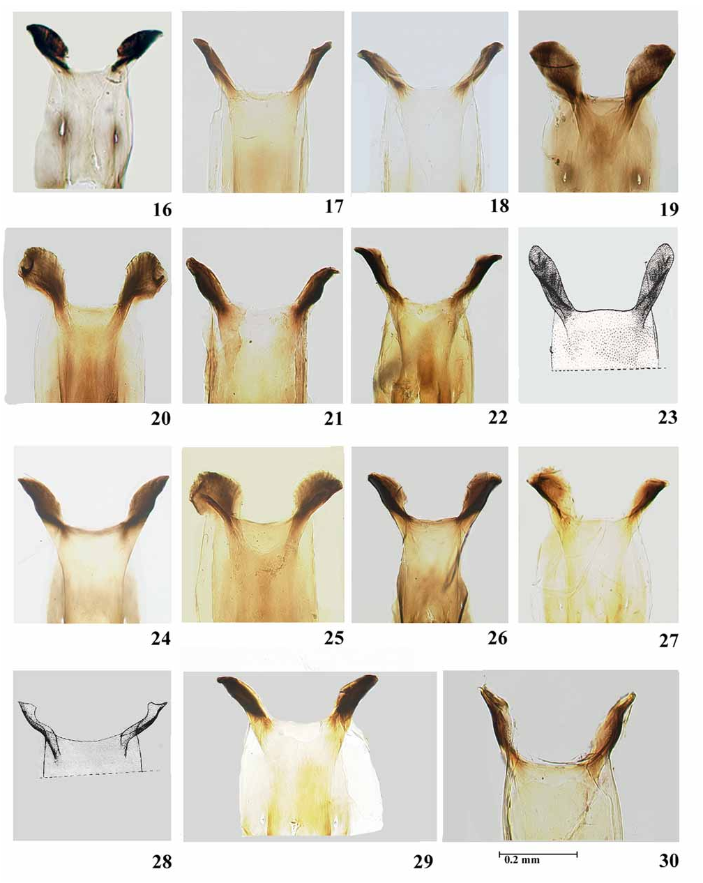
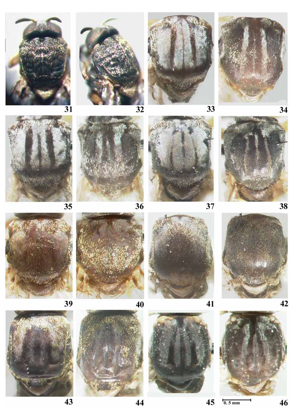
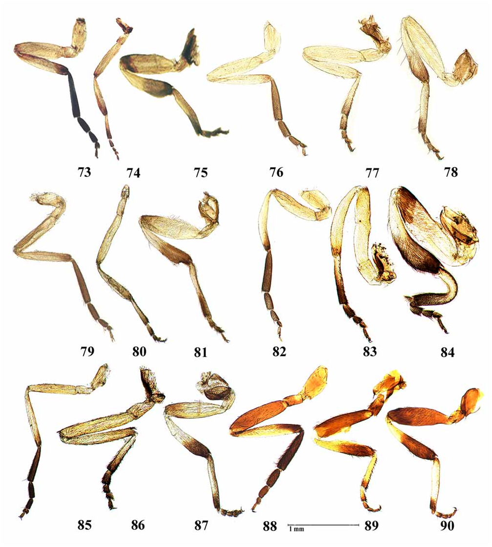
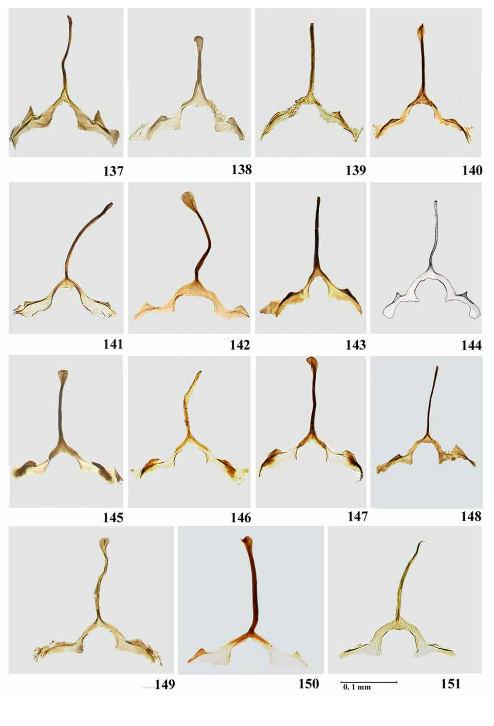
( Figs. 1 View PLATE 1 , 16 View PLATE 2 , 31, 32 View PLATE 3 , 73–77 View PLATE 6 , 106 View PLATE 8 , 121 View PLATE 10 , 137 View PLATE 11 , 152, 153 View PLATE 12 , 180 View PLATE 14 , 194 View PLATE 15 , 206 View PLATE 16 , 219 View PLATE 17 , 232 View PLATE 19 , 245 View PLATE 21 )
A recently described species from the state of São Paulo, Brazil. Our description is based on the original description and figures of this species in Hamada & Pepinelli (2004) because we were unable to obtain specimens.
Simulium bifenestratum Hamada & Pepinelli, 2004: 45 View in CoL . HOLOTYPE ♂ (reared), BRAZIL: São Paulo State, Bocaina mountain range, São José do Barreiros county, Verde GoogleMaps stream, 22º43’S 44º37’W; 16.v.2003, (Hamada, N. & Pepinelli, M.) (INPA).
Female. General scutum colour dark grey; body length 3.2–3.5 mm (n=2); lateral length of thorax 1.0– 1.25 mm (n=2). Wing length 3.3 mm (n=1), width 1.5 mm (n=1).
Head— eyes dark red. Frons, clypeus and occiput dark brown with silver pruinosity; nudiocular area small, as in Fig.1 View PLATE 1 . Antennal segments with silver pubescence; pedicel, scape and proximal area of first flagellomere brownish yellow, remaining flagellomeres dark brown. Mouthparts dark brown. Cibarium with- out teeth, with sclerotised cornuae ( Fig.16 View PLATE 2 ).
Thorax— [ Hamada & Pepinelli (2004) stated that that the number of reared females obtained was low (n=2), and that the emerged females were not well-preserved and so scutal characters were indistinct.] Scutum greyish black with pale golden hairs distributed unevenly, except in median region where groups of hairs form a line; one pair of small indistinct subrectangular silver spots on anterior margin adjacent to median line of hairs ( Figs. 31, 32 View PLATE 3 ). Scutellum light brown, with black and golden hairs; postnotum brown. Costa of wing with sparse distribution of spines and hairs. Subcosta with line of setae, but bare towards apex. Radius with row of setae intermixed with spines; basal section of Radius with hairs. Leg coloration and proportions as in Figs. 73–75 View PLATE 6 . Foreleg with coxa, trochanter, femur, and basal three-fourths of tibia light brown, remainder black. Mid leg with coxa dark brown, trochanter, femur and most of tibia light brown; distal end of tibia dark brown; basitarsus light brown with distal third dark brown; other segments dark brown. Hind leg with coxa dark brown; trochanter light brown; femur, basal half of tibia, and basitarsus light brown; with distal articulation of femur, distal half of tibia, basitarsus and remainder of tarsus dark brown. Tarsal claws with basal thumb-like lobe. Femora and tibiae with filiform setae.
Abdomen— sclerites dark brown; membranous areas light brown. Basal fringe with long, thin, golden hairs. Tergite II with silver pruinosity; tergites VI–IX shiny. Eighth sternite weakly sclerotised with numerous setae on posterior margin; gonapophyses subtriangular with posterior apex lobed, membranous with internal margin weakly sclerotised, presence or absence of basal setae not evident from original authors’ photograph ( Fig. 106 View PLATE 8 ). Cercus hemispherical; paraproct subrectangular with extension beyond ventral margin of cercus triangular and twice as long as cercus; cercus and paraproct covered by hairs ( Fig. 121 View PLATE 10 ). Genital fork with triangular anterior process developed and posterior process undeveloped ( Fig. 137 View PLATE 11 ). Spermatheca subspherical, with internal setae, not forming a pattern; spermathecal duct and area of attachment unpigmented.
Male. General body colour black. Body length: 3.4 mm (n=1); lateral thorax length: 1.0 mm (n=1). Wing, length 3.1 mm (n=1), width 1.35 mm (n=1).
Head— holoptic, coloration as in female.
Thorax— scutum, irrespective of light direction, black, with densely distributed golden yellow hairs ( Fig. 152, 153 View PLATE 12 ). Scutellum dark brown, postnotum black. Wing veins as in female. Leg coloration as in female.
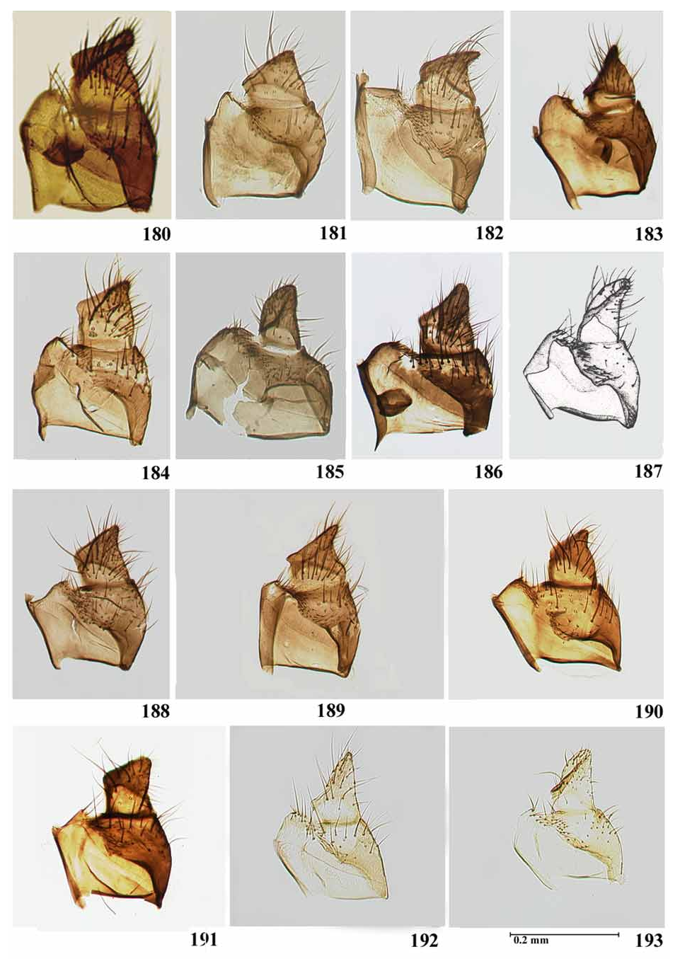
Abdomen— tergites black; basal fringe with thin, long, black hairs; lateral areas of tergites II, V–VII with silver pruinosity. Gonocoxite and gonostyle black; gonocoxite subrectangular; gonostyle shorter than gonocoxite and trapezoidal with apex curved dorsally [no information in original description about presence or absence of spine], ridge parallel to distal margin ( Fig. 180 View PLATE 14 ). Ventral plate subtriangular, covered in setae and with sclerotised basal arms ( Fig. 194 View PLATE 15 ). Median sclerite pear-shaped and elongate with apical incision ( Fig. 206 View PLATE 16 ). Paramere with spines in apical half; basal arms of paramere developed ( Fig. 219 View PLATE 17 ).
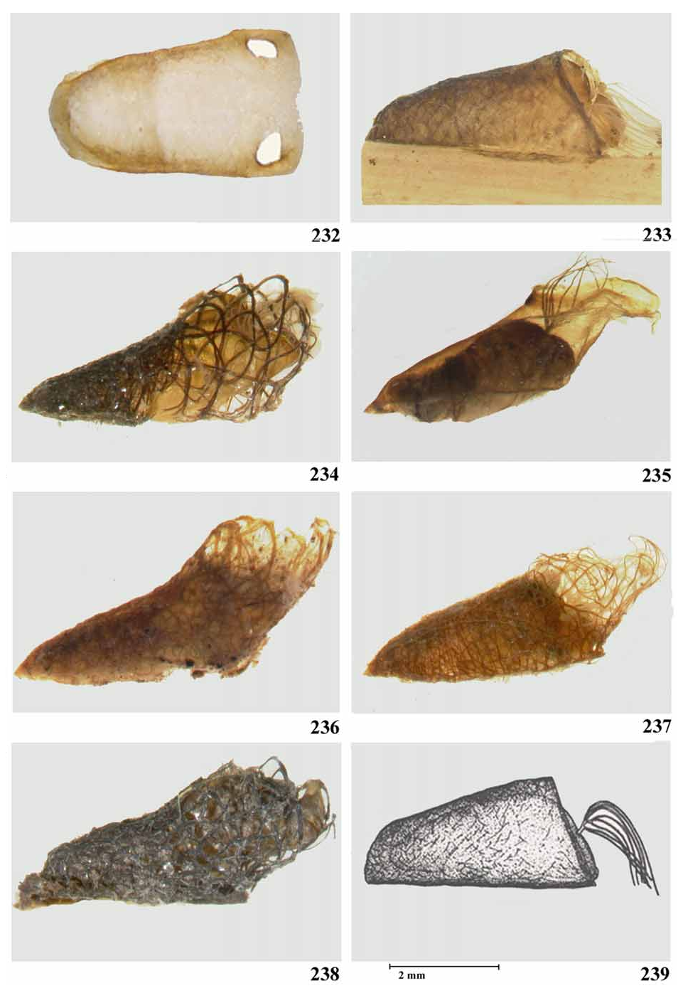
Pupa. Body length 3–4.3 mm (mean= 3.7 mm, n=5). Length along dorsal surface of cocoon 3.9–4.5 mm (mean= 4.2 mm, n= 5); ventral surface of cocoon 1.8–2.2 mm (mean=2.0 mm, n=5).
Cocoon— slipper-shaped, very hard and thick, with two lateral, anterior openings ( Fig. 232 View PLATE 19 )
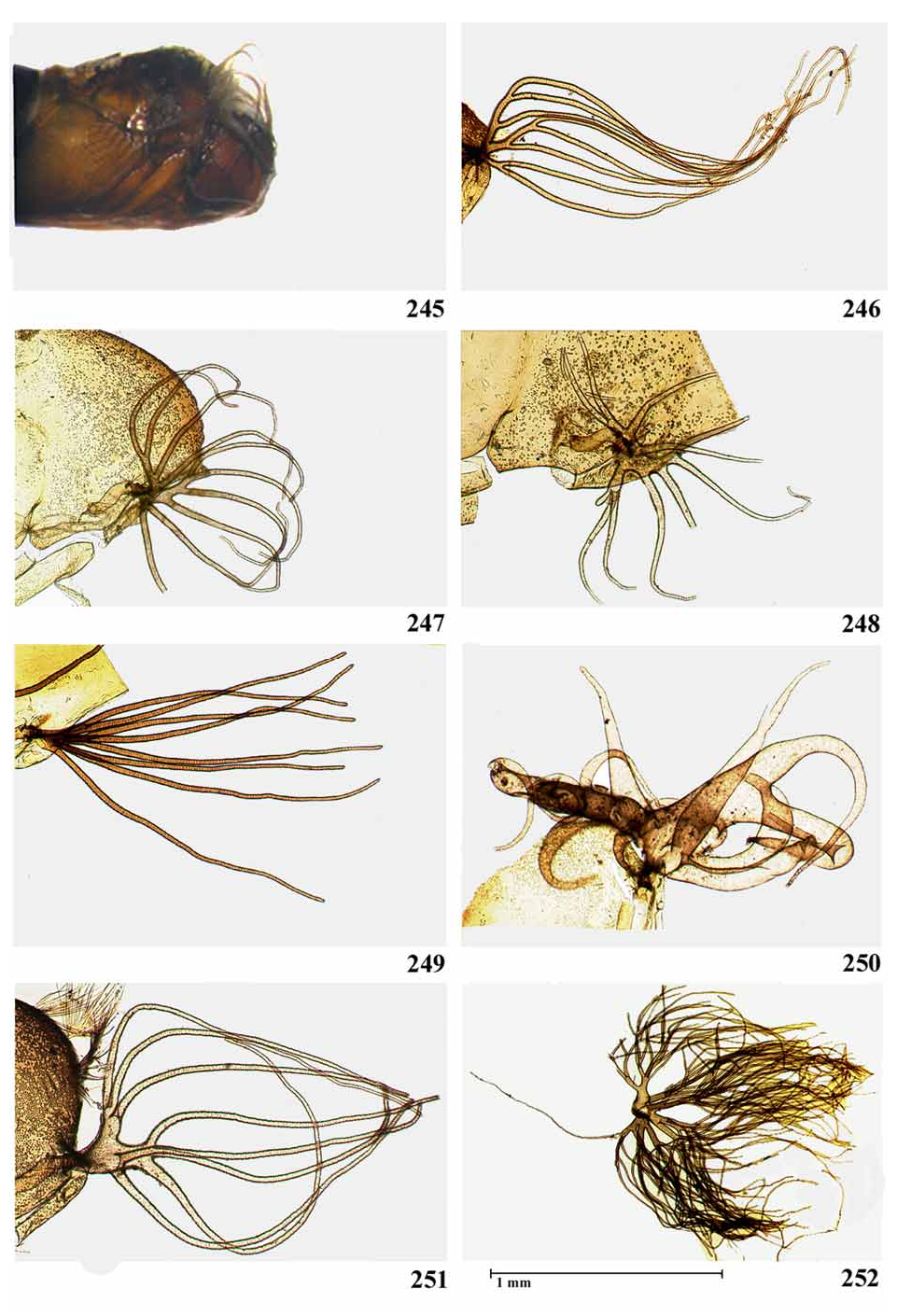
Gills— pale with 10, thicker at base, arranged as rosette initially in vertical plane and then curving over dorsum of cephalothorax ( Figs. 245 View PLATE 21 ).
Head— frontoclypeu s projecting downwards, with two pairs of frontal trichomes, each with 1–2 branches, one pair of facial trichomes with 2–3 branches; large rounded tubercles covered with small protuberances. Marginal area of frontoclypeus elevated, especially lateral region, forming a structure similar to a crown.
Thorax— with large tubercles on anterior to median region, many of which are pointed on dorsal side; middle to posterior region with tubercles of various sizes with or without pointed projections; five pairs of trichomes with 3–9 branches, and one pair of simple, lateral trichomes.
Abdomen— tergite I with one pair of simple or bifurcated setae sublaterally and 2–3 pairs of simple setae on median region; tergite II with three pairs of stout setae and three thinner pairs sublaterally; tergites III and IV each with four anteriorly directed pairs of hooks on posterior margin; one small seta between outermost hooks; tergites I–IV with small, unpigmented, elongated spots, forming an open V-shaped pattern in median region; tergite V with one pair of stout setae laterally and three pairs of small simple trifid submedian setae; tergite VI with two pairs of simple bifid submedian setae; tergites VIII and IX with stronger spines on central, anterior margin; tergite IX with one pair of short terminal spines; tergites VI–IX with comb-like groups of fine, posteriorly directed spine-combs on anterior margins. Sternites III–IX with anterior, median group of spine combs; sternites IV–V with two plates divided by median membranous area; sternite IV with three pairs of submedian to lateral setae and one pair of stout hooks (simple or bifid); sternite V with two pairs of lateral setae and two pairs of stout hooks (bifid or trifid); sternites VI–VII with four plates, each divided by membranous areas; each plate bearing one hook (simple-trifid).
Larva. A description of the larval stage may be found in Hamada & Pepinelli (2004).


No known copyright restrictions apply. See Agosti, D., Egloff, W., 2009. Taxonomic information exchange and copyright: the Plazi approach. BMC Research Notes 2009, 2:53 for further explanation.
|
Kingdom |
|
|
Phylum |
|
|
Class |
|
|
Order |
|
|
Family |
|
|
Genus |
Simulium (Chirostilbia) bifenestratum Hamada & Pepinelli
| Hernández, Luis Miguel, Shelley, Anthony John, Dias, Antonio Paulino Andrade De Luna & Maia-Herzog, Marilza 2008 |
Simulium bifenestratum
| Hamada, N. & Pepinelli, M. 2004: 45 |