
Serrapinnus potiguar, Jerep & Malabarba, 2014
|
publication ID |
https://doi.org/ 10.1590/1982-0224-20130218 |
|
DOI |
https://doi.org/10.5281/zenodo.4775845 |
|
persistent identifier |
https://treatment.plazi.org/id/039ED57B-BA0D-606F-87D5-C20B2A6DC047 |
|
treatment provided by |
Carolina |
|
scientific name |
Serrapinnus potiguar |
| status |
sp. nov. |
Serrapinnus potiguar View in CoL , new species
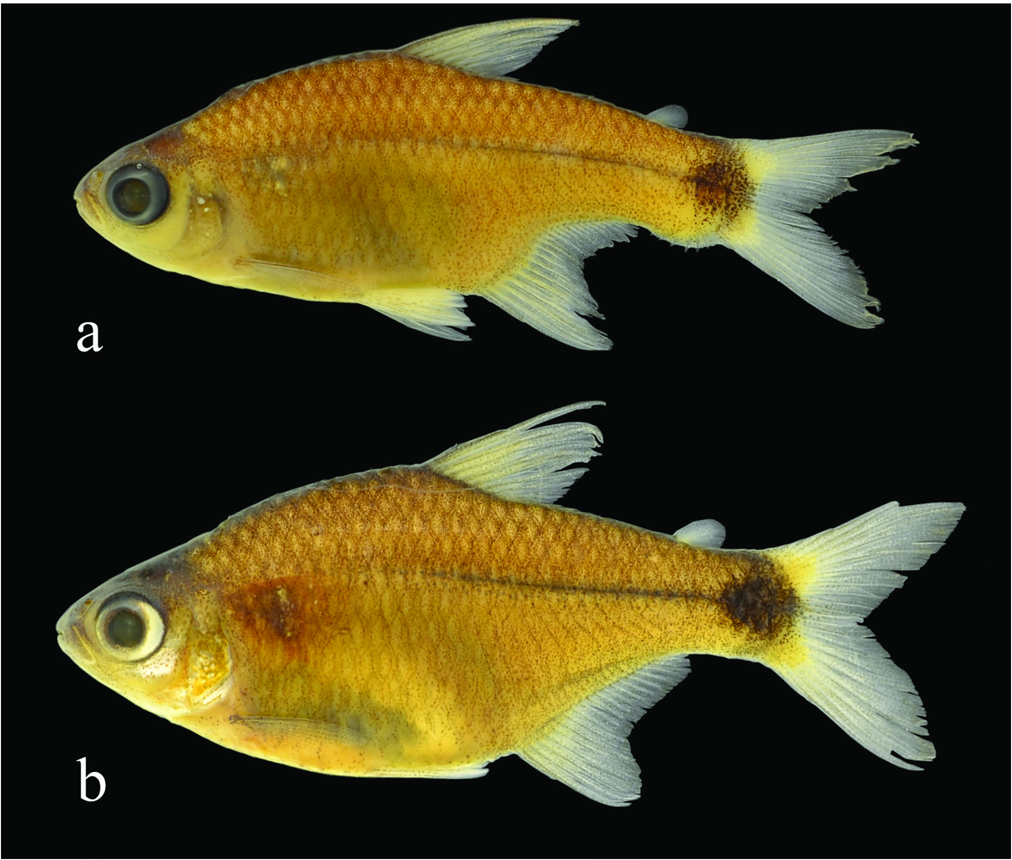
Figs. 1-5 View Fig View Fig View Fig View Fig View Fig
Serrapinnus sp.A. - Dias & Fialho, 2009: 242-248 [trophic category and feeding behavior].
Holotype. MCP 48054, 26.6 mm SL, male, Brazil, Rio Grande do Norte, Taipu, rio Ceará-Mirim, district of Umari , 05°37’47’’S 35°37’09’’W, 15 Oct 2000, H. Gurgel. GoogleMaps
Paratypes. All from Rio Grande do Norte State, Brazil: MCP 34076, 5 View Materials , 25.8-27.8 mm SL, Taipu , district of Umari , rio Ceará-Mirim , 5°37’47’’S, 35°37’9’’W, 24 Jul 2001, L. R. Malabarba & H. Gurgel. GoogleMaps MCP 43320, 9 View Materials , 17.5-29.5 mm SL, same as holotype. UFRGS 9216 View Materials , 13 View Materials , 22.6-29.5 mm SL (5, 25.6-29.1 mm SL), rio Ceará-Mirim , 5°37’46.9’’S 35°37’9.1’’W, 23 Feb 2002. UFRGS 9219 View Materials , 27 View Materials , 18.5- 28.4 mm SL + 10 c&s 25.6-29.1 mm SL (5 measured, 26.2-28.7 mm SL), rio Ceará-Mirim, H. Rangel, Sep 2001. GoogleMaps UFRGS 9226 View Materials , 55 View Materials , 22.4-32.3 mm SL (20, 26.5-32.3 mm SL), rio Ceará-Mirim, 5°37’46.9’’S 35°37’9.1’’W, H. Rangel, 27 Apr 2002, H. Rangel GoogleMaps .
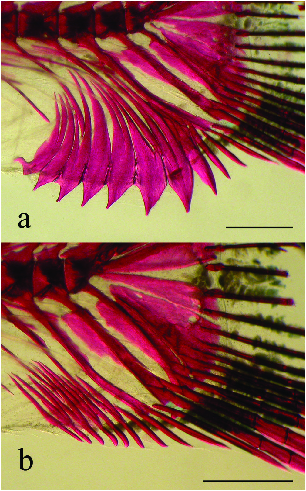
Diagnosis. Serrapinnus potiguar is distinguished from all congeners by the shape and arrangement of the ventral procurrent caudal-fin rays on mature males, where the hypertrophied elements are scimitar-shaped and arranged in a semi-circle through the ventral profile of the caudal peduncle ( Fig. 2a View Fig ) (vs. hypertrophied elements rod-shaped or pointed distally, arranged parallel to each other). Additionally S. potiguar can be diagnosed by the presence of 5 cusps in the premaxillary and dentary teeth (vs. 7 to 9 in S. calliurus , S. micropterus , and S. piaba , 9 to 11 in S. sterbai , and 10 to 12 in S. gracilis and S. littoris ), terminal mouth (vs. slightly superior mouth in S. microdon ), an incomplete lateral line (vs. complete lateral line in S. heterodon ), dorsal fin mostly hyaline (vs. dorsal fin with an anterior and proximal dark blotch in S. notomelas ), and absence of a black spot in the posteroventral region of the abdomen (vs. presence of a black spot in that region in S. kriegi ).
Description. Morphometric data on Table 1 View Table 1 . Body slightly short and compressed. Greatest body depth at vertical through dorsal-fin origin. Snout slightly rounded. Dorsal profile of head convex from snout to vertical through posterior border of nares, slightly convex from that point to distal tip of supraoccipital bone. Predorsal profile convex from posterior end of supraoccipital to dorsal-fin origin. Dorsal-fin origin located at vertical at midlength of standard length. Dorsal profile from last dorsal-fin ray to adipose-fin base slightly convex in females and strongly ventrally arched on preserved mature males. Caudal-peduncle dorsal profile slightly concave from adipose-fin origin to caudal-fin origin. Caudal-peduncle ventral profile straight to slightly concave in females, pronounced convex with exposed bony spines due to hypertrophy of ventral procurrent caudal-fin rays on males. Caudal peduncle slightly longer than deep. Ventral profile of head slightly convex from mouth to pelvic-fin origin; straight from that point to anal-fin origin in females and concave in mature males. Anal-fin base straight to slightly concave in females, convex anteriorly and deeply concave posteriorly on mature males. Head pointed on lateral view. Mouth terminal, at horizontal line through middle of pupil. Maxilla angled posteroventrally, posterior tip reaching vertical through anterior border of eye and surpassing ventrally the horizontal through ventral border of eye. Teeth pedunculated, distally expanded, with similar shape and cusp number. Four (1), 5(7), or 6(2) premaxillary teeth aligned in one single row, bearing 5 cusps. Midcentral cusp longer and wider than lateral ones. Three (3) or 4(7) maxillary teeth bearing 3 to 5 cusps. Six (8) to 7(2) large dentary teeth bearing 5 cusps, followed posteriorly by one smaller tooth with 3 to 4 cusps and one or two conical teeth. Central cusp larger than lateral cusps. All dentary tooth cusps slightly directed lingually ( Fig. 3 View Fig ).
Dorsal-fin origin on midlength of standard length. Dorsal-fin rays ii,9*(31). First unbranched dorsal-fin ray about half length of second unbranched dorsal-fin ray, following branched rays gradually decreasing in size posteriorly. Adipose-fin origin slightly posterior to vertical through base of last anal fin. Anal-fin origin posterior to vertical through base of last dorsalfin ray. Anal-fin rays ii(5), iii*(17), or iv(7), 17(1), 18(10), 19*(16), or 20(4). Anal-fin distal profile rounded on anterior lobe and concave posteriorly on females; pointed on anterior lobe and deeply concave posteriorly on males. Last unbranched and first two branched anal-fin rays longer, remaining rays decreasing in size posteriorly. Distal tip of anterior anal-fin rays of mature males overlapping laterally last anal-fin rays, commonly reaching ventral procurrent caudal-fin rays when caudal peduncle deeply arched. Males with acute, retrorse hooks on posterior border and posterior branches of anal-fin rays, posterolaterally arranged on last unbranched to 8 th, 9 th or 10 th branched rays, rarely on 12 th ( Fig. 4 View Fig ). Two to three unpaired hooks per ray segment of lepidotrichia. Hooks mostly distributed along middle third of anal-fin ray’s length. Hookbearing rays with segments and branches progressively fused according to degree of maturation on males. Hypertrophied soft tissue associated to hook bearing anal-fin rays. Pectoral-fin rays i*(31), 10*(16), or 11(12), rarely 9(1) or 12(2). Unbranched pectoral-fin ray falling short of pelvic-fin origin on females, and extending beyond that point on adult males. Pelvic-fin origin anterior to vertical through dorsal-fin origin. Pelvic-fin rays i,7*(30), rarely i,8(1). Unbranched and branched pelvic-fin rays with acute hooks on males, 1 pair per segment of lepidotrichia, ventromedially placed on rays, associated with hypertrophied soft tissue. Principal caudal-fin rays 16(1), 17(1), 18(1), 19*(25), or 20(1). Procurrent caudal-fin rays 9(1), 10(4), 11(2), or 12(2) dorsal; 11(5), 12(3), or 13(2) ventral. Ventral procurrent caudalfin rays hypertrophied on mature males, trespassing muscles and skin on ventral margin of caudal-peduncle, resulting in semicircular profile. Hypertrophied ventral procurrent ray scimitar-shaped: anteriorly bent, proximally acute, expanding distally, abruptly ending in pointed distal tip ( Fig. 2a View Fig ).
Scales cycloid. Pored scales on lateral line 6*(1), 7(6), 8*(12), 9(10), or 10(2); scales on longitudinal line 30(1), 31(4), 32*(6), 33(13), 34(6), or 35(1); predorsal scales 10*(5), 11(16), or 12(10); scale rows from lateral line to dorsal-fin origin 6(28) or 7*(3); scale rows from lateral line to pelvicfin origin 4*(30) or 5(1); circumpeduncular scale rows 12(2), 13(16), or 14*(13). Scales along anal-fin base 9(1), 10(2), 11*(13), 12(9), 13(5), or 14(1).
Supraneurals 4(6) or 5(4); abdominal vertebrae 15(10); caudal vertebrae 18(9) or 19(1). Gill rakers 8(1), 9(2), or 10(2) on lower branch; 3(1) or 4(4) on upper branch.
Color in alcohol. Overall body coloration yellowish to dun. Head darker over dorsal region of neurocranium, snout and maxilla, due to higher concentration of dark chromatophores. Dark brown chromatophores sparsely scattered over infraorbitals and opercular apparatus. Opercular apparatus, some infraorbitals and branchiostegal rays silver in some individuals. Ventral region of head and abdomen light. Humeral region with triangular darkened area due to pseudotympanum muscular hiatus. Longitudinal line of dorsum darker than lateral of body. Body scales with higher concentration of black chromatophores on distal margin, resulting in reticulated coloration pattern more conspicuous on upper portion of body. Longitudinal black subcutaneous line on lateral of body extending from vertical through dorsalfin insertion to caudal peduncle spot. Black chromatophores aligned and following line of miosepta on body above anal fin. Caudal spot black, slightly rounded, occupying most of caudal peduncle termination, not reaching upper or lower margins of caudal peduncle. Dorsal, anal, pectoral, pelvic, and caudal fins mostly hyaline, with dark chromatophores scattered on interradial membrane. Unbranched dorsal-fin rays darker than branched rays due to higher concentration of black chromatophores. Adipose fin mostly hyaline, with few black chromatophores distributed near dorsal margin. Caudal fin with clear yellowish areas on base of caudal-fin lobes, bordering caudal-fin spot posteriorly.
Distribution. Serrapinnus potiguar is known to inhabit the rio Ceará-Mirim basin, Rio Grande do Norte State, northeastern Brazil.
Etymology. The epithet potiguar refers to “potiguar”, a term traditionally used in Brazil to refer to someone born in the Rio Grande do Norte State. An adjective.
Ecological notes. The type locality of Serrapinnus potiguar in the rio Ceará-Mirim (05°37’47’’S 35°37’09’’W) was described by Dias & Fialho (2009) as clear and transparent water, sandy bottom high depth 1.2 m. The water current varied from slow to Color in life. Dark pigmentation as described on alcohol preserved specimens. Body light olive brown slightly translucent. Pectoral fin slightly orange or red; dorsal, anal, pelvic and caudal fins intense orange or red ( Fig. 5 View Fig ). Distal tip of longest anal- and pelvic-fin rays white or hyaline. Middle caudal-fin rays hyaline.
Sexual dimorphism. Bony hooks were only observed on pelvic and anal-fin rays of sexually dimorphic males, as well as hypertrophied and ventrally exposed procurrent caudalfin rays ( Fig. 2a View Fig ). Sexually dimorphic males also present those anal-fin rays that bear bony hooks hypertrophied and expanded in the sagittal plane ( Fig. 4 View Fig ). On fixed and preserved specimens the caudal peduncle is generally found arched ventrally only on adult males ( Fig. 1a View Fig ). Furthermore, sexually mature males have gill glands, located on the anteriormost portion of the lower branch of the first gill arch, extending posteriorly through six to eight of gill filaments.
moderate and in that stretch the river presented a large amount of floating and submerse vegetation. In that locality S. potiguar is sympatric and syntopic with the cheirodontines Compsura heterura , S. heterodon and S. piaba ; and its diet was mainly composed by vegetal matter and algae, but also by insects and microcrustaceans (Dias & Fialho, 2009).


| MCP |
Pontificia Universidade Catolica do Rio Grande do Sul |
No known copyright restrictions apply. See Agosti, D., Egloff, W., 2009. Taxonomic information exchange and copyright: the Plazi approach. BMC Research Notes 2009, 2:53 for further explanation.
|
Kingdom |
|
|
Phylum |
|
|
Class |
|
|
Order |
|
|
Family |
|
|
Genus |