
Scorpaena regina, Wibowo & Johnson & Motomura, 2019
|
publication ID |
https://doi.org/ 10.11646/zootaxa.4706.2.5 |
|
publication LSID |
lsid:zoobank.org:pub:86EF57D4-01F7-437C-8DAD-88D2549C17F3 |
|
persistent identifier |
https://treatment.plazi.org/id/861487C2-FFB4-FFBF-FF0A-75021D7EF8A5 |
|
treatment provided by |
Plazi |
|
scientific name |
Scorpaena regina |
| status |
sp. nov. |
Scorpaena regina sp. nov.
New English name: Eastern Queen Scorpionfish
Figures 1–6 View FIGURE 1 View FIGURE 2 View FIGURE 3 View FIGURE 4 View FIGURE 5 View FIGURE 6 ; Tables 1–3 View TABLE 1 View TABLE 2 View TABLE 3
Holotype. QM I. 37447, 63.4 mm SL, Tangalooma Wrecks, Moreton Bay , Queensland, 27°10ʹS, 153°22ʹE, 3–9 m, J. Johnson, 6 June 1994. GoogleMaps
Paratypes. 58 specimens, 22.5–64.5 mm SL, all from the east coast of Queensland (Qld), Australia. AMS I. 34323-001, 42.0 mm SL, northwest of Quoin Island GoogleMaps , 22°33ʹ42.0ʹʹS, 150°47ʹ40.8ʹʹE, AMS Party GoogleMaps ; AMS I. 34324- 026, 28.4 mm SL, off Dome Island GoogleMaps , 3 m, AMS Party GoogleMaps ; AMS I. 34326-013, 35.2 mm SL, south of Quoin Island GoogleMaps , 22°34ʹ01.2ʹʹS, 150°47ʹ52.2ʹʹE, AMS Party GoogleMaps ; AMS IA. 3841 (2 specimens), 22.5–32.8 mm SL, off Whitsunday GoogleMaps Pas- sage, 20°18ʹS, 149°54ʹE, M. Ward ; CSIRO H 7372-01 View Materials , 33.5 mm SL, east of Great Keppel Island GoogleMaps , 23°16ʹS, 151°09ʹE, 25 m, D. Gledhill, FRV Gwendoline May, 24 Apr. 2004 ; KAUM–I. 132015, 63.5 mm SL, KAUM–I. 132016, 53.8 mm SL, KAUM–I. 132017, 45.5 mm SL, KAUM–I. 132018, 41.3 mm SL, Curtin Artificial Reef, Moreton Bay GoogleMaps , 27°07ʹS, 153°21ʹE, 21 m, J. Johnson, 5 Apr. 1995 ; QM I. 21339, 49.4 mm SL, southeast of Lady Elliot Island , 46–55 m, trawl, G. Lowe, 9 Mar. 1982; QM I. 23083, 48.9 mm SL, off Swain Reefs, 21°13.8ʹS, 150°43.1ʹE, 47 m, trawl, Queensland Fisheries Service, 16 Sept. 1986 GoogleMaps ; QM I. 29123, 47.7 mm SL, off Whaling Jetty, Tangalooma, Moreton Bay GoogleMaps , 27°11ʹS, 153°22ʹE, 15 m, J. Johnson & J. Short, 28 Apr. 1994 ; QM I. 29155 (4), 26.4–55.7 mm SL, Tangalooma Wrecks, Moreton Bay GoogleMaps , 27°10ʹS, 153°22ʹE, 3–6 m, J. Johnson & J. Short, 28 Apr. 1994 ; QM I. 29562, 46.3 mm SL, Gneering Shoals, off Mooloolaba, 26°40ʹS, 153°12ʹE, 21 m, J. Johnson, 30 Jan. 1995 ; QM I. 30365 (2) 39.2–46.1 mm SL, Myora Reef, Moreton Bay , 27°28ʹS, 153°25ʹE, 3–6 m, J. Johnson, 5–7 Mar. 1996 GoogleMaps ; QM I. 31139, 55.1 mm SL, off Southport Seawall, 27°56ʹS, 153°26ʹE, 2–4 m, J. Johnson, 29 May 1998 ; QM I. 34098 (5), 38.9–44.1 mm SL, off Burnett Heads, 24°31ʹS, 152°44ʹE, 30 m, trawl, Queensland Fisheries Service, 8 Oct. 2000 GoogleMaps ; QM I. 34099, 49.8 mm SL, off Burnett Heads, 24°36ʹS, 152°32ʹE, 20 m, trawl, Queensland Fisheries Service, 9 Oct. 2000 GoogleMaps ; QM I. 34172, 54.1 mm SL, off Keppel Islands GoogleMaps , 23°07ʹS, 151°11ʹE, 29 m, trawl, Queensland Fisheries Service, 3 Oct. 2000 ; QM I. 36171, 42.1 mm SL, Broad Sound Channel, 22°10.5ʹS, 150°27.9ʹE, 33 m, dredge, Sea- bed Biodiversity Project Team, 10 May 2004 ; QM I. 36308 (2), 34.5–36.3 mm SL, east of Palm Islands GoogleMaps , 18°42.3ʹS, 146°48.3ʹE, 30 m, dredge, Seabed Biodiversity Project Team, 30 Apr. 2004 ; QM I. 36311, 37.9 mm SL, east of Cur- tis Island, 23°39.9ʹS, 151°24.3ʹE, 23 m, dredge, Seabed Biodiversity Project Team, 20 May 2004 ; QM I. 36344 (2), 37.0– 38.8 mm SL, southeast of Great Keppel Island GoogleMaps , 23°15.9ʹS, 151°9.9ʹE, 28 m, dredge, Seabed Biodiversity Proj- ect Team, 21 May 2004 ; QM I. 36370 (11), 24.7–40.3 mm SL, west of North West Island GoogleMaps , 23°21.3ʹS, 151°29.1ʹE, 35 m, dredge, Seabed Biodiversity Project Team, 22 May 2004 ; QM I. 36557, 32.6 mm SL, northwest of Percy Isles GoogleMaps , 21°14.1ʹS, 150°07.5ʹE, 55 m, dredge, Seabed Biodiversity Project Team, 29 Sept. 2004 ; QM I. 36563, 46.1 mm SL, east of Redbill Island GoogleMaps , 20°59.7ʹS, 150°16.5ʹE, 57 m, dredge, Seabed Biodiversity Project Team, 29 Sept. 2004 ; QM I. 36718, 36.8 mm SL, off Northumberland Islands GoogleMaps , 21°27.9ʹS, 149°47.7ʹE, 23 m, dredge, Seabed Biodiversity Project Team, 28 Sept. 2004 ; QM I. 36773 (2), 34.0– 53.3 mm SL, west of North West Island GoogleMaps , 23°17.7ʹS, 151°35.7ʹE, 37 m, dredge, Seabed Biodiversity Project Team, 20 Sept. 2004 ; QM I. 36815 (3), 37.0– 49.3 mm SL, west of Lady Musgrave Island, 23°53.1ʹS, 152°06.3ʹE, 41 m, dredge, Seabed Biodiversity Project Team, 21 Sept. 2004 ; QM I. 36836, 34.5 mm SL, northeast of Stanage Bay, 22°03.9ʹS, 150°05.1ʹE, 19 m, dredge, Seabed Biodiversity Project Team, 5 Oct. 2004 ; QM I. 37446, 61.1 mm SL, Bandicoot Wreck, Amity Point, 27°24ʹS, 153°26ʹE, 2–9 m, J. John- son, 14 Mar. 1995 ; QM I. 40982 (3), 34.8–64.5 mm SL, same data as holotype
.
Diagnosis. A species of Scorpaena with the following combination of characters: dorsal-fin soft rays 9; pectoral-fin rays 13–17 (mode 16); scale rows in longitudinal series 39–46 (41 or 42); pored lateral-line scales 21–24 (23); scales above lateral line 5–7 (6), below 11–14 (12); scale rows between sixth dorsal-fin spine base and lateral line 5 or 6; scale rows between last dorsal-fin spine base and lateral line 5 or 6; pre-dorsal scale rows 4–7 (4 or 5); gill rakers on upper limb 4 or 5, lower limb 9–12 (10) [7–9 (8) and 1–3 (2) rakers on ceratohyal and hypobranchial, respectively], total rakers 13–17 (14 or 15; rarely 16 or 17, 3 and 1 of all paratypes, respectively); ctenoid scales covering lateral surface of body; exposed (or embedded in thin skin) cycloid scales covering anteroventral surface of body and pectoral-fin base; lateral surface of maxilla without a longitudinal ridge; lateral surface and dorsal margin of lacrimal without spines (two paratypes with lateral lacrimal spine and vertical spine on dorsal margin of lacrimal, respectively); anterior lacrimal spine simple (one paratype with a small spinous point on posterior margin); posterior lacrimal spine simple, directed posteroventrally throughout life; median interorbital ridge and coronal spine absent; occipital pit and supplementary preopercular spine present; pterotic spine simple; posterior tip of pectoral fin not reaching to vertical through first anal-fin spine base; first anal-fin spine base located slightly posterior to vertical through last dorsal-fin spine base; prepelvic-fin length 36.3–43.0 (mean 39.8) % of SL; 9th and 10th dorsal-fin spine lengths 8.9–13.3 (11.1) % of SL and 6.5–10.1 (8.4) % of SL, respectively; caudal peduncle depth 10.5–12.3 (11.3) % of SL; space between upper and lower opercular spines covered by thin skin with small sensory pores; underside of lower jaw smooth, without tentacles; supraocular tentacle length variable, longest approximately equal to orbit diameter; several distinct slender tentacles (associated with pored lateral-line scales) scattered on lateral surface of body; largest recorded specimen 64.5 mm SL; and depth of distribution at 2– 57 m.
Description. Data for holotype presented first, followed by paratype data in parentheses (if different). Morphometrics and meristics given as percentages of SL in Table 1 View TABLE 1 . Head spination illustrated in Figure 3 View FIGURE 3 . Dorsal fin with 12 spines and 9 soft rays; all soft rays branched; fourth (sometimes fifth) spine longest, slightly shorter than upper-jaw length; fourth to eleventh spines progressively shorter; second (sometimes third) soft ray longest, slightly longer than longest dorsal-fin spine; posterior branch of last soft ray joined by membrane to caudal peduncle for approximately four-fifths (two-thirds) its length. Anal fin with 3 spines and 5 soft rays; all soft rays branched; second soft ray longest; posterior branch of last soft ray joined by narrow membrane to caudal peduncle for approximately one-seventh its length. Pectoral fins each with 16 rays [left side 13–17, usually 16, one specimen with 13 rays; right side 11–17, usually 16, three specimens with 11–13 rays]; single uppermost ray and 11 lower rays unbranched, remaining 4 branched [1 or 2 upper rays and 8–12 (usually 10) lower rays unbranched in larger paratypes over 33.1 mm SL; all rays unbranched (7 specimens) or 2–4 upper rays and 11–12 lower rays unbranched (7 specimens) in smaller paratypes less than 32.8 mm SL]; 7th (or 8th) ray longest, shorter than head length; lower unbranched rays thickened; posterior margin of fin rounded. Pelvic fin with 1 spine and 5 branched soft rays; second soft ray longest, longer than upper-jaw length; last soft ray joined by membrane to abdomen for approximately two-thirds its length. Caudal fin with 13 principal rays, posterior margin of fin slightly rounded. Gill rakers 13–17, short and spinous, length of longest raker on first gill arch shorter than gill filaments around angle of gill arch; fourth gill slit closed by membrane. Branchiostegal rays 7. Vertebrae 24. Swimbladder absent.
Body moderately compressed anteriorly, progressively more compressed posteriorly. Nape and anterior body arched. Body relatively shallow, depth less than head length. Numerous small to tiny papillae on upper half of head, including upper outer margin of eye membrane, interorbital space, occipital pit, and behind orbit extending to an area between upper and lower opercular spines. A short, slender, fleshy tentacle (supraocular tentacle), its length approximately equal to pupil diameter (largest, right side tentacle damaged in holotype; length variable, longest tentacle approximately equal to eye diameter) on posterior end of supraocular spine base; supraocular tentacle lacking (with several) short branches along margin. A short but distinct slender tentacle (parietal tentacle) on posterior end of parietal spine base, shorter than nuchal spine length (usually extending beyond tip of nuchal spine when laid back). A short slender tentacle between tympanic and pterotic spines. A short slender tentacle (preocular tentacle) on posterior end of preocular spine base, its length one-seventh (to one-third) of supraocular tentacle length. A pair of narrow tentacles on anterior margins of lacrimal (frontal view). Several short distinct tentacles on upper and anterior outer margins of eye membrane.A short tentacle, with several short branches along distal margin, on upper posterior edge of low membranous tube associated with anterior nostril, its length approximately equal to preocular tentacle length. Anterior lacrimal spine associated with a short slender tentacle, length of latter approximately equal to (or less than) that of anterior nostril tentacle; one (or two) much smaller tentacles posteriorly on base of large tentacle. Posterior lacrimal spine associated with a short, broad, fleshy tentacle, the latter longer than anterior nostril tentacle; a much smaller tentacle posteriorly on base of large tentacle; posterior lacrimal spine tentacle linked posteriorly to head by fringed skin. A short slender tentacle (or flap) usually on posterior end of supplemental preopercular spine (damaged in holotype); a thin skin flap near tips of 3rd–5th preopercular spines. A short slender tentacle centrally on cheek. Tentacles absent from occipital pit, mid-interorbital space, maxilla (some paratypes with 1 or 2 tiny tentacles on dorsolateral surface), lips, underside of lower jaw, and opercle. Several slender tentacles associated with pored lateral scales, scattered on lateral surface of body, length variable, longest less than length of supraorbital tentacle. Distinct tentacles absent on spines, soft rays, and all fin membranes. Pectoral-fin axil without skin flaps.
Well exposed, weakly ctenoid scales (changing from cycloid with growth) covering area enclosed by opercular margin, and tips of upper and lower opercular spines. Well exposed ctenoid scales on lateral surface of trunk, becoming cycloid and sometimes embedded in thin skin ventrally. Body scales not extending onto fin rays or membranes, except basal caudal fin. Exposed (or embedded in thin skin) cycloid scales covering pectoral-fin base and ventral body surface, including between pelvic fins. Lateral line above opercle tip or pectoral fin not sloping strongly downward. Numerous small sensory pores on upper half of head, including posterior of head to occipital pit, upper and lower suborbital ridge, and behind orbit on to space between upper and lower opercular spines. Underside of dentary with three well developed sensory pores, first pore slightly posterior to vertical through anterior lacrimal spine tip, second between anterior and posterior lacrimal spine tips, third on posterior margin of dentary. A pair of pores behind lower jaw symphysial knob. A pore on each side of symphysial knob.
Mouth large, slightly oblique, forming an angle of ca. 25 (25–30) degrees to horizontal axis of head and body. Posterior margin of maxilla beyond a vertical through posterior margin of pupil (just reaching posterior margin of orbit). Upper edge of posterior maxilla swollen laterally, forming a distinct ridge; central part of maxilla slightly convex (relatively flat in small individuals), but not forming a ridge. Width of symphysial gap separating premaxillary teeth bands slightly less than width of each band. Upper jaw with a band of short, incurved, conical teeth, with pointed tips. About 9 tooth rows at front of upper jaw, band narrowing posteriorly. Teeth band of upper jaw similar in extent to that of lower jaw. Lower jaw with a band of short, incurved, conical teeth, with pointed tips; most teeth shorter than those of upper jaw. About 4 (3–5) rows of small teeth anteriorly on vomer, reducing to 2 rows posteriorly, forming a V-shaped patch. Width of vomerine plate approximately equal to length of palatine plate. About 3 (3–5) teeth rows on palatine. Underside of lower jaw without ridges.
Dorsal profile of snout steep, forming an angle of about 55 (50–60) degrees to horizontal axis of head and body. Nasal spine simple, sharp, directed dorsally, its length greater than anterior nostril diameter. Ascending process of premaxilla not intruding into interorbital space, its posterior margin just reaching level with posterior margin of posterior nostril in dorsal view. Median interorbital ridge absent. Other interorbital ridges well developed, anteriorly and posteriorly divergent in dorsal view, originating posterior to nasal spines, initially separated by a moderately deep channel but conjoined level with posterior end of postocular (sometimes tympanic) spine bases, forming a distinct broad ridge (more distinct with growth) to anterior angular edge of occipital pit; least distance between ridges slightly anterior to vertical midline through eye. Interorbital space moderately deep, ca. one-third of orbit above dorsal head profile. Preocular spine simple, directed dorsoposteriorly; tip extending beyond level of upper margin of pupil in lateral view; flattened anteriorly and posteriorly; anterior surface without a median vertical ridge. Supraocular spine simple, its tip slightly beyond vertical midline of eye in lateral view; its length approximately equal to that of postocular spines. Postocular spine simple, slightly canted laterally; base wider than tympanic spine base, joined to (sometimes separated by) interorbital ridge, separated from tympanic spine base. Tympanic spine simple, with narrow base, strongly pointed, slightly canted laterally; base separated from interorbital ridge (sometimes joined) or parietal spine base. Interorbital, coronal and pre-tympanic spines absent. A distinct transverse ridge anterior to occipital pit (becoming more distinct with growth) formed from interorbital ridges, slightly curved posteromedially in dorsal view. Occipital pit relatively shallow, central area slightly convex. A low transverse ridge (formed from posterior bases of nuchal spines) posteriorly in occipital pit between bases of parietal and nuchal spines. Occipital pit bordered laterally only by tympanic and parietal spines; no ridges on sides of pit anterior to tympanic spines (ridge sometimes present) or between tympanic and parietal spines in dorsal view. Parietal spine simple, its base curving strongly into occipital pit. Nuchal spine simple, joined with parietal spines at base. Sphenotic with a small spine (2 spines; rarely 3, in 3 paratypes). Postorbital smooth, without pointed spines (rarely with 1 or 2 small pointed spines, in 4 paratypes). Pterotic spine simple (with 2 points in one paratype), located below parietal and nuchal spines. An indistinct oblique low ridge (with a small spine in one paratype) in region surrounded by parietal, nuchal, pterotic and lower posttemporal spines. Upper posttemporal spine small, simple, pointed (with 2 points and point absent in 1 and 5 paratypes, respectively), slightly oblique, much shorter than lower posttemporal spine. Lower posttemporal spine simple, its base length similar to that of pterotic spine. Supracleithral spine simple, flattened, pointed. Cleithral spine flattened, strongly pointed with a low median ridge.
Lateral surface of lacrimal with a distinct ridge centrally, but lacking spines. Anterior lacrimal spine simple (one small spinous point on posterior margin in one paratype), directed forward, its tip reaching dorsal margin of upper lip. Posterior lacrimal spine simple, directed ventroposteriorly, its tip reaching upper lip; longer than anterior lacrimal spine. Suborbital ridge with three spines (two spines in one paratype), first spine midway below midline and posterior margin of pupil, second spine extending slightly beyond orbit, third spine at end of suborbital ridge. Space between ventral margin of eye and suborbital ridge very narrow. Suborbital pit absent. Preopercle with 5 spines; uppermost spine largest, linked to lateral surface of opercle by fringed skin, with a supplementary preopercular spine on its base; second spine with a narrow base and low median ridge; third to fifth spines without a distinct median ridge. Preopercle, between uppermost preopercular spine and upper end of preopercle, without serrae or spines. Upper opercular spine simple, with a low median ridge, relatively thin skin covering entire spine, except tip; lower opercular spine simple, with a distinct median ridge, not covered by thick skin. Space between upper and lower opercular spines without ridges or scales, covered with thin skin. Posterior tip of upper opercular spine not reaching opercular margin; posterior tip of lower opercular spine just reaching (or short of) opercular margin.
Origin of first dorsal-fin spine above lower posttemporal spine. Posterior margin of opercular membrane reaching a vertical through anterior (or posterior) margin of fourth dorsal-fin spine base. Posterior tip of pectoral fin not reaching a vertical through first anal-fin spine base. Origin of pelvic-fin spine slightly posterior to origin of pectoral fin. Posterior tip of depressed pelvic fin extending beyond anus, but not reaching first anal-fin spine base. Origin of first anal-fin spine slightly posterior to origin of last dorsal-fin spine.
Color of preserved specimens. Blackish and yellowish color forms were apparent. Blackish form ( Fig. 1A View FIGURE 1 , 26 specimens): head brownish dorsolaterally, yellowish with irregular dark markings ventrally; broad dark bars radiating from outer margin of eye, lowest bar most obvious, descending to cheek; upper and lower lips mottled with narrow whitish and brownish bars. Body brownish, with four irregular blackish saddles on lateral surface; anteriormost saddle extending from nape to 7th dorsal-fin base, descending obliquely to below pored-lateral line scales; second short, descending from 9th to 11th dorsal-fin base to above pored-lateral line scales; third descending from dorsal-fin soft ray base to posterior half of anal-fin base; posteriormost narrow, on caudal peduncle; interspaces between saddles with indistinct whitish blotches, each smaller than eye diameter; many small indistinct yellowish (or whitish) spots (size slightly larger than body scale) scattered on lower lateral half of body. All tentacles on head and body generally whitish. Spinous portion of dorsal fin whitish (or translucent) with irregular dark markings (a black blotch in males on posterior portion of dorsal fin, usually from 7th to 10th spines); membrane covering all spines with small mottled brownish bars. Soft-rayed portion of dorsal fin whitish (or translucent), a bar descending obliquely from about middle of first ray to last ray base and a blotch descending obliquely from first ray from above level of last dorsal-fin spine to fifth ray (sometimes continuing to last ray). Pectoral fin grey to yellowish (or whitish), some small dark spots on distal half of rays; insertion brownish with whitish upper and lower blotches. Pelvic and anal fins yellowish, with small scattered brownish spots along rays. Caudal peduncle with whitish upper and lower blotches anteriorly on fin base, diameter of each less than pupil diameter. Caudal fin whitish distally; blackish basally, divergent dorsally and ventrally at about one-third of its length; a large blackish blotch posteriorly on caudal fin.
Yellowish form ( Fig. 1B View FIGURE 1 , 33 specimens): head and body mostly yellowish; all fins translucent, all bars, blotches, spots, and scattered melanophores indistinct or faded.
Distribution and habitats. Currently known only from the east coast of Queensland, Australia, ranging from Palm to Stradbroke islands ( Fig. 2 View FIGURE 2 ). Collection data of blackish specimens indicates a coral reef habitat, usually at depths less than 20 m, whereas yellowish specimens were collected from deeper sandy bottom areas below 30 m depth.
Etymology. The species name regina , meaning queen, is derived from the type locality of the species (Queensland, Australia).
Remarks. Dissection of the right side of the abdomen of three (54.3–61.1 mm SL) and five (27.9–64.5 mm SL) paratypes of S. regina sp. nov., with and without a black blotch on the membrane of the spinous portion of the dorsal fin, respectively, showed that only three specimens larger than 49.4 mm SL without the black spot on the dorsal fin have eggs (two specimens, 55.1–64.5 mm SL, have an expanded gonad with relatively large-sized ova), indicating that it is a small species and only males have a black blotch on the distal margin of the membrane of the spinous portion of the dorsal fin between the 7th and 10th dorsal-fin spines.
Two preserved specimen color morphs (melanistic and yellowish, as found in S. regina sp. nov.) within Indo- Pacific species of Scorpaena have previously been reported in S. neglecta Temminck & Schlegel, 1843 and S. onaria Jordan & Snyder, 1900 ( Motomura et al. 2005b; Wibowo & Motomura 2019a). In all three species, the color forms are associated with discrete depth distributions of the specimens.
Comparisons. Scorpaena regina sp. nov. can be distinguished from all other Indo-Pacific species of Scorpaena except the southwestern Pacific species S. bulacephala and the Hawaiian endemic S. colorata , by the exposed cycloid scales covering the anteroventral surface of the body and pectoral-fin base, the lateral surface and dorsal margin of the lacrimal without spines, and the lateral surface of the maxilla without longitudinal ridges. However, the new species differs from the latter two by having fewer pectoral-fin rays [13–17 (usually 16; rarely 13 or 17, in one and three paratypes, respectively) (vs. 17–18 in S. bulacephala and 16–18, usually 17, in S. colorata ) ( Eschmeyer & Randall 1975: 276, table 2; Motomura et al. 2005a, 2011a; Table 2 View TABLE 2 )]; relatively fewer scale rows below the lateral line [11–14 (12; rarely 14, in two paratypes only) (vs. 13–16 in S. bulacephala and 14–16 in S. colorata ) (Motomura et. al. 2005a; Table 2 View TABLE 2 )]; fewer total gill rakers [13–17 (14 or 15; rarely 17, in one paratype only) (vs. 17–20) ( Motomura et al. 2005a; Table 2 View TABLE 2 )]; a simple anterior lacrimal spine (one paratype with a small spinous point on the posterior margin) (vs. anterior lacrimal spine with 1–3 spines occurring at its posterior base) ( Eschmeyer & Randall 1975; Motomura et al. 2005a, 2011a; Fig. 3 View FIGURE 3 ); the posterior pectoral-fin tip (seventh or eighth rays longest) not reaching a vertical through first anal-fin spine (vs. ninth longest, its tip extending beyond a vertical through first anal-fin spine base; Motomura et al. 2005a); a smaller body size (maximum recorded length 64.5 mm SL) (vs. larger body, up to 87.5 mm SL in S. bulacephala and 90.5 mm SL in S. colorata ; Eschmeyer & Randall 1975, Motomura et al. 2005a); and shallower habitation depth [2–57 m (vs. offshore depths of 86–150 m in S. bulacephala and 78–219 m in S. colorata ; Eschmeyer & Randall 1975, Motomura et al. 2005a, 2011a)]. Although the yellowish form of preserved specimens of S. regina sp. nov. is reminiscent of preserved specimens of S. bulacephala and S. colorata , the blackish form of preserved specimens of the new species is very distinct from those of S. bulacephala and S. colorata (see Motomura et al. 2005a; this study).
Scorpaena regina sp. nov. can also be distinguished from S. bulacephala (co-occuring off the eastern coast of Australia) by the first anal-fin spine base location (slightly posterior to a vertical through the last dorsal-fin spine base, vs. slightly anterior to the last dorsal-fin spine in the latter; Motomura et al. 2005a), and several morphometrics e.g., relatively shorter prepelvic-fin, and 9th and 10th dorsal-fin spine lengths [36.3–43.0 (mean 39.8) % of SL, 8.9–13.3 (11.1) % of SL, and 6.5–10.1 (8.4) % of SL, respectively] [vs. 40.2–48.0 (46.0) % of SL, 13.4–16.6 (14.9) % of SL, and 9.5–14.0 (12.3) % of SL, respectively], and relatively greater caudal-peduncle depth [10.5–12.3 (11.3) % of SL, vs. 9.0–10.2 (9.5) % of SL] ( Motomura et al. 2005a, table 1; Fig. 4 View FIGURE 4 ). Moreover, the two species also differed in tentacle distribution on the body, several being scattered on the dorsal, lateral and ventral surfaces, as well as on the pored lateral-line scales in S. regina sp. nov., but present only on the pored lateral-line scales in S. bulacephala ( Motomura et al. 2005a) .
Ontogenetic changes. Analysis of developmental stages ranging from 22.5–64.5 mm SL, disclosed that [as previously reported in S. bulacephala , S. cardinalis Solander & Richardson in Richardson, 1842 and S. jacksoniensis Steindachner, 1866 ( Motomura et al. 2005a, 2011b)] the number of branched pectoral-fin rays in S. regina sp. nov. tended to increase with growth ( Fig. 5 View FIGURE 5 ). Furthermore, scales enclosed by the posterior tips of the upper and lower opercular spines and opercular margin also changed with growth, from cycloid to ctenoid, all in the smallest specimen being cycloid, cycloid and ctenoid in larger specimens to 40.0 mm SL, and ctenoid only in specimens exceeding 40.0 mm SL [similar changes in these scales also reported for S. bulacephala and S. onaria by Motomura et al. (2005a, b)].
Forty-two separate measurements taken for S. regina sp. nov. indicated that some changed remarkably relative to SL with growth, the body width, snout and 7th to 10th dorsal-fin spine lengths, and space between the ventral orbital margin and suborbital ridge becoming remarkably greater, whereas head, occipital pit and caudal fin lengths, and orbit diameter became remarkably shorter with growth (selected characters presented in Fig. 6 View FIGURE 6 ). Similar growthrelated changes have previously been reported in S. bulacephala , S. cardinalis , S. jacksoniensis , S. miostoma Günther, 1877 , S. neglecta , and S. onaria ( Table 3 View TABLE 3 ). However, allometric changes in upper jaw, postorbital and predorsal-fin lengths, head and occipital pit widths, maxilla depth, and some fin rays, all previously reported in the latter species, were not apparent in S. regina sp. nov. In summary, head length and 7th to 10th fin spine length changes in S. regina sp. nov. contrasted with other species of Scorpaena , the former becoming proportionally greater (in S. onaria and S. jacksoniensis ) and the latter, proportionally shorter with growth ( S. cardinalis , S. jacksoniensis , S. miostoma , and S. neglecta ) ( Table 3 View TABLE 3 ). In addition, the absence of allometric changes in predorsal-fin length and head width in S. regina sp. nov. more or less mirrored the limited changes in those proportions in S. miostoma and S. neglecta , respectively ( Table 3 View TABLE 3 ).


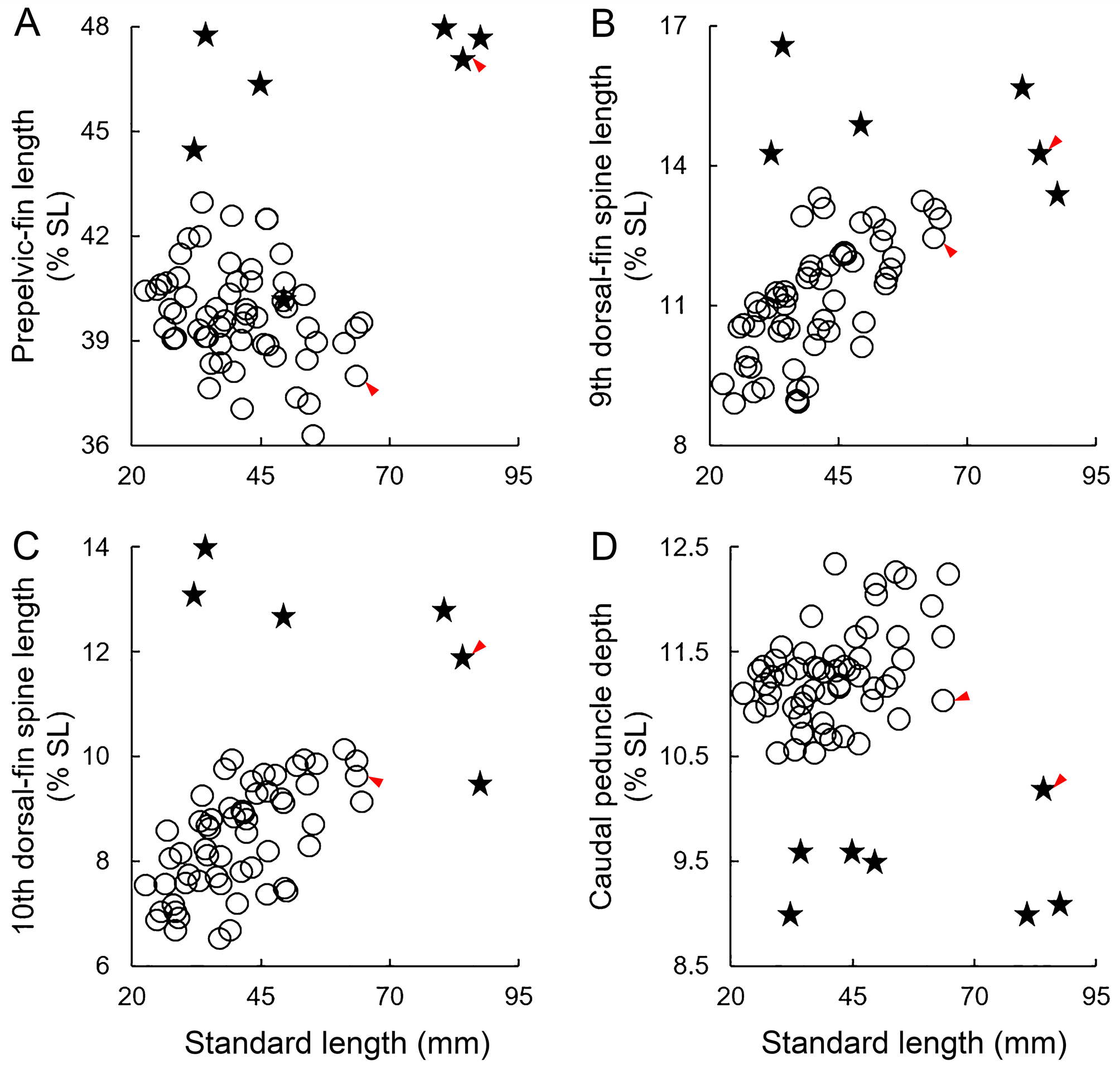
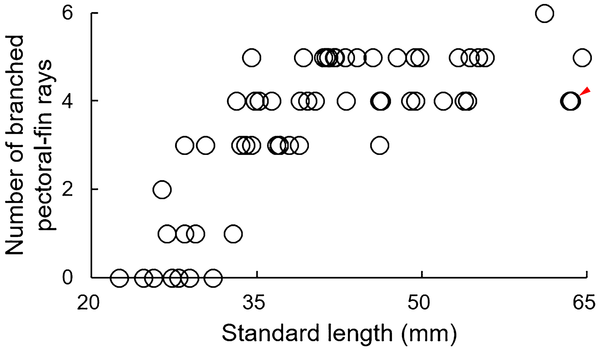
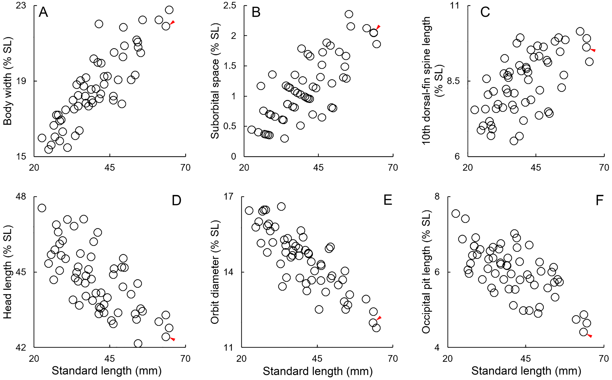
TABLE 2. Frequency distributions of numbers of pectoral-fin rays, scale rows below lateral line, and total gill rakers in S. regina sp. nov., S. bulacephala and S. colorata.
| n | Pectoral-fin rays (left side of body) | |||||||||||
|---|---|---|---|---|---|---|---|---|---|---|---|---|
| 13 | 14 | 15 | 16 | 17 | 18 | |||||||
| S. regina sp. nov. | 59 | 1 | 5 | 50 H | 3 | |||||||
| S. bulacephala | 9 | 8 | 1 | |||||||||
| S. colorata | 13 | 1 | 11 | 1 | ||||||||
| n | Scale rows below lateral line | |||||||||||
| 11 | 12 | 13 | 14 | 15 | 16 | |||||||
| S. regina sp. nov. | 52 | 11 | 27 | 12 H | 2 | |||||||
| S. bulacephala | 6 | 2 | 1 | 1 | 2 | |||||||
| S. colorata | 8 | 4 | 3 | 1* | ||||||||
| n | Total gill rakers | |||||||||||
| 13 | 14 | 15 | 16 | 17 | 18 | 19 | 20 | |||||
| S. regina sp. nov. | 59 | 1 | 31 H | 23 | 3 | 1 | ||||||
| S. bulacephala | 7 | 3 | 1 | 1 | 2 | |||||||
| S. colorata | 13 | 3 | 4 | 5 | 1 | |||||||
| QM |
Queensland Museum |
| CSIRO |
Australian National Fish Collection |
No known copyright restrictions apply. See Agosti, D., Egloff, W., 2009. Taxonomic information exchange and copyright: the Plazi approach. BMC Research Notes 2009, 2:53 for further explanation.
|
Kingdom |
|
|
Phylum |
|
|
Class |
|
|
Order |
|
|
Family |
|
|
Genus |