
Pungalina waldockae, Richardson, Barry J., 2016
|
publication ID |
https://doi.org/ 10.11646/zootaxa.4114.5.1 |
|
publication LSID |
lsid:zoobank.org:pub:8F950473-E021-4704-9DA7-9AA9A259C5C3 |
|
DOI |
https://doi.org/10.5281/zenodo.5694081 |
|
persistent identifier |
https://treatment.plazi.org/id/03E487E9-FFD0-E60E-FF59-8FAEE497FD74 |
|
treatment provided by |
Plazi |
|
scientific name |
Pungalina waldockae |
| status |
sp. nov. |
Pungalina waldockae View in CoL sp. nov.
Figs 134–149 View FIGURES 134 – 141 View FIGURES 142 – 149
Type material. Holotype: 1F, Goldfields Survey, W.A. 122.32°E, 31.8°S, Aug. 1980, W.F. Humphries ( WAM T58738); Paratypes: WESTERN AUSTRALIA: 1M, Parmelia, 115.82°E, 32.25°S, Mar. 1991, A.E. de Jong ( WAM T58740); 1M, Elashgin Nature Reserve, 117.43°E, 31.33°S, 7 Sep. 1999, J.M. Waldock & I. Studley ( WAM T58742); 1F, 1imm., Buningonia Spring, 123.53°E, 31.45°S, W.F. Humphries ( WAM T58739); 1M, Yangebup, 115.82°E, 32.12°S, 11 Apr. 1985, D. Mead-Hunter ( WAM T58758); 1M, Parmelia, 115.82°E, 32.25°S, 18 Dec. 1990, A.E. de Jong ( WAM T58741); 1M, Australind, 115.70°E, 33.30°S, 1982, B. Baehr (QM S96212 View Materials ).
Etymology. The name is in honour of Ms J. Waldock, a fellow student of the Australian Salticidae , who has provided patient, sage advice and arranged many loans of specimens from the collections of the WAM.
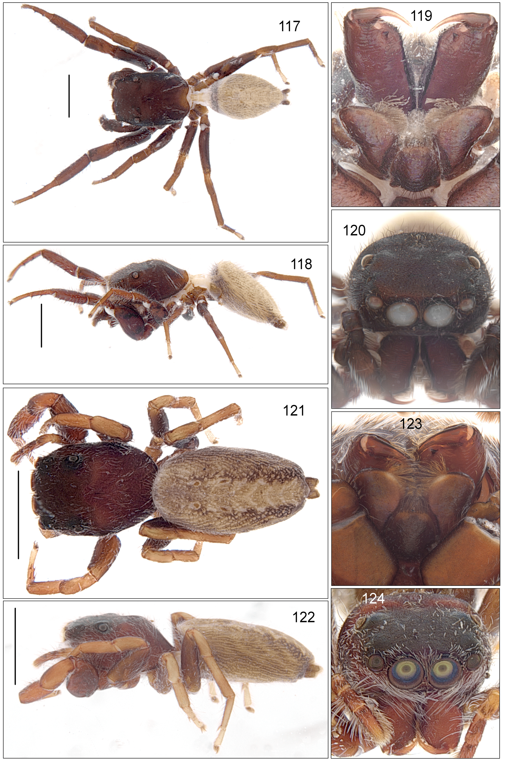
Diagnosis. Externally ( Figs 134, 135 View FIGURES 134 – 141 ), very similar to P. semiferruginea (L. Koch) . The abdomen in the female is light brown with a distinctive brown pattern, different to that in P. semiferruginea . In P. waldockae the abdomen in the male is black with four pairs of white dots; in P. semiferruginea ( Figs 117, 118 View FIGURES 117 – 124 ) the abdomen is brown with a pattern similar to that in the female. The epigyne ( Figs 145–148 View FIGURES 142 – 149 ) includes a pair of large, atria with, sclerotised margins forming a smooth curve, unlike the uneven edge seen in P. semiferruginea ( Fig. 131 View FIGURES 125 – 133 ). There is a large median pocket in the epigastic fold in P. semiferruginea , unlike the smaller pocket seen in P. waldockae . The copulatory openings are smaller in P. waldockae and only open into the posterior edge of the atrium. In P. semiferruginea they are broad and extend partially along the median guide. There is only a small pocket in the epigastic fold in P. waldockae . The palps differ markedly. The embolus ( Figs 142–145 View FIGURES 142 – 149 ) is strongly built in both species; however, it consists of a straight narrow distal half in P. semiferruginea arising from a broad base and with heavily sclerotised bumps along the posterior edge ( Fig. 129 View FIGURES 125 – 133 ) In P. waldockae the embolus is thick throughout its length with a bifurcate distal, clock-wise curved, end.
Description. Male: Paratype: Cephalothorax ( Figs 134, 135 View FIGURES 134 – 141 ) rounded with a large bulge anterior to the PLE, very dark brown to black, covered with scattered pennate white hairs. Series of striae made of white hairs on the posterior face of the cephalothorax. Clypeus black or very dark brown, narrow, without a fringe. Chelicerae ( Figs 136, 137 View FIGURES 134 – 141 ) dark brown, straight. Two promarginal teeth and one large straight unident retromarginal tooth placed close to the retromargin. Endites and labium dark brown grading to mid-brown. Sternum dark brown. Abdomen ovate. Dorsal abdomen mid-brown with a dense pattern of black markings. Thick fringe of white hairs around the anterior and lateral margins. Four pairs of white spots along the length of the abdomen ( Fig. 134 View FIGURES 134 – 141 ). Spinnerets dark brown. Ventral abdomen similar colours to dorsal abdomen with two longitudinal lines of lighter coloured dots. All legs dark brown or black, with sparse white fringe on part of the femur and patella. L1 robust, L2, L3 and L4, grading to lighter build. Palp ( Figs 142–145 View FIGURES 142 – 149 ): dark brown to black, tibial apophysis very small, broad and rectangular. Tegulum dark brown, relatively broad with a large ventral bulge, a small proximal lobe and a medium sized lateral lobe on the posterior edge. Embolus thick, very heavily built, arising on the posterior edge of the tegulum and curving in a quarter-circle clock-wise curve. The distal end, blunt and strongly bifurcate. Proximal arm heavier and longer than the distal arm. Dimensions: General: CL 4.02, EFL 1.36, CW 3.10, AEW 2.04, AMEW 1.36, PEW 2.29, AL 3.53, P1+T1 3.84. L1 8.98 (2.72 + 1.80 + 2.04 + 1.55 + 0.87), L2 6.93 (2.11 + 1.42 + 1.36 + 1.24 + 0.80), L3 6.56 (2.23 + 1.18 + 1.11 + 1.30 + 0.74), L4 7.86 (2.60 + 1.24 + 1.73 + 1.49 + 0.80).
Female: Holotype: Cephalothorax ( Figs 138, 139 View FIGURES 134 – 141 ) rounded with a distinct bulge posterior to the PLE, very dark brown to black, covered with scattered pennate white hairs. Series of striae made of white hairs on the posterior face of the cephalothorax. Clypeus ( Figs 140, 141 View FIGURES 134 – 141 ) black or very dark brown, narrow, with a fringe of grey hairs. Chelicerae dark brown, straight. Two promarginal teeth and one large straight unident retromarginal tooth placed on or very close to the retromargin. Endites and labium dark brown grading to mid-brown. Sternum dark brown. Abdomen ovate. Dorsal abdomen mid-brown with a dense pattern of darker brown markings. Fringe of lighter coloured hairs around the anterior margin. Four pairs of lighter spots along the length of the abdomen ( Fig. 138 View FIGURES 134 – 141 ). Spinnerets dark brown. Ventral abdomen similar colours to dorsal abdomen with two longitudinal lines of lighter coloured dots. All legs dark brown or black proximally grading to mid brown distally. L1 not as robust as in the male, L2, L3 and L4, grading to a lighter build. Epigyne ( Figs 146–148 View FIGURES 142 – 149 ): consisting of a pair of oval atria with sclerotised margins. Narrow copulatory openings placed parallel to the median line just inside the heavily sclerotised posterior margin of the atria. Insemination ducts pass laterally, alongside the proximal edges of the atria before passing posteriorly and entering the spermathecae in the middle of the distal surface. The spermathecae are placed laterally, posterior to the lateral edges of the atria. Fertilization ducts medium sized and placed on the median edge of the spermathecae. Spermathecae placed very close to the epigastric fold, which is sclerotised and with a median pocket. Dimensions: CL 2.54, EFL 1.11, CW 1.98, AEW 1.73, AMEW 1.11, PEW 1.73, AL 4.58, P1+T1 1.86: L1 4.46 (1.42 + 0.93 + 0.99 + 0.62 + 0.50), L2 3.90 1.30 + 0.80 + 0.74 + 0.62 + 0.43), L3 3.96 (1.36 + 0.68 + 0.68 + 0.74 + 0.50), L4 5.33 (1.73 + 0.80 + 1.18 + 1.05 + 0.56).
Distribution and biology. Found across southern Western Australia in a range of habitats ( Fig. 149 View FIGURES 142 – 149 ). As a consequence, likely IUCN Red List Category LC. Collected in litter in woodlands and forests.


| WAM |
Western Australian Museum |
No known copyright restrictions apply. See Agosti, D., Egloff, W., 2009. Taxonomic information exchange and copyright: the Plazi approach. BMC Research Notes 2009, 2:53 for further explanation.