
Pulvinaria photiniae Kuwana, 1914
|
publication ID |
https://doi.org/ 10.11646/zootaxa.5071.1.3 |
|
publication LSID |
lsid:zoobank.org:pub:8ABB2F10-6E91-402F-A3BA-69156A33B77D |
|
DOI |
https://doi.org/10.5281/zenodo.5725419 |
|
persistent identifier |
https://treatment.plazi.org/id/03B187AB-A837-FFCC-FF6F-0C69FE71D362 |
|
treatment provided by |
Plazi |
|
scientific name |
Pulvinaria photiniae Kuwana, 1914 |
| status |
|
Pulvinaria photiniae Kuwana, 1914 View in CoL
( Fig. 11 View FIGURE 11 )
[Japanese name: Ushikoroshi-wata-kaigaramushi]
Pulvinaria photiniae Kuwana 1914: 6 View in CoL ; Takahashi, 1956: 29; Kawai 1972: 17; Kawai 1980: 154; Ben-Dov 1993: 275; Choi & Lee 2017: 98
Eupulvinaria photiniae ( Kuwana, 1914) View in CoL ; Borchsenius 1953: 288
Material examined. Lectotype (here designated): Pulvinaria / Photiniae / Kuw. / Pholinia / villosa / ウシコ¤ シ[Ushikoroshi: Pourthiaea villosa ] / = エノキ[Enoki: Celtis sinensis ] / Type / 5/1912, 1 adult female on a slide together with 1 paralectotype; the lectotype is the individual on left ( OMNH); Paralectotype (here designated): same data as Lectotype ( OMNH).
Other material. JAPAN: Tokyo, Nerima-ku, Syakujii Park , on Viburnum dilatatum , 16.v.1972, coll. S. Kawai , 2 adult females mounted on a slide (1 KTUA); Chiba Prefecture, Matsudo-shi, Matsudo , on Celtis sinensis , 24.iv.2001, coll. H. Tanaka, 8 adult females mounted singly (4 ELKU, 4 EUMJ); Chiba Prefecture, Matsudo-shi, Matsudo, on Celtis sinensis , 1.v.2003, coll. H. Tanaka, 3 adult females mounted singly (2 ELKU, 1 EUMJ) .
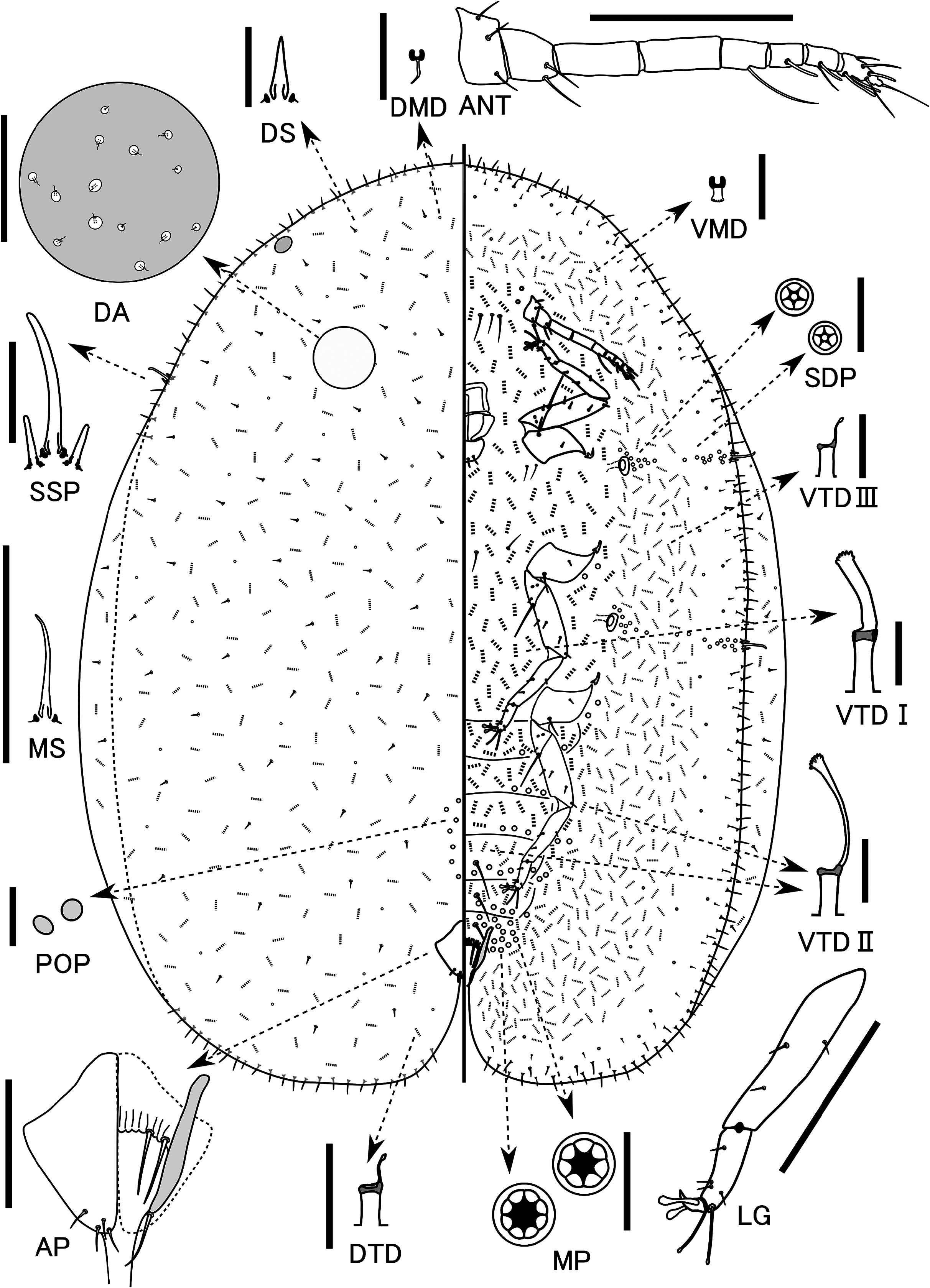
Redescription. Live appearance: Body of adult female elongate oval, usually moderately convex; dorsum dark brown with a yellow longitudinal stripe on midline and a considerable amount of powdery wax before oviposition; ovisac short, about 1–2 times as long as body; posterior part of body uplifted strongly by ovisac.
Slide-mounted adult female (n=15). Note: The Lectotype specimen is not in good condition, so for some characters it was not possible to provide measurements. Body elongate oval, 4.0 (2.5–4.1) mm long, 3.5 (1.3–3.5) mm wide, stigmatic cleft discernible but shallow; anal cleft approximately 1/5–1/7 of body length.
Dorsum. Derm membranous, dermal areolations well developed. Dorsal setae spiniform, frequent, scattered throughout dorsum, each [not visible in Lectotype] (5–10) µm long, with a well-developed basal socket. Preopercular pores each oval to circular, [not visible in Lectotype] (2–3) µm in diameter, barely sclerotised and often difficult to see and count, with (4–29) anterior to anal plates. Tubular ducts and microducts frequent throughout, each one situated within an areolation ( Fig. 11 View FIGURE 11 DA); tubular ducts significantly more numerous than microducts, ratio of numbers of tubular ducts to that of microducts about 2.3 (1.5–4.0): 1. Dorsal submarginal tubercles absent. Anal plates together quadrate; each plate 95–103 (95–150) µm long, 70–75 (50–97) µm wide, with a well-developed supporting bar, a slightly convex posterolateral margin and 4 apical setae. Ano-genital fold with [not visible in Lectotype] (0–3) pairs of setae along anterior margin and (1 or 2) pairs laterally. Anal ring bearing 6 (5 or 6, mostly 6) setae. Eyespots present in marginal area.
Margin. Marginal setae each with a well-developed basal socket and usually apex simple pointed, rarely apex branched; each seta 25–65 (14–65) µm long, each side with [cannot be counted in Lectotype] (10–20) setae between anterior and posterior stigmatic clefts. Stigmatic clefts shallow, each with 3 (0–3, mostly 3) stigmatic spines, median spine 75 (29–92) µm long, approximately 2–4 times longer than a lateral spine.
Venter. Derm membranous. Multilocular pores each 6–8 (6–9) µm wide, with 7–9 (5–12, mostly 8 or 9) loculi, present around genital opening and on medial areas of preceding 4 or 5 abdominal segments; a small group also present lateral to each meso- and metacoxa and occasionally lateral to each procoxa. Spiracular disc pores each 4–6 µm wide, mostly each with 5 loculi, present in bands 1–5 pores wide between margin and each spiracle; anterior bands each containing [cannot be counted in Lectotype] (22–56) pores, posterior bands each with [cannot be counted in Lectotype] (27–70) pores. Microducts scattered throughout venter. Tubular ducts of 3 types: type I each with large outer ductule (about 2–5 µm wide and 6–14 µm long), a stout inner ductule (about 2–5 µm wide and 10–18 µm long) and a well-developed flower-shaped terminal gland, present in posterior medial area of head, medial areas of all thoracic and anterior abdominal segments, also in inner submarginal areas of head, thoracic and anterior abdominal segments; type II tubular ducts each with a rather small outer ductule (1–4 µm wide and 6–14 µm long) ending in a shallow cup-shaped invagination leading to a narrower inner ductule (<1–2 µm wide and 10–20 µm long) with a well-developed terminal gland, mostly occurring in medial areas of posterior abdominal segments and inner submarginal areas of head, thoracic and abdominal segments; type III ducts similar to type II, but each with a short, filamentous inner ductule (<1 µm wide and 1–5 µm long) and a minute terminal gland, present intermixed with type I and type II ducts in inner submarginal areas of head, thorax and abdomen and in a broad submarginal band on head, thorax, and abdomen, forming an complete submarginal ring. Posteriormost 3 abdominal segments each with 1 pair of long ventral setae present on medial areas. With 2 or 3 (2–5) pairs of long setae between antennae, 1 (0–2) pairs of long setae on area mesad of each procoxa and occasionally 0–1 pairs of long setae on medial areas of thoracic and anterior abdominal segments; other setae short and fine, distributed throughout venter. Spiracles normal for the genus; peritreme widths: anterior spiracle 50 (38–66) µm, posterior spiracle 60 (50–80) µm. Legs well developed, each with a completely articulated tibio-tarsal joint and an articulatory sclerosis; claw without a denticle; both claw digitules rather broad and slightly shorter than thin tarsal digitules. Hind trochanter + femur 300–307 (275–350) µm long, hind tibia 205–208 (200–245) µm long, and hind tarsus 100–108 (96–110) µm long. Antennae each 8 (7–8, mostly 8) segmented, 382–393 (357–489) µm long. Labium 63 (55–106) µm long, 125 (91–140) µm wide.
Host plants in Japan. Cannabaceae : Celtis sinensis ( Kawai 1972, Kuwana 1914, Takahashi 1956), Hydrangeaceae : Deutzia crenata ( Kawai 1972, 1980), Rosaceae : Pourthiaea villosa ( Kawai 1972, 1980, Kuwana 1914, as Photonia villosa ), and Viburnaceae : Viburnum dilatatum ( Kawai 1972, 1980).
Remarks. Pulvinaria photiniae is similar to P. kuwacola in having: (i) eyespots located in the marginal area of the dorsum, and (ii) ventral type III tubular ducts forming a complete submarginal ring. However, it differs from P. kuwacola as follows (character states of P. kuwacola in brackets): (i) multilocular pores mostly each with eight or nine loculi (mostly seven loculi in each pore), and (ii) dorsal tubular ducts more numerous than dorsal microducts (dorsal microducts more numerous than dorsal tubular ducts). Pulvinaria photiniae is also similar to P. nipponica in having (i) eyespots located in the marginal area of the dorsum, and (ii) ventral type III tubular ducts forming a complete submarginal ring. However, it differs from P. nipponica in having some preopercular pores ( P. nipponica lacks preopercular pores). The important diagnostic morphological character states for P. photiniae and a comparison with the type species of the genus, P. vitis , are summarised in Table 1 View TABLE 1 . Pulvinaria photiniae can be separated from other Pulvinaria species described in this study and P. vitis by the presence of dorsal tubular ducts, absence of dorsal submarginal tubercles, the condition of dermal areolation, location of eyespots, body shape and size, multilocular pore distribution, type III ventral tubular duct distribution, number of loculi in each multilocular pore, number of preopercular pores, and ratio of number of dorsal tubular ducts: number of dorsal microducts.
The adult female morphology of P. photiniae redescribed here mostly agrees well with the redescription by Takahashi (1956). However, the present description differs slightly from that of Takahashi (1956) as follows (character states of Takahashi’s redescription in parenthesis): (i) marginal setae between anterior stigmatic and posterior stigmatic clefts on each side numbering 10–20 (13–15); (ii) spiracular disc pores between each spiracle and stigmatic cleft numbering 22–70 in each band (approximately 40 in each band); (iii) preopercular pores numbering 4–29 (referred to as dorsal median pores, approximately 30–54); and (iv) marginal setae each 14–65 μm long (34–43 μm). These morphological discrepancies are probably due to intraspecific morphological variation or the quality of the microscopes used.
No known copyright restrictions apply. See Agosti, D., Egloff, W., 2009. Taxonomic information exchange and copyright: the Plazi approach. BMC Research Notes 2009, 2:53 for further explanation.
|
Kingdom |
|
|
Phylum |
|
|
Class |
|
|
Order |
|
|
Family |
|
|
Genus |
Pulvinaria photiniae Kuwana, 1914
| Tanaka, Hirotaka & Kamitani, Satoshi 2021 |
Eupulvinaria photiniae ( Kuwana, 1914 )
| Borchsenius, N. S. 1953: 288 |
Pulvinaria photiniae
| Choi, J. & Lee, S. 2017: 98 |
| Ben-Dov, Y. 1993: 275 |
| Kawai, S. 1980: 154 |
| Kawai, S. 1972: 17 |
| Takahashi, R. 1956: 29 |
| Kuwana, S. I. 1914: 6 |