
Physodeutera (Toxoma) sulcoprothoracica (W. Horn, 1913 )
|
publication ID |
https://doi.org/ 10.11646/zootaxa.5060.2.1 |
|
publication LSID |
lsid:zoobank.org:pub:F6BFFABC-7F88-473C-9517-D031485140DD |
|
DOI |
https://doi.org/10.5281/zenodo.5633551 |
|
persistent identifier |
https://treatment.plazi.org/id/03B47722-112A-FFF7-DCF8-DBF528E9FE07 |
|
treatment provided by |
Plazi |
|
scientific name |
Physodeutera (Toxoma) sulcoprothoracica (W. Horn, 1913 ) |
| status |
|
Physodeutera (Toxoma) sulcoprothoracica (W. Horn, 1913) View in CoL
( Figs 90–106 View FIGURES 90–92 View FIGURES 93–101 View FIGURES 102–105 View FIGURE 106 )
Prothyma (Physodeutera) viridicyanea sulcoprothoracica W. Horn, 1913: 15 View in CoL .
Prothyma (Megalomma) viridicyanea sulcoprothoracica: W. Horn 1934: 14 View in CoL .
Megalomma (Diarrhiza) viridicyanea sulcoprothoracica: Jeannel 1946: 191 .
Physodeutera (Diarrhiza) viridicyanea sulcoprothoracica: Rivalier 1967: 276 View in CoL .
Physodeutera (Toxoma) sulcoprothoracica: Moravec 2000: 212 View in CoL .
Physodeutera (Toxoma) sulcoprothoracica: Moravec 2002a: 120 View in CoL , figs 322, 323–329, 726–727.
Type locality. “Diego Suarez”.
Type material. Lectotype (designated by Moravec 2000), ♀ in SDEI, labelled: “Diego Suarez” [dark green, printed] // “Donckier” [printed] // “peut-être / la vraim / viridicyaneum / Brullé” [handwritten, nearly illegible] // “f. sulcoprotho- / racica / mihi” [blue, handwritten] // “Type! / Dr. W. Horn” [printed] // “ Syntypus ” [red, printed] // “Coll. W. Horn // DEI, Eberswalde” [printed] // “ SDEI Coleoptera / # 301924”. Paralectotype. 1 ♀ in MNHN, labelled: “Diego Suarez” [handwritten]; “voisin viridicyaneum / qui a les élytres entierement punctuis / le labre plus grand / noirâtre / rouge au milieu et aussi bords” [handwritten by Fleutiaux, partly illegible]; “Collection / Fleutiaux” [printed]; “Co-type W. Horn vidit pr. 14 / Collection Fleutiaux” [handwritten (nearly illegible) / printed]; “ Prothyma (Physodeutera) / sulcoprothoracica m. / Dr. W. Horn det. 1914” [handwritten/printed]; “Muséum Paris / Coll. E. Fleutiaux” [printed]; “Cotype / W. Horn” [printed]. The two type specimens are provided with labels: “ Lectotype (or Paralectotype respectively) / Prothyma viridicyanea / sulcoprothoracica / W. Horn, 1914 / design. by J. Moravec, 2000 ” [red, printed], and “ Physodeutera (Toxoma) / sulcoprothoracica / (W. Horn, 1914) stat. nov. / det. Jiří Moravec 2000 ” [printed].
Other material examined. 1 ♂ in CCJM (ex APCA): “ Ambre ”; “ Mt. d’Ambre ” . 1 ♀ in MNHN: “ Diego Suarez” .
Differential diagnosis. Physodeutera (Toxoma) sulcoprothoracica differs from the other species treated above in having a larger and dully cyaneous body, notopleural sutures of pronotal disc distinctly visible from above and distantly from lateral margins of dorsally visible proepisterna, notably distinct, velvety black shadowy zone on elytra with almost effaced punctures within the area and also on posterior declivity ( Figs 90 View FIGURES 90–92 , 102–104 View FIGURES 102–105 ), more voluminous aedeagus with uneven ventral margin ( Figs 100–101 View FIGURES 93–101 ) and different sclerites within the internal sac, particularly the voluminous, uniquely shaped (bird-like) upper dorsal piece with small acute apex, which is well-visible both in left and right lateral aspect ( Figs 100–101 View FIGURES 93–101 ) but has not been observed in the other species. Due to its similar (though somewhat less distinct) velvet-black shadowy zone on the elytral disc, Ph. (T.) sulcoprothoracica may resemble Physodeutera (T.) subtilevelutina (W. Horn, 1934). However, apart from its different body coloration, Ph. (T.) subtilevelutina is immediately distinguished by its elytral maculation consisting of elongate humeral-lateral lunule and sublateral-median macula, as well as by very different shape of its aedeagus (see Moravec 2002a).
The discovery of the male ( Moravec 2000, 2002a) has revealed that the aedeagus and internal sac also differ from those in the subgenus Diarrhiza and that Ph. (T.) sulcoprothoracica is a distinct species of the subgenus Toxoma . Females of these two subgenera can easily be recognized by the shape of the penultimate palpomeres of labial palpi, which are only moderately dilated in Toxoma , whereas those in Diarrhiza possess widely inflated lateral margins.
Redescription. Body large and rather robust ( Fig. 90 View FIGURES 90–92 ), length 9.60–10.8 (female LT 10.75) mm, width 3.00– 3.50 mm.
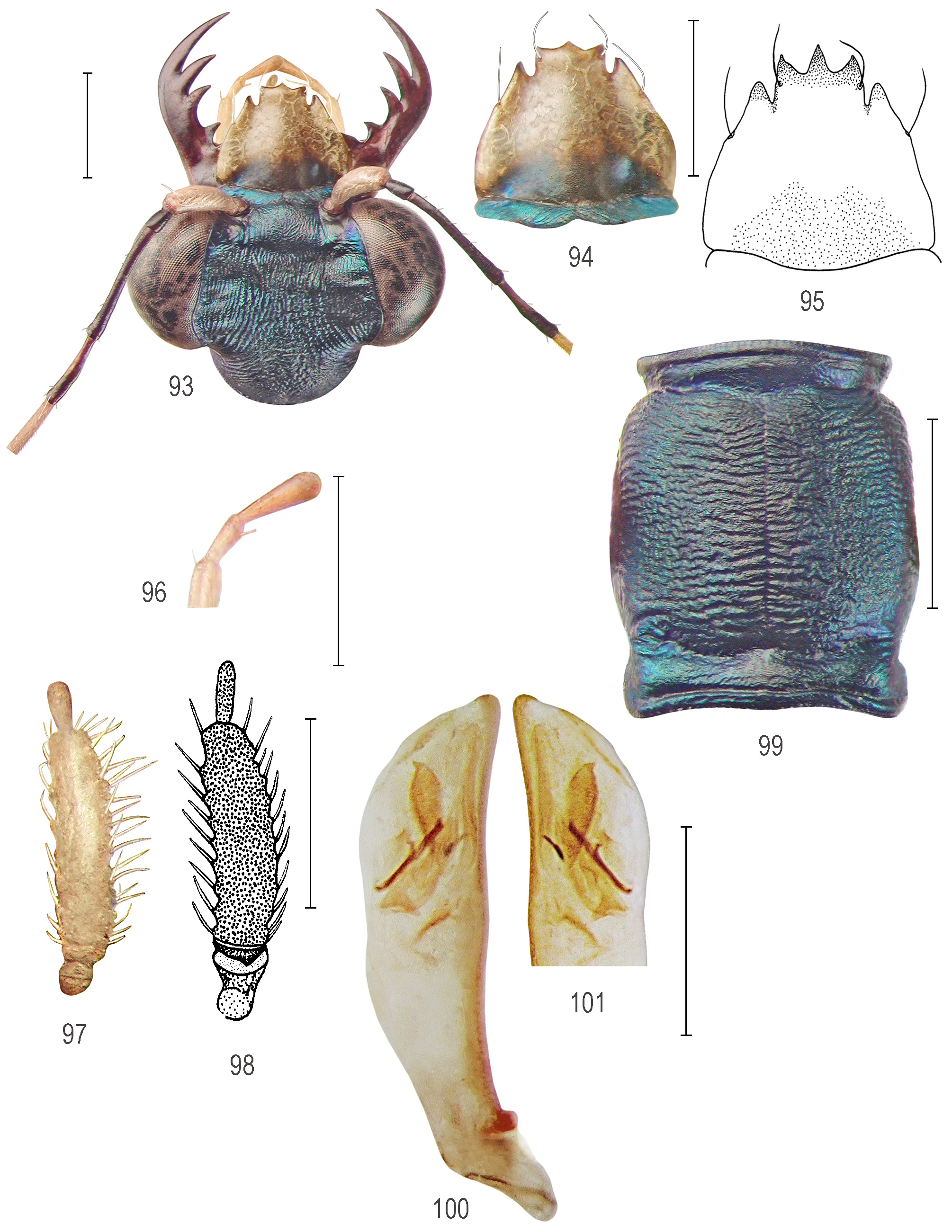
Head ( Fig. 93 View FIGURES 93–101 ) with large eyes, length 3.10–3.30 mm.
Frons dark metallic cyaneous, convex, almost smooth; supraantennal plates indistinct, inconspicuously bordered, merging with surface sculpture; frons-vertex fold blunt, fluently passing into vertex.
Vertex deep cyaneous, with faint violet lustre, with deep and large transverse anterior impression reaching eyes; surface densely parallel-longitudinally striate-rugulose, rugae divergent posteriorly when passing onto temples; large juxtasutural areas with dense parallel rugae interrupted by two shallow yet distinct posterolateral impressions; posteromedian area finely wavy-rugulose; posteromedian and occipital areas finely irregularly rugulose.
Genae deep blue with violaceous reflections, finely wrinkled.
Clypeus black-blue, smooth with few wrinkles in middle.
Labrum with four setae, ivory-testaceous to testaceous with metallic black-blue basomedian area, 1.05 mm long, 1.30 mm wide, basolateral margins arcuate, then conically attenuated towards indistinct lateral indentations and prominent, subacute anterolateral teeth and elongate, anteriad-prolonged tridentate median lobe; male labrum ( Fig. 94 View FIGURES 93–101 ) with anteromedian lobe possessing right-angled but pointed teeth; female labrum ( Fig. 95 View FIGURES 93–101 ) almost as long as wide, length 1.40–1.45 mm, width 1.50 mm, possessing longer anteromedian lobe with acute or three mucronate teeth, median tooth of which slightly longer.
Mandibles ( Fig. 93 View FIGURES 93–101 ) almost symmetrical, normally shaped with four teeth and basal molar, brown with mahogany tinge, inner teeth gradually smaller towards basal molar, fourth tooth distinctly distant from third tooth.
Palpi in male testaceous, only terminal palpomeres brownish-darkened in male ( Fig. 96 View FIGURES 93–101 ), while last two palpomeres of maxillary palpi brown-darkened in female; penultimate palpomeres of labial palpi moderately dilated with subparallel lateral margins ( Figs 97–98 View FIGURES 93–101 ), their ventral (smooth) side ochre-testaceous in male, brownish darkened in female.
Antennae sexually dimorphic in coloration; in male with scape testaceous, antennomeres 2–3 black, 4 black with basal half paler, brownish (brown on right antenna), 5–7 testaceous, gradually darkened, 8–11 brownishsmoky; in female antennomeres 1–4 black-brown (except for paler ventral area of scape and base of antennomeres 3 and 4), antennomeres 5–6 testaceous, remaining ones darkened.
Thorax. Pronotum ( Fig. 99 View FIGURES 93–101 ) moderately elongate, length 2.00– 2.10 mm, width 1.65–1.85 mm, dark cyaneous with faint, deep violet lustre; anterior and posterior sulci well pronounced; anterior lobe narrower than posterior lobe and disc, with distinct anterior rim, surface shallowly irregularly rugulose; disc with lateral margins including dorsally visible proepisternal margins moderately convex, almost subparallel in female, slightly anteriad-attenuated in male, notopleural sutures markedly visible from above, mutually parallel-running or slightly mutually constricted in middle; median line narrow, but distinct; surface of disc densely transversely wavy-rugose; posterior lobe with distinct posterior rim, dorsolateral bulges pulvinate-elevated, almost smooth, fluently passing to irregularly wrinkled median area; proepisterna iridescent blue-green, indistinctly parallel-striate, striae particularly present along notopleural sutures; prosternum, mesosternum and metasternum iridescent blue-green, finely wrinkled (metasternum smooth); mesepisterna and metepisterna shiny blue-green; female mesepisternal coupling sulci indistinct, in form of opened shallow pit in middle.
Elytra ( Figs 102–104 View FIGURES 102–105 ) elongate, 5.90–6.50 mm long; humeral impressions short yet rather deep, basodiscal convexity moderate, discal impression shallow yet large, apical impressions moderate; lateral margins slightly dilated in middle and at arcuate anteapical angles (more distinctly so in female), with sparse and short white setae on juxtaepipleural area, apices rounded in both sexes, indistinctly emarginated towards indistinct blunt sutural spine; elytral punctation with denser and coarser punctures on humeral, discal and lateral areas, occasionally anastomosing in chains, sometimes their elevated anterior margins forming blunt crests (the sculpture appears rasp-liked when observed under different light-angle); posterior half of elytral disc almost smooth as the punctures are almost effaced within black-velvety shadowy zone and on posterior declivity; elytral coloration dark metallic cyaneous with diffusing bronze and violet lustre and velvet-black shadowy zone passing from lateral margins across middle of elytral disc (the intensity of the dark zone changes depending on angles of illumination); elytral maculation consisting of large, white to yellowish humeral macula in male, which is reduced, brownish, barely visible in female.
Abdomen. Abdominal ventrites metallic black-blue with faint bronze lustre, their surface glabrous as in other species.
Legs. Coxae pale brownish with testaceous apices except for nearly testaceous male procoxae, female procoxae metallic brownish-blue; trochanters dirtily testaceous; male pro- and mesofemora distinctly bicoloured, blackbrown dorsally, testaceous or light-brownish ventrally; metafemora black with testaceous base; female legs much darker, femora almost entirely black or black-brown with mahogany tinge.
Aedeagus ( Figs 100–101 View FIGURES 93–101 ) notably stout, length 2.55 mm, width 0.65 mm; dorsally with uneven margin while ventral outline almost straight in middle, apically moderately bent ventrad; apical portion almost fluently passing towards short, regularly rounded apex; structure of internal sac consisting of thin, straight (stick-like) arciform piece while the satellite piece is only rudimental; other sclerites consist of basal and dorsal plates, thin longitudinal ventral-upper piece, and additional, uniquely shaped dorsal-upper tooth that is voluminous in middle (bird-like shaped).
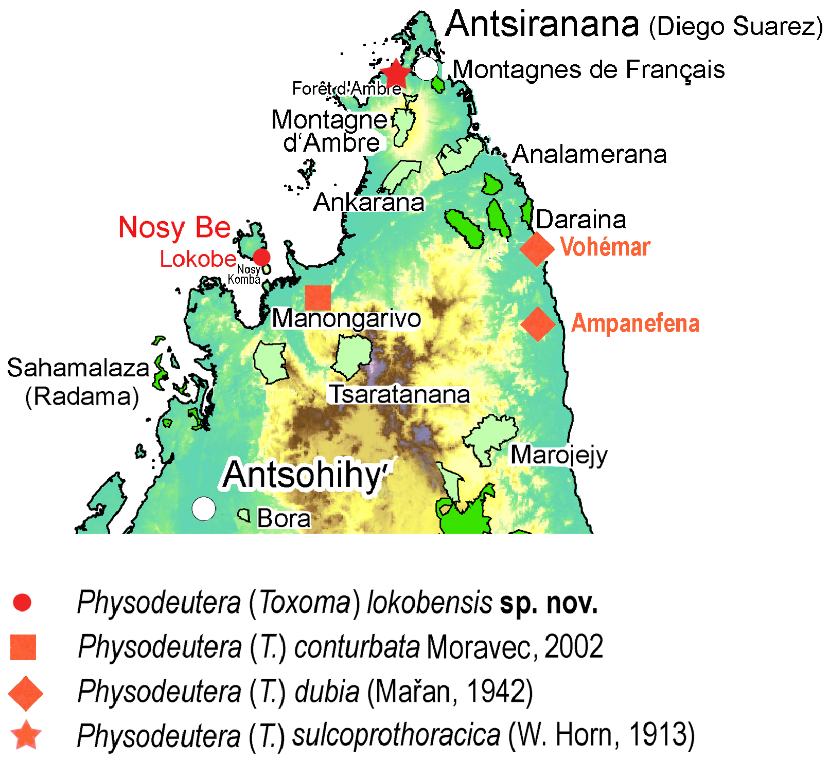
Distribution ( Fig. 106 View FIGURE 106 ). Known only from the type locality in the prefecture of Diego Suarez (northern Madagascar); the only male (CCJM ex APCA) comes from the Montagne d’Ambre of the same area.
Remarks. This species was originally described by Horn (1913) as Prothyma (Physodeutera) viridicyanea sulcoprothoracica (the issuing date was previously interpreted as 1914, also in Moravec 2002a) and this status was consecutively maintained by Jeannel (1946) and Rivalier (1967) (as subspecies of Physodeutera (Diarrhiza) viridicyanea). However, Ph. (T.) sulcoprothoracica only superficially resembles Ph. (D.) viridicyanea and clearly differs from it by numerous characters, which are distinctive of the subgenus Toxoma , especially the moderately dilated but not inflated penultimate palpomeres of the labial palpi, aedeagus with reduced sclerites within the internal sac and the differently shaped and coloured labrum. Also the reduced epimero-proepisternal furrow of the prothorax (laterally separating posterior lobe from proepisterna), which was used as one of the diagnostic characters in the description of “ssp.” sulcoprothoracica by W. Horn (1913), is a diagnostic character of the subgenus Toxoma .
In the original description, only two females from Antsiranana (Diego Suarez), North Madagascar (Montagne d’Ambre) were mentioned, and also Jeannel (1946), who treated it as Megalomma (Diarrhiza) viridicyanea sulcoprothoracica, mentioned only two females from the collection of Fleutiaux (probably the same type females on which Horn (1913) based the description and which are now deposited in SDEI). The only hitherto known male of Ph. (T.) sulcoprothoracica is that in CCJM (ex APCA), first described and illustrated in line drawings by the first author ( Moravec 2002a), accompanied with a colour photograph of its habitus (fig. 726); the colour photographs of the male are presented here. Only the elytron of the female lectotype (SDEI) is illustrated here ( Fig. 103 View FIGURES 102–105 ); for other illustrations of the lectotype see Moravec (2002a).


| MNHN |
Museum National d'Histoire Naturelle |
No known copyright restrictions apply. See Agosti, D., Egloff, W., 2009. Taxonomic information exchange and copyright: the Plazi approach. BMC Research Notes 2009, 2:53 for further explanation.
|
Kingdom |
|
|
Phylum |
|
|
Class |
|
|
Order |
|
|
Family |
|
|
Genus |
Physodeutera (Toxoma) sulcoprothoracica (W. Horn, 1913 )
| Moravec, Jiří & Trýzna, Miloš 2021 |
Physodeutera (Toxoma) sulcoprothoracica: Moravec 2002a: 120
| Moravec, J. 2002: 120 |
Physodeutera (Toxoma) sulcoprothoracica:
| Moravec, J. 2000: 212 |
Physodeutera (Diarrhiza) viridicyanea sulcoprothoracica:
| Rivalier, E. 1967: 276 |
Megalomma (Diarrhiza) viridicyanea sulcoprothoracica:
| Jeannel, R. 1946: 191 |
Prothyma (Megalomma) viridicyanea sulcoprothoracica: W. Horn 1934: 14
| Horn, W. 1934: 14 |
Prothyma (Physodeutera) viridicyanea sulcoprothoracica W. Horn, 1913: 15
| Horn, W. 1913: 15 |