
Phrynus giseae, Joya, 2021
|
publication ID |
https://doi.org/ 10.11646/zootaxa.4948.2.1 |
|
publication LSID |
lsid:zoobank.org:pub:6E073009-5EB5-4C00-89C6-0728E7FA544B |
|
DOI |
https://doi.org/10.5281/zenodo.4651564 |
|
persistent identifier |
https://treatment.plazi.org/id/A66B1A7C-EF43-D336-FF0F-F89A0EE2F8F7 |
|
treatment provided by |
Plazi |
|
scientific name |
Phrynus giseae |
| status |
sp. nov. |
Phrynus giseae View in CoL sp. nov.
Figures. 10–12 View FIGURE 10 View FIGURE 11 View FIGURE 12 , 17–18 View FIGURE 17 View FIGURE 18 . Table. 4, 6.
Holotype (Female): MEXICO, Oaxaca, Municipio San Dionysius (16.427722, -94.796055): 5 km at North-West of San Dionysius , 20 March 2011, G. Montiel, G. Contreras, V. Cordova, D. Barrales, female ( CNAN-T01450 ). GoogleMaps
Paratypes (6 females, 4 males, 2 juveniles): MEXICO. Chiapas: Municipio Arriaga (16.269166, -93.876666): 4 km at north of San Dionysius , 163 masl, 1 September 2005, O. Francke, M. Cordova, A. Jaimes, A. Valdez, H. Montaño, three females, two males, two juveniles ( CNAN-T01451 ) GoogleMaps . Oaxaca: Municipio Nejapa de Madero, Portillo de Nejapa (16.54651, -95.94847): Forest of Pine-oak , 1305 masl, collected during the day, 15 September 2009, A. Valdez, C. Santibañes two females, one male ( CNAN-T01452 ) GoogleMaps . Juchitán (16.583055, -98.672777): North-West , under stones, thorn forest, 3 April 1967, W. Peck, one male, one female ( CASENT 9057691 ) GoogleMaps .
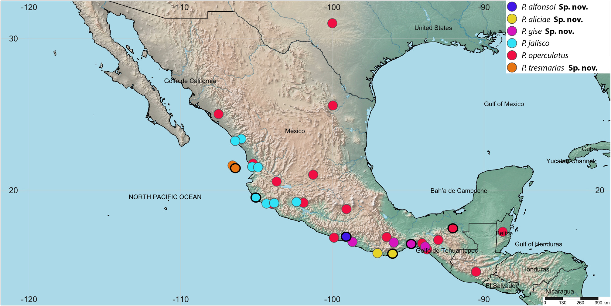
Distribution. MEXICO: States of Chiapas and Oaxaca ( Fig. 18 View FIGURE 18 ).
Etymology. The species is dedicated to Gisella Castilla, whom I affectionately call “Gise”, a name in apposition. Thanks for all these years of supporting and trusting me.
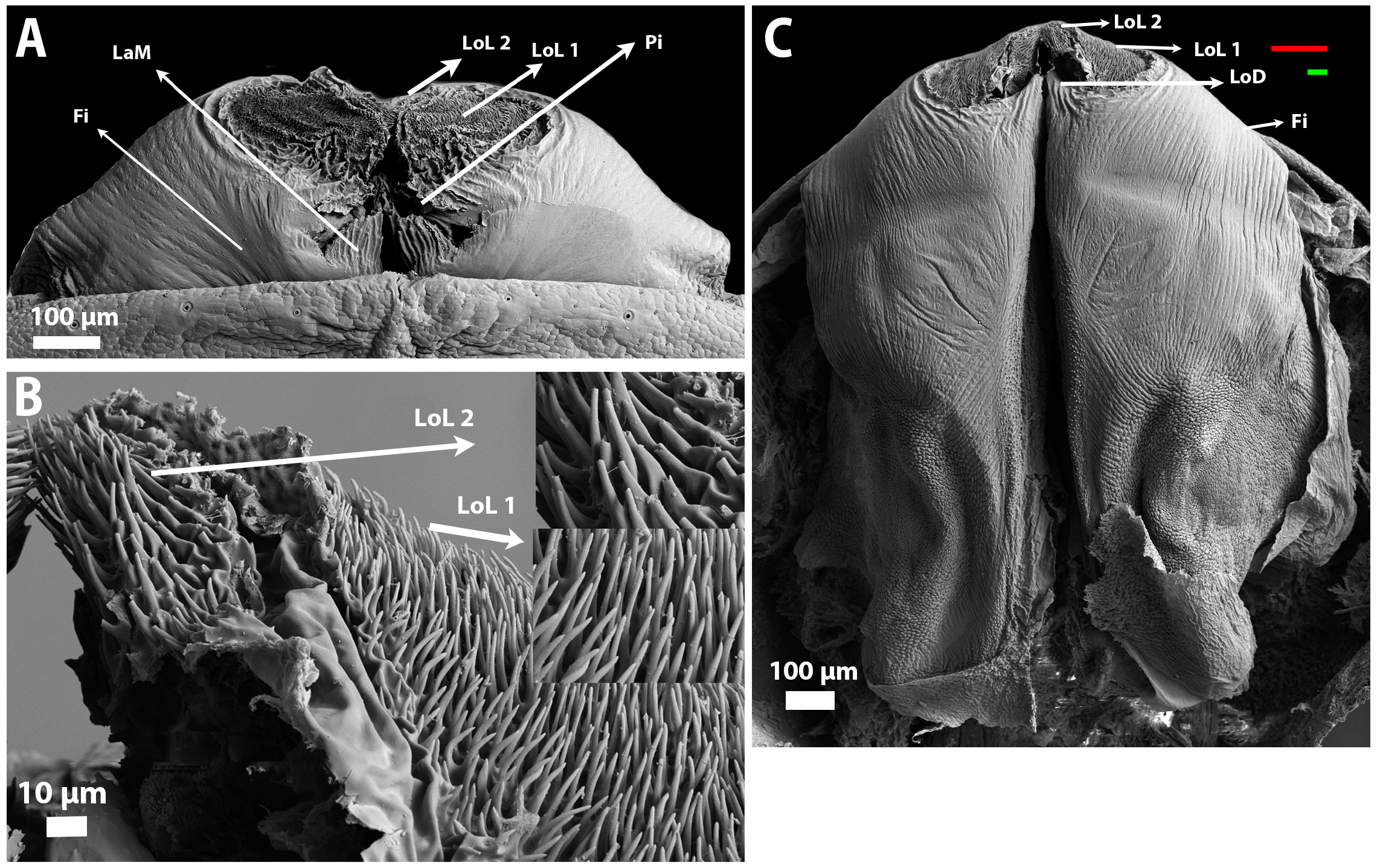
Diagnosis. Phrynus giseae sp. nov. presents one conspicuous tooth in the ectal row of the base of chelicerae, with an acuminated apex ( Fig. 10F View FIGURE 10 ). Without an inconspicuous spine in the dorsomedial area of the tarsus, Pd2 half of the size of Pd4, Fv3 smaller than Fv5, Td1 is small, similar in size to Td4 ( Fig. 11 View FIGURE 11 ). Female genitalia with gonopod bases elliptical and a little elongated; sclerites of gonopods are triangular, with light coloration, and placed in a convergent way ( Fig. 17D View FIGURE 17 ). In the male genitalia, the projections over LoL1 and 2 are conical, short, and thick. The LoD is thin and a few pronounced ( Fig. 12 View FIGURE 12 , Table 6).
Species comparisons: Phrynus giseae sp. nov., is similar to P. operculatus , P. jalisco , and P. aliciae sp. nov., however, the female sclerites of the gonopods in all these species are bigger, more robust, and stick out from gonopod base ( Fig. 17 View FIGURE 17 ). Also, in these three species, the Pd2 has a similar size to Pd4, in P. giseae sp. nov. the Pd2 is evidently smaller ( Fig. 11D View FIGURE 11 ). The ornamentations over the LoL 1and 2 in the male genitalia are different from other species as well as the size of LoD ( Fig. 12B, C View FIGURE 12 ).
In Phrynus giseae sp. nov., the spine Pd1 is small, and Pd1a and Pd1-a are absent or as tubercles ( Fig. 11D View FIGURE 11 ). That is similar in P. tresmarias sp. nov., and small specimens of P. operculatus . Tv1 and Tv3 are subequal in size ( Fig. 11E View FIGURE 11 ) similar to P. jalisco , whereas in P. operculatus , P. aliciae sp. nov. and P. tresmarias sp. nov., Tv1 is larger ( Fig. 2E View FIGURE 2 , 8E View FIGURE 8 , 14E View FIGURE 14 ). Td1 is small ( Fig. 11F View FIGURE 11 ) differing from P. operculatus .
Description. Female (CNAN-T01450): Total length 11.9 mm. Carapace and opisthosoma color brown, pedipalps and legs are brown with red tonalities. Dimensions of prosoma, opisthosoma, pedipalps segments, and all leg femora are provided in Table 4.
Carapace. The frontal margin is straight, without lobes. Carapace presents a group of granules widely spaced; lateral and posterior margins with a lighter border and dorsal area with lines of lighter coloration, posterior margin concave. Lateral and anteromedial eyes clearly visible, ocular tubercle black ( Fig. 10A View FIGURE 10 ).
Sternum. Tri-segmented; all segments poorly sclerotized, the area around segments is sclerotized too. Tritosternum projected anteriorly, elongated, conical, with four setae on the basal region, four on the medial region, two on the distal region, and two more in the apex. Second segment (tetrasternum) oval with two setae on the medial region. Third segment (pentasternum) oval, slightly smaller than the second segment, with two setae on the medial region. Metasternum longitudinally divided, with two setae on the posterior margin of each half ( Fig. 10B View FIGURE 10 ).
Abdomen. Oblong, color dark brown. Presents lines of light coloration on each tergite. The carapace is as wide as the abdomen ( Fig. 10C View FIGURE 10 ).
Chelicera. The mesal row of the basal segment of chelicerae with three teeth, the first is bilobed, placed in the proximal portion, lobe 1b bigger than 1a, followed by one tooth shorter in medial portion, and the third, bigger than the others, placed in the distal region ( Fig. 10D View FIGURE 10 ). The ectal row with one conspicuous tooth (tooth 2) with an acuminated apex, placed in the medial region. There is a small keel on the most proximal region (tooth 1) ( Fig. 10F View FIGURE 10 ). The mobile segment of chelicerae with four teeth, the first and third are the biggest.
Pedipalp. Trochanter: Prolateral face with four spines; spines Tr1 and Tr3 placed in medial region, Tr2 placed near to the ventral margin. Tr3 and Tr4 subequal in length. Spine lengths: Tr2>Tr1>Tr4ŻTr3. Dorsal oblique series of five setiferous tubercles. Dorsomedial area without spines but with one tubercle ( Fig. 10E View FIGURE 10 ). Femur: Ventral face with five major spines, Fv4 and Fv7 are small, and between Fv3–Fv4 there is one small spine Fv3a, between Fv5–Fv6 there are two very small spines, between Fv6–Fv7 there is one small spine, distal to Fv7 there is one small tubercle. Spine lengths: Fv1>Fv2>Fv5>Fv3>Fv6>Fv7>Fv4>Fv3a ( Fig. 11A View FIGURE 11 ). Dorsal face with five conspicuous spines, spine Fd4 is absent; there is a small tubercle between Fd6–Fd7, one tubercle distal to Fd7, Fd1 and Fd2 share the same base, Fd3 separated from Fd2, but close to each other. Spine lengths: Fd2>Fd3>Fd5>Fd6>Fd1>Fd7 ( Fig. 11B View FIGURE 11 ). Patella: Ventral face with five major spines. With spines Pv1-a and Pv1a; there is a small tubercle between Pv5–Pv6. Lengths: Pv2>Pv5>Pv1>Pv4>Pv6>Pv7>Pv3>Pv1-a=Pv1a ( Fig. 11C View FIGURE 11 ). Dorsal face with six major spines, proximal to Pd1 there is a very small tubercle, Pd8 is absent. Spine lengths: Pd5>Pd3>Pd4>Pd2>Pd6>Pd7>Pd1 ( Fig. 11D View FIGURE 11 ). Tibia: Ventral face with three major spines, one small spine between Tv2–Tv3. Spine lengths: Tv2>Tv3>Tv1 ( Fig. 11E View FIGURE 11 ). Dorsal face with two major spines, between Td2–Td3 there is one tubercle, two small spines over Td3, distal to Td3 there are two very small spines, and another a little bigger here named Td4, Td1 has less than a third of the length of Td3. Spine lengths: Td2>Td3>Td1>Td4 ( Fig. 11F View FIGURE 11 ). Tarsus-metatarsus: Internal face with two lines of dorsomedial bristles; suture between the tarsus and metatarsus is not visible. Tarsus without an inconspicuous spine on the proximal end of the dorsomedial surface.
Legs. Femora brown. Femora lengths: I>III>II>IV ( Table 4). Leg I: Tibia with 30 segments and tarsus with 61 segments in the right leg; the left leg is missing. Leg IV: Basitibia with three segments. Basitibia-distitibia lengths: BT1>DT>BT3>BT2. Basitarsus and Telotarsus subequal in size. Tarsus tetramerous.
Female genitalia. Genital operculum pentagonal, it extends to the second segment of the opisthosoma. Gonopod bases elliptical, dorsal surface poorly sclerotized on the medial region, and more sclerotized on the lateral external margins; on the most proximal area, there is a sclerotized line joining both gonopod bases, coloration brown in the most proximal region, and white on the medial area and internal margins, brown coloration on the external margins. Sclerites of the gonopods triangular and placed convergent, the base is more than three times wider than the apical region, apex acuminated, color light brown, darker at the medial region ( Fig. 17D View FIGURE 17 , Table 6).
Variation. Type series measurements and the number of segments in the basitibia IV, Tibia I and tarsus I, are summarized in Table 4. Sternum: Setae over all segments of the sternum are variable in number and position. Pedipalp: Trochanter: The number and size of dorsal tubercles are variable from five to seven. Femur: Ventral face: the presence of the tubercles is variable, in larger specimens the tubercles are more visible and other tubercles can be present, between Fv3 and Fv4 can be present a Fv4a. Dorsal face: the size and presence of tubercles mentioned in the holotype are variable in the paratypes, Fd4 and Fd5a can be present but small. Patella: Ventral face: Tubercles were more conspicuous in larger individuals; other small tubercles can be present between spines. Dorsal face: in some individuals, there is a tubercle anterior to spine Pd1, spine Pd1a is always present but can be small. In larger individuals, the spine Pd8 is present, smaller than the others. Legs. The number of segments of the tibia and tarsus of leg I, was variable among the individuals, the number of tibial segments varies between 29 and 31, and the tarsal segments between 60 and 65. In observed specimens, the number of segments did not depend on size or sex ( Table 4).
Male. Observed males are similar in size to females ( Table 4) spination pattern as in females, genital operculum has the posterior margin oval and covers part of the third segment of the opisthosoma.
Male genitalia. (CNAN-T01451): LaM longer than Pi. LoL 1 bigger than LoL 2, both densely covered with minute projections, in LoL 1, projections are tubular, and a little thick, placed close together, the insertion base is not visible; in LoL 2 projections are similar to LoL 1, the insertion base is also not visible. In dorsal view, the LoD is short, little pronounced, and shorter than LoL1 ( Fig. 12 View FIGURE 12 , Table 6).


No known copyright restrictions apply. See Agosti, D., Egloff, W., 2009. Taxonomic information exchange and copyright: the Plazi approach. BMC Research Notes 2009, 2:53 for further explanation.