
Phaenicocleus schwendingeri, Štys, Pavel & Ř, Petr Ba Ň A, 2009
|
publication ID |
https://doi.org/ 10.5281/zenodo.186281 |
|
DOI |
https://doi.org/10.5281/zenodo.6224270 |
|
persistent identifier |
https://treatment.plazi.org/id/03C26853-FF90-1947-FF55-1C95A46757EC |
|
treatment provided by |
Plazi |
|
scientific name |
Phaenicocleus schwendingeri |
| status |
sp. nov. |
P. schwendingeri View in CoL n. sp.
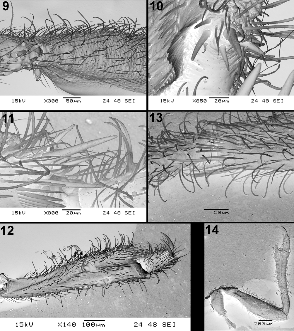
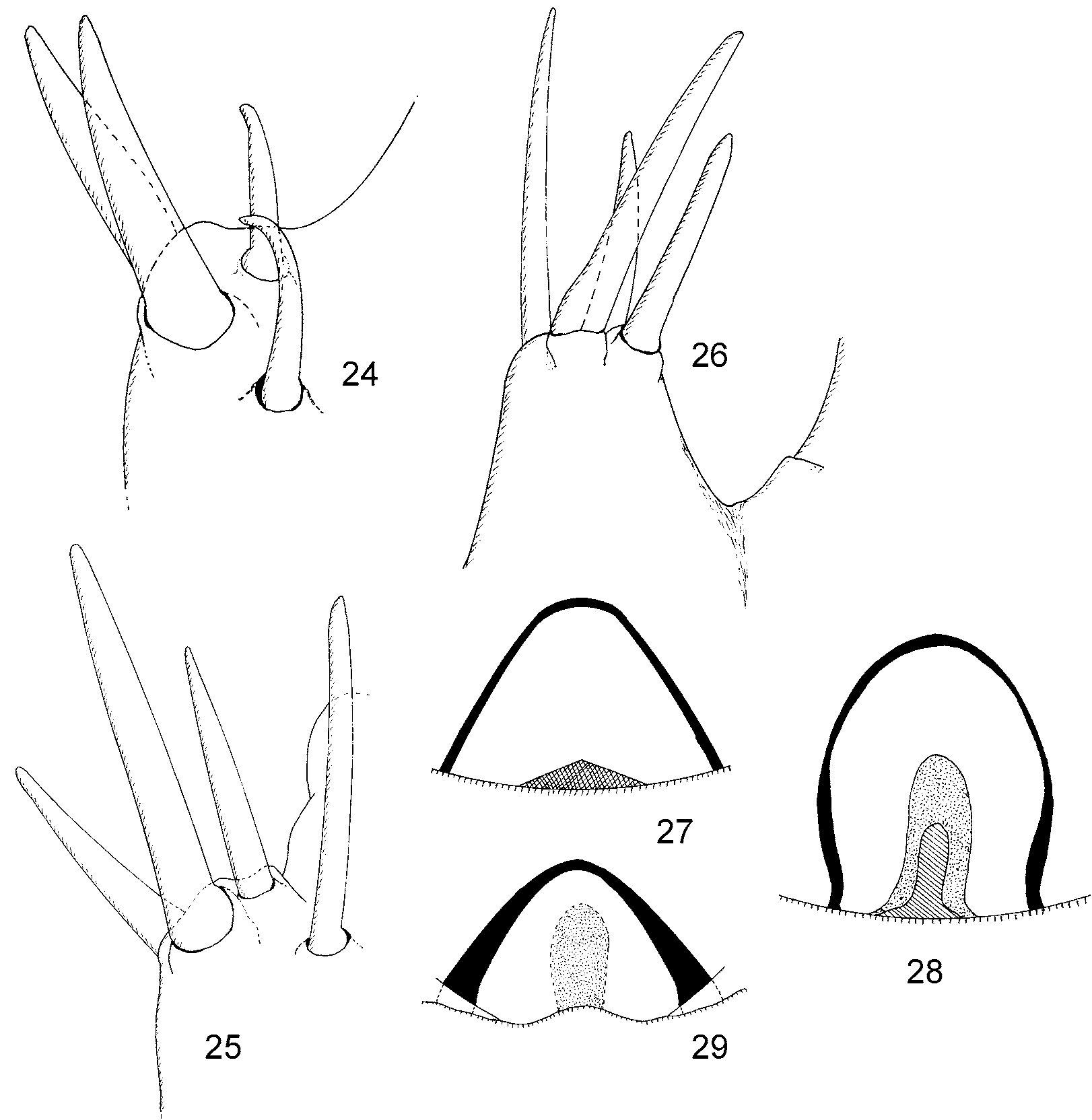
( Figs. 14 View FIGURES 9 – 14. 9 – 13 , 17, 20, 23 View FIGURES 15 – 23 , 26, 29 View FIGURES 24 – 29. 24 – 26 )
Etymology: patronymical adjective; dedicated to Peter Schwendinger (Museum of Natural History, Geneva), our colleague and friend.
Type material. Holotype, ɗ, ‘11a SABAH (West Coast Residency): / Kinabalu Park, Mt Kinabalu, 2600m, / á proximité de Layang-Layang / forêt brumeuse, tamisage de mousses / et de feuilles mortes très humides / 2.v.1987, leg. D.Burckhardt et I.Löbl‘. Preserved in alcohol in a glass tube, together with locality label and red label: ‘ HOLOTYPE / Phenicocleus schwendingeri n. sp. / Štys & Baňař det. 2009’. Specimens is dissected – head with prothorax separate from the rest of body, both forelegs and right hindleg separately. The left wing with an aberrant venation (see below and Discussion) is missing, being unfortunately lost during preparation of a slide mount. Holotype is deposited in the collections of Museum of Natural History, Geneva.
Habitat. Collected in mountain foggy forest on Mt. Kinabalu.
Method of collecting. The specimens was collected by sieving moss and very damp leaf litter.
Total length: 4.95 mm. For other measurements see Table 1.
Distribution of setigerous tubercles on body: head without setigerous tubercles excepting ca 15 small ones on ventral side of the postocular constriction and posterior lobe. Pronotum: dorsum of collum with minute setigerous tubercles all over, venter with 2 lateral groups of 4 large rounded tubercles each; midlobe without tubercles, lateral margins of posterior lobe with minute tubercles.
Lateroventral margin of buccular bridge (ventral view) with one large black tubercle directed into the gulf between the buccular bridge and antennifer.
Distribution of setigerous tubercles on forelegs. Forecoxa on distal half of ventral face with 10 sharp tubercles, foretrochanter on ventral face with 14 such tubercles, situated approximately in a curved doublerow. Basalmost part of ventral face of forefemur with 2 setigerous tubercles only. Foretibia and foretarsus without tubercles.
Coloration. Body (inclusive appendages, forewings and its veins) concolorous, uniformly brown.
Head. Cuticle on dorsum smooth. Maximum width of eye 0.58 times width of dorsal synthlipsis, 1.22 times width of ventral synthlipsis. Posterior lobe transverse, laterally rounded, slightly pear-shaped, widest behind the middle, 1.41 times as wide as long; median indicated by an indistinct shallow concavity without linear impression but with a median line slightly darker than surroundings. Ocelli very large, situated rather mesally, interocellar distance the same as shortest distance ocellus – eye.
Antennal formula (longest segment first) III = IV, II, I.
Labium. Ratio length segment II: III 1.04, II: (III + IV combined) 0.63.
Pronotum. Collum with two not very prominent, transverse bulges, medially separated by a shallow, indistinctly delimited concavity, and not protruding laterad; however, lateral outline of collum strikingly rounded, postcollar constriction strikingly deep. Lateral outline of midlobe rounded, but free in its anterior half only, posterior part being embraced by an anterolateral extension of hindlobe. Posterior margin of midlobe laterally concave, with three convexities discally. Combined lateral outlines of midlobe and hindlobe nearly straight (with a shallow concavity at the site of termination of free part of midlobe, postrolateral angles of strikingly ample hindlobe broadly rounded, its posterior margin only shallowly and broadly obtusangularly excised. Midlobe with a percurent linear impression starting in anterior 1/3 and passing across a sharply delimited small central fossette provided with a central puncture; the impression continues across all the hindlobe, being there in its anterior third doubled ( Fig. 17 View FIGURES 15 – 23 ). Midlobe – width: medial length 2.9; hindlobe - ditto 5.9.
Proepimeral lobes closing the fore acetabula from behind. Prosupracoxale reaching euprosternal unpaired posteromesal rectangular elevation ( Fig. 20 View FIGURES 15 – 23 ). Posterior transverse bars of median eumesosternal apodeme simply arcuate, the neighbouring parts of sternum uniformly convex. Eumetasternum with a median, percurrent, linear ridge, not quite reaching apex of sternum ( Fig. 23 View FIGURES 15 – 23 ).
Foreleg ( Fig. 14 View FIGURES 9 – 14. 9 – 13 ) slender, ventral faces of forecoxa, trochanter, and ventral face, ventral half of forefemur with numerous, lens-like, nonsetigerous tubercles, these sparsely distributed for sharp setigerous tubercles see above. Foretrochanter shorter than forecoxa. Forefemur 4.3 times as long as wide, slender, maximum width in middle of its length. Foretibia slender, more than five times as long as wide, without cuticular tubercles, unusually dorsoventrally curved, slightly S-shaped. Apex of foretibia protruding as a conspicuous process. Apicitibial armature consists of four spiniform setae ( Fig. 26 View FIGURES 24 – 29. 24 – 26 ), two ventral, one anterior subventral and one dorsal. Bristle comb very long, consisting of approximately 45 setae, two dorsalmost and two ventralmost much stouter and longer. Foretarsus subequal to foretibia maximum width, posttarsus formed from two well developed claws, subequal in length. Tarsal armature consists from one short spiniform seta only, typical for genus, but seta longer and thinner as in other species.
Right forewing of the holotype normally developed, left wing with an incomplete vein suggesting presence of an incompletely closed basal cell as well (for its significance see Discussion).
Guide ( Fig. 29 View FIGURES 24 – 29. 24 – 26 ) inversely V-shaped, its arms in posterior view strikingly thick proximally; its internal sclerite long, tongue-shaped, not much distinctly or hardly sclerotized.
Associated enicocephalids in the sample. Undescribed genus and species of Enicocephalinae (to be described later), 1 adult female with shed wings and strongly incrassate hind femora.
Differential diagnosis and comparative notes. See the key and illustrations of diagnostic characters. This is a species easily recognizable by its smooth texture of its cuticle, its sparse distribution of lens-like tubercles on forefemur, its small amount of setigerous tubercles, its relatively short labial segment II, its uniform coloration, its slender forelegs, particularly the femora, and a peculiar architecture of the pronotum (see Discussion).

No known copyright restrictions apply. See Agosti, D., Egloff, W., 2009. Taxonomic information exchange and copyright: the Plazi approach. BMC Research Notes 2009, 2:53 for further explanation.